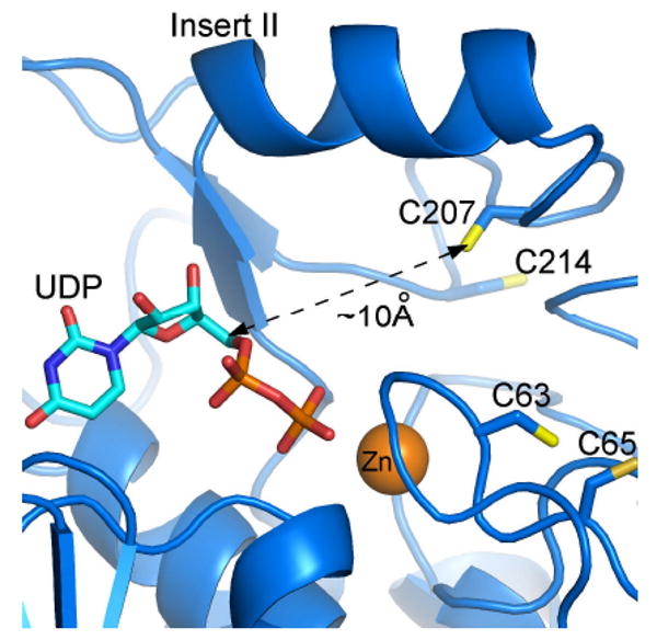
Figure 7.

Potential binding mode of UDP and location of cysteine residues in the E. coli LpxC active site. This model is based upon an A. aeolicus LpxC – UDP complex, as determined by x-ray crystallography (2IER). The zinc ion is shown as an orange sphere. This figure was prepared using PyMOL.
