
Fig. 7.
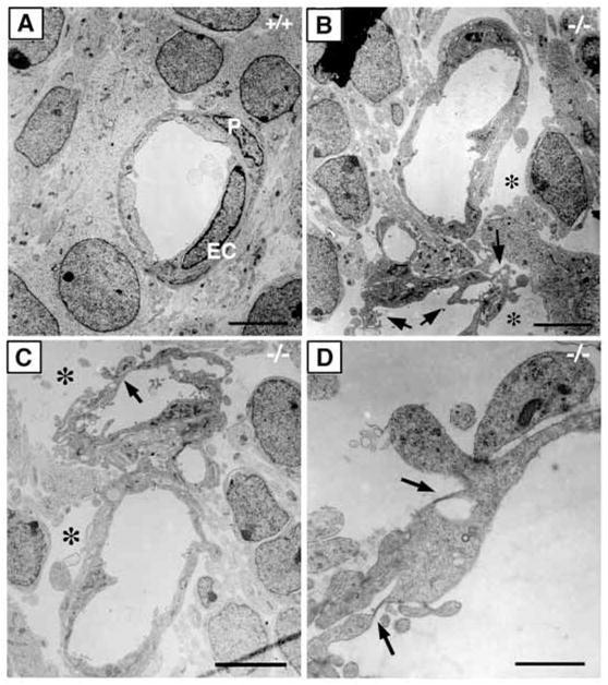
Abnormal endothelial cell morphology in class B integrin β8- deficient brains. (A–D) Electron micrographs of capillary structure in E12.5 wild-type (A) and integrin β8-deficient mutant (B–D) brains. In contrast to the wild type (A), the endothelial cells in the mutant display abundant active membrane protrusions (B, arrows) and large empty spaces surrounding the capillaries (B,C, *). In addition, fenestrations (D, arrows) are often seen in the mutant endothelium. P, pericyte; EC, endothelial cell. Scale bars: 5 μm in A–C; 1 μm in D.