Abstract
Thermal injury produces a profound hypermetabolic and hypercatabolic stress response characterized by increased endogenous glucose production via gluconeogenesis and glycogenolysis, lipolysis, and proteolysis. The liver is the central body organ involved in these metabolic responses. It is suggested that the liver, with its metabolic, inflammatory, immune, and acute phase functions, plays a pivotal role in patient survival and recovery by modulating multiple pathways following thermal injury. Studies have evaluated the role and function of the liver during the postburn response and showed that liver integrity and function are essential for survival, and that hepatic acute phase proteins are strong predictors for postburn survival. This review discusses these studies and delineates the pivotal role of the liver in patients following severe thermal injury.
INTRODUCTION
A thermal injury represents one of the most severe forms of trauma and occurs in over two million people per year in the United States of America (1). According to the World Health Organization (WHO), an estimated 322,000 deaths per year worldwide are related to thermal injury (2). Over 440,000 children receive medical attention for burn injuries each year in the United States (3). With approximately 1,100 children dying of burn-related injuries in the United States every year (4), severe burns represent the third most common cause of death in the pediatric patient population (5), and account for a significant number of hospital admissions in the United States (6,7). A severe burn, therefore, represents a devastating injury affecting nearly every body organ system and leads to significant patient morbidity and mortality (8). Burns produce a profound hypermetabolic stress response characterized by increased glucose production via glycogenolysis and gluconeogenesis, lipolysis, and protein catabolism (7–9). The hypermetabolic stress response is driven by the inflammatory response, which encompasses hormones, cytokines, and acute phase proteins (10–12). Clinical studies have shown that sustained or increased hypermetabolic, inflammatory, and acute phase responses can be life threatening with the uncontrolled and prolonged action of counterregulatory stress hormones (cortisol, catecholamines, glucagon), proinflammatory cytokines (IL-1, IL-6, TNF-α), and acute phase proteins contributing to multiorgan failure, hypermetabolism, hypercatabolism, morbidity, and mortality (11–13).
Over the last two decades, burn research focused on areas such as hypermetabolism, resuscitation, wound healing, pulmonary support, and infection (7). Advances in these areas improved post-burn outcomes, but severe burn is still associated with significant morbidity and mortality. Our group (14–17) proposed that an integral part of the postburn response has not been determined; therefore, we focused our research on the role of the liver. The liver, with its metabolic, inflammatory, immune, and acute phase functions, plays a pivotal role in patient survival and recovery by modulating multiple pathways (13). The role of the liver during the postburn response is essentially unknown and we, therefore, initiated a variety of studies to determine the function and role of the liver during the postburn response. This review aims to discuss the liver and its role during the postburn response.
LIVER ANATOMY, FUNCTION, AND PHYSIOLOGY
Anatomy
The liver constitutes approximately one-fiftieth of total body weight (2%) and weighs approximately 1,500 g in the adult. Its size reflects the complexity of its functions. The American system (lobar anatomy) divides the liver anatomically, based on the distribution of the intrahepatic branches of the hepatic artery, portal vein, and bile ducts. The right lobe is divided into an anterior section and a posterior section, and the left lobe into a medial section and lateral section. The French system divides the liver into VIII segments (Soupault and Couinaud’s segmental system). The afferent blood supply to the liver arises from two sources: the hepatic artery, which carries oxygenated blood and accounts for approximately 25% of the hepatic blood flow; and the portal vein, which drains the splanchnic circulation and accounts for 75% of the hepatic blood flow (18).
Physiology
The liver has a unique spectrum of functions. The liver regulates the amount of energy; stores, distributes, and disposes various nutrients; and synthesizes, transforms, and metabolizes many endogenous substrates and pollutants. The liver consists of five physiologic/anatomic units that are interrelated (18,19).
The circulatory system
A dual blood supply nourishes the liver and acts as a vehicle for material absorbed from the intestinal tract to be utilized in the metabolic pool. Blood vessels are accompanied by lymphatics and nerve fibers that contribute to the regulation of blood flow and intrasinusoidal pressure.
The biliary system
These serve as channels of exit for material secreted by the liver cells, including bilirubin, cholesterol, and detoxified drugs. This system originates with the Golgi apparatus adjacent to the microvilli of the bile canaliculi, and eventually terminates in the common bile duct.
The reticuloendothelial system (RES)
This system has 60% of its cellular elements in the liver and includes the phagocytic, Kupffer cells, and endothelial cells.
The functioning liver cells (hepatocytes)
These cells are capable of a wide variety of activities. The metabolic pool in the liver serves the needs of the entire body. The cells perform both anabolic and catabolic activities, as well as secrete and store metabolites. The large amount of energy required for these transformations result from the conversion of adenosine triphosphate to adenosine diphosphate.
Liver as a hormone producing and secreting unit
Various hormones are synthesized and secreted by the liver, such as insulin-like growth factor-I (IGF-I), insulin-like growth factor binding proteins (IGFBPs), and hepatocyte growth factor (HGF). Further, the liver interacts with various other hormonal systems thus playing a central role in the hormonal axes.
Circulatory System
Most of the body’s metabolic needs are regulated in some way by the liver. The liver expends approximately 20% of the body’s energy and consumes 20% to 25% of the total utilized oxygen, which is due to the remarkable hepatic architecture and the blood supply. The hepatocellular organelles in plasma membranes permit specific functions, and, at the same time, interrelate with an extracellular matrix which facilitates metabolic exchange between blood and hepatocytes (18,19). The liver not only conducts a large number of functions, but also manufactures many substances which serve other organs or tissues. The liver collects such substrates to meet the fuel requirements of other tissues in response to multiple metabolic signals. It is the only organ producing acetoacetate for use by muscle, brain, and kidney, but not itself. The energy-related functions of the liver are regulated by hormones, other agonists, and substrates coming to and from the liver. The liver receives blood from the arterial and portal circulation, processes nutrients, metabolizes toxins and wastes, and stores, transforms, and distributes them to the vascular, biliary, or lymphatic circulations. Mean total hepatic blood flow has been estimated to be 100 to 130 mL/kg/min; 70% to 75% of total hepatic blood flow comes from the portal vein, while the remainder comes from the hepatic artery. There is a reciprocal increase in hepatic arterial blood flow in response to a reduction in the portal flow, but the reverse does not occur (13,18).
To a large extent, portal venous flow into the liver is regulated by extrahepatic factors such as the rate of flow from the intestines and spleen. Food, bile salts, secretin, cholecystokinin, pentagastrin, epinephrine, vasoactive intestinal peptide, and glucagon all increase portal blood flow. The liver serves as a physiologic reservoir of blood with 25% to 30% of its volume composed of blood. During acute blood loss, 300 mL or more can be released into the systemic circulation without adverse effects on liver function. Conversely, in the state of right-sided heart failure, up to 1000 mL of blood can be stored in the liver without affecting its function (18–20).
Biliary System
Bile secretion is an active process that is relatively independent of total liver blood flow, except in conditions of shock. Bile is formed at two sites: the canalicular membrane of the hepatocyte; and the bile ductules or ducts. Total unstimulated bile flow in a 70-kg man has been estimated to be 0.41 to 0.43 mL/min; 80% of the total daily production of bile (approximately 1500 mL) is secreted by hepatocytes and 20% is secreted by the bile duct epithelial cells. The principal organic compounds in bile are the conjugated bile acids, cholesterol, phospholipids, and protein. As bile passes through the biliary ductules or ducts, it is modified by secretion or absorption of epithelial cells. The highest cells in the biliary ductules are called cholangiocytes and have functions and architecture in common with both hepatocytes and ductular cells. The best characterized hormone to stimulate bile secretion is secretin. The bile is then being secreted into the gallbladder, which only functions to concentrate and store bile during fasting. Approximately 90% of the water in gallbladder bile is absorbed in 4 h. Cholecystokinin appears to be the principle physiologic stimulator of gallbladder concentration. Cholinergic stimulation causes contraction of the gallbladder and relaxation of the sphincter of Oddi, forcing the bile into the intestines. Most of the bile salt is absorbed into the enterohepatic circulation. The liver extracts the bile acids and transports them back to the canalicular membrane where they are resecreted back into the biliary system. Total bile pool size in humans is 2 to 5 g and undergoes this circulation 2 to 3 times per meal and 6 to 10 times a day, depending on the dietary habit. In addition, 0.2 to 0.6 g are lost in the stool per day, and this quantity is replaced by newly synthesized bile acids (20).
Bilirubin is a breakdown product of heme and is almost completely excreted in the bile. With hepatocellular disease or extrahepatic biliary obstruction, free bilirubin may accumulate in blood and tissues. Approximately 75% of bilirubin is derived from senescent red blood cells. Bilirubin circulates bound to albumin, which protects tissue from its toxicity. It is rapidly removed from the plasma by the liver through a carrier transport system. In the hepatocyte, bilirubin is conjugated with glucuronide and secreted in bile. Conjugated bilirubin may form a covalent bond with albumin, so called delta bilirubin. In the intestine, bilirubin is reduced by bacteria to mesobilirubin and stercobilirubin, collectively termed urobilinogen. These are both excreted in the stool. A part of urobilinogen is oxidized to urobilin, which is a brown pigment and gives stool its normal color (18–21).
Reticuloendothelial System (RES)
The Kupffer cells of the liver are part of the RES. The RES is part of the immune system and consists of the phagocytic cells located in reticular connective tissue, primarily monocytes and macrophages. The RES is divided into primary and secondary lymphoid organs. The liver is a secondary lymphoid organ. The function of the secondary lymphoid structures is to survey all entering or circulating antigen and to mobilize an immune response against foreign antigen upon its discovery.
METABOLIC SYSTEM
Acute Phase Response
The acute phase response is a cascade of events initiated to prevent tissue damage and to activate repair processes (13,22). The acute phase response is initiated by activated phagocytic cells, fibroblasts, and endothelial cells, which release proinflammatory cytokines leading to the systemic phase of the acute phase response (13,22). The systemic reaction affects: the hypothalamus, which leads to fever, release of steroid hormones by the pituitary-adrenal axis; the liver, which causes the synthesis and secretion of acute phase proteins; the bone marrow, which promotes further hemopoietic responses; and the immune system, which allows the activation of the RES and the stimulation of lymphocytes (13,22). However, a crucial step in this cascade of reactions involves the interaction between the site of injury and the liver, which is the principle organ responsible for producing acute phase proteins and modulating the systemic inflammatory response. The acute phase response usually encompasses positive acute phase proteins, whose expression is increased (C-reactive protein, α2-macroglobulin, haptoglobin, etc.) and negative acute phase proteins, whose expression is decreased (albumin and prealbumin, transferrin, retinol-binding protein, etc.).
Carbohydrate Metabolism
The liver has a central role in energy metabolism. It provides glucose as a readily available source of energy to the central nervous system, red blood cells, and adrenal medulla. During the fed state, results of intestinal carbohydrates digestion (glucose: 80%; galactose and fructose: 20%) are delivered to the liver, with galactose and fructose rapidly converted into glucose. Glucose absorbed by the hepatocyte is converted directly into glycogen for storage—up to a maximum of 65 g of glycogen per kg of liver mass. Excess glucose is converted to fat. Glycogen is also produced by skeletal muscles, but this is not available for use by any other tissues. During the fasting state, glycogen is the primary source of glucose. However, after 48 hours of fasting, liver glycogen is reduced and the body mobilizes fat and proteins to meet the metabolic need. In the muscle, mainly alanine is mobilized which then is converted to glucose by the liver (23,24).
Glycogenesis, glycogenolysis, and the conversion of galactose into glucose all represent hepatic functions, which ensure adequate glucose synthesis and, therefore, hypoglycemia is rare and only associated with extensive hepatic disease. Hyperglycemia, however, is common with severe liver disease due to deficient glycogenesis. Lactate is ordinarily converted to pyruvate and subsequently back into glucose by anaerobic metabolism and only in the liver. This shuttling of glucose and lactate between liver and peripheral tissue is carried out in the Cori cycle. The brain does not participate in the cycle and a continuous source of glucose for the brain must come at the expense of muscle proteins. In liver disease, the metabolism of glucose is often deranged. Frequently, in patients with cirrhosis the portal-systemic shunting causes decreased exposure of portal blood to the hepatocytes, producing an abnormal result of the oral glucose tolerance test. In fulminate hepatic failure, however, there is extensive loss of hepatocyte mass and function, and hypoglycemia supervenes as gluconeogenesis fails (21).
Lipid Metabolism
There are three sources of free fatty acids (FFA) available to the liver: fat absorbed from the gut, fat liberated from adipocytes in response lipolysis, and fatty acids synthesized from carbohydrates and amino acids. These fatty acids are etherified with glycerol to form triglyceride. The export of triglycerides (TG) is dependent on the synthesis of very low density lipoproteins. In cases of an excess supply of fatty acid, there is lipid accumulation in the liver because there is an imbalance of triglyceride relative to very low density lipoproteins. This is seen in obesity, corticosteroid use, pregnancy, diabetes, and total parenteral nutrition. Simple protein malnutrition or protein:calorie imbalance may also result in fatty change of liver, based on decreased export of TG because of limited supply of precursors for hepatic synthesis of lipoproteins (21,25).
Synthesis of both the phospholipid and cholesterol takes place in the liver, and the latter serves as a standard for the determination of lipid metabolism. The liver is the major organ involved in the synthesis, esterification, and excretion of cholesterol. In the presence of parenchymal damage, both the total cholesterol and the percentage of esterified fraction decreased. The biliary obstruction results in the rise in cholesterol, and the most pronounced elevations are noted in the primary biliary cirrhosis and the cholangiolitis accompanying toxic reactions to the phenothiazine derivatives (20,21).
Protein Metabolism
At least 17 of the major plasma proteins are synthesized and secreted by the liver. The liver is the only organ that produces serum albumin and α-globulin, and it synthesizes most of the urea in the body. Production of various serum proteins is an important index of liver function. Albumin is the most abundant serum protein, its synthesis accounting for 11% to 15% of total hepatic synthesis. Synthesis of albumin is influenced by nutrition, thyroxin, insulin, glucagons, cortisol, and cytokines produced during the systemic inflammatory response (16,26).
Hepatic cells are responsible for the synthesis of albumin, fibrinogen, prothrombin, and other factors involved in blood clotting. A reduction of serum albumin is one of the most accurate reflections of liver disease and the effects of medical therapy. Because the half-life of albumin is 21 days, impairment of hepatic synthesis must be present for at least 3 weeks before liver synthetic abnormalities can be reflected in relative decrease in serum albumin concentrations. The correlation between total protein and disease of the liver is not as close as that between the serum albumin level and liver disease, since albumin is produced only by hepatic cells and a reduction is frequently compensated for by the increase in the level of globulin (21).
Vitamin Metabolism
The liver has many important roles of uptake, storage, and mobilization of vitamins. Most important are the fat-soluble vitamins A, E, D, and K. The absorption of these vitamins is dependent on bile salts. The vitamins appear in the thoracic duct 2 to 6 hours after oral administration. Vitamin A is exclusively stored in the liver, and excess ingestion of vitamin A may be associated with significant liver injury. A role for storage of vitamin A in the Ito cells (fat-storing cells) has been suggested. The initial step in vitamin D activation occurs in the liver where vitamin D3 is converted to 25-hydroxycholecalciferol. Vitamin K is essential for the γ-carboxylation of the vitamin K-dependent coagulation factors II, VII, IX, and X. These factors are inactive without γ-carboxylation (see paragraph below) (20,21). A vitamin that has gained attention is vitamin E, because it has been shown to be a very potent antioxidant which could reduce oxidative stress following trauma or thermal injury.
Coagulation
The liver produces multiple coagulation factors which can be altered or become defective in the state of liver disease. In the state of jaundice, vitamin K resorption is decreased, resulting in a decreased synthesis of prothrombin, or, when the liver is severely damaged or in a state of hepatocellular dysfunction, prothrombin is not synthesized at all. The diagnosis of pathological prothrombin synthesis is made by the prothrombin time (PT). PT as well as international normalized ratio (INR) are measures of the extrinsic pathway of coagualtion. Decreases in factors V, VII, IX and fibrinogen also have been noted in hepatic disease (20,21).
Hormonal System
Multiple hormones are either synthesized by, secreted by, or interact with the liver. Angiotensinogen is made and secreted into the bloodstream by the liver. The liver also is actively involved in vitamin D synthesis by hydroxylation of cholecalciferol to 25-hydroxycholecaciferol by the enzyme 25-hydroxylase. IGF-I and IGFBPs, synthesized and secreted in the liver, are important hormones that play a role in the growth and development. The main stimulus for IGF-I production is growth hormone (GH). Lastly, HGF, a major hepatic regenerative growth factor, is synthesized in the liver (27).
HEPATIC CHANGES IN RESPONSE TO A BURN AND THE HEPATIC ACUTE PHASE RESPONSE
Liver Damage and Morphological Changes
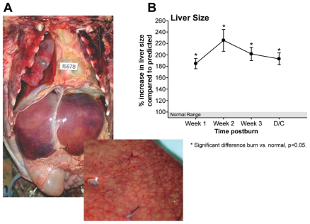
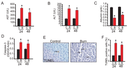
After a thermal injury, a variable degree of liver injury is present and it is usually related to the severity of the thermal injury. By which mechanisms a thermal injury induces fatty changes is not entirely defined and we suggest that fatty changes are associated with hepatic apoptosis (described in detail below). Fatty changes and hepatomegaly (Figure 1), a very common finding are, per se, reversible and their significance depends on the cause and severity of accumulation (28). However, autopsies of burned children who died have shown that fatty liver infiltration was associated with increased bacterial translocation, liver failure, and endotoxemia (29). Immediately after burn, the damage of the liver may be associated with an increased hepatic edema formation. We (15) have shown that the liver weight and liver/bodyweight significantly increased 2 to 7 days after burn when compared with controls. As hepatic protein concentration was significantly decreased in burned rats, we suggest that the liver weight gain is due to increased edema formation rather than increases in the number of hepatocytes or protein levels. An increase in edema formation may lead to cell damage, with the release of the hepatic enzymes. Liver enzymes, such as aspartate aminotransferase (AST) and alanine aminotransferase (ALT) are the most sensitive indicators of hepatocyte injury. Both AST and ALT are normally present in low concentrations. However, with cellular injury or changes in cell membrane permeability, these enzymes leak into circulation. Of the two, the ALT is the more sensitive and specific test for hepatocyte injury as AST also can be elevated in the state of cardiac arrest or muscle injury. Serum glutamate dehydrogenase also is a marker and is elevated in the state of severe hepatic damage. Serum alkaline phosphatase (ALKP) provides an elevation of the patency of the bile channels at all levels, intrahepatic and extrahepatic. Elevation is demonstrated in patients with obstruction of the extrahepatic biliary tract or calculi. In general, serum levels are elevated in hepatobiliary disease. Thermal injury causes liver damage by edema formation, hypoperfusion, proinflammatory cytokines, or other cell death signals with the release of the hepatic enzymes. Serum AST, ALT, and ALKP are elevated between 50% to 200% from baseline when compared with normal levels (Figure 2). Jeschke et al. (16,26) observed that serum AST and ALT peaked during the first day postburn and ALKP during the second day postburn. During hepatic regeneration, all enzymes returned to baseline during acute hospitalization. A limitation of the use of hepatic enzymes is that they could be elevated because of other reasons. For example, ALKP can be elevated in patients with mass bone resorption and in growing children. Therefore, alterations in hepatic enzymes need to be evaluated carefully.
Figure 1.
(A) Massive hepatomegaly (upper Figure 1A) and hepatic fatty infiltration (lower Figure 1A) of a burn victim at autopsy. (B) Liver size increases throughout acute hospitalization by over 200% in 242 surviving burn patients. Reprinted with permission from Jeshcke MG, et al. (2008) Pathophysiologic Response to Severe Burn Injury. Ann Surg. 248:387–401.
Figure 2.
Hepatic dysfunction of the rat burn model mimics the postburn human disease state. (A) Serum aspartate amino transferase (AST) 24 and 48 h after thermal injury. (B) Serum alanine amino transferase (ALT) 24 and 48 h after thermal injury. (C) Serum albumin 24 and 48 h after thermal injury. (D) Caspase-3 activity in liver lysates as determined by successive Western blotting with active caspase-3 and actin antibodies. The data is expressed as a ratio of the two band intensities. (E) TUNEL staining of a liver section before (Control) and 24 h after thermal injury (Burn). (F) Quantified TUNEL positive cells 24 and 48 h after thermal injury. Time after injury in h is indicated. C, control; B, burn. Data presented are mean ± SEM. *P < 0.05 (Burned animals n = 8 and controls n = 4 per time point). *Significant difference between burn and control, P < 0.05. Reprinted with permission from Jeschke MG, et al. (2008) Calcium and ER Stress Mediate Hepatic Apoptosis after Burn Injury. J Cell Mol Med. Accepted for publication. [Epub ahead of print 2009 Jan 14].
Liver damage has been associated with increased hepatocyte cell death (15). In general, cell death occurs by two distinctly different mechanisms: programmed cell death (apoptosis) or necrosis (30). Apoptosis is characterized by cell shrinkage, DNA fragmentation, membrane blebbing, and phagocytosis of the apoptotic cell fragments by neighboring cells or extrusion into the lumen of the bowel without inflammation. This is in contrast to necrosis, which involves cellular swelling, random DNA fragmentation, lysosomal activation, membrane breakdown, and extrusion of cellular contents into the interstitium. Membrane breakdown and cellular content release induced inflammation with the migration of inflammatory cells and release of proinflammatory cytokines and free radicals, which leads to further tissue breakdown. Pathological studies found that 10% to 15% of thermally injured patients show signs of liver necrosis at autopsy (28). The necrosis is generally focal or zonal, central or paracentral, sometimes microfocal, and related to burn shock and sepsis. The morphological differences between apoptosis and necrosis are used to differentiate the two processes.
A cutaneous thermal injury induces liver cell apoptosis associated with caspase activation (see Figure 2) (15). This increase in hepatic programmed cell death is compensated for by an increase in hepatic cell proliferation, suggesting that the liver attempts to maintain homeostasis. Despite the attempt to compensate increased apoptosis by increased hepatocyte proliferation, the liver cannot regain hepatic mass and protein concentration, as we found a significant decrease in hepatic protein concentration in burned rats. It has been shown that a cutaneous burn induces small bowel epithelial cell apoptosis (31,32). In the same study, the authors showed that small bowel epithelial cell proliferation was not increased, leading to a loss of mucosal cells and hence mucosal mass (31,32). Similar findings were demonstrated in the heart (33,34). In burn-induced cardiocyte apoptosis, however, cardiocyte proliferation remained unchanged causing cardiac impairment and dysfunction (33,34).
The mechanisms whereby a cutaneous burn induces programmed cell death in hepatocytes are not defined. Studies suggested that, in general, hypoperfusion and ischemia-reperfusion are associated to promote apoptosis (35–38). After a thermal injury, it has been shown that the blood flow to the bowel decreases by nearly 60% of baseline and stays decreased for approximately 4 hours (31). It can be surmised that the hepatic blood flow also decreases, thus causing programmed cell death. In addition, proinflammatory cytokines such as IL-1 and tumor necrosis factor (TNF)-α have been described to be an apoptotic signal (30,39–42). After a thermal injury, serum and hepatic concentration of proinflammatory cytokines such as IL-1α/β, IL-6, and TNF-α are increased (14,43–45) and we, therefore, suggest that two possible mechanisms are involved in increased hepatocyte apoptosis: decreased splanchnic bloodflow; and elevation of proinflammatory cytokines, initiating intracellular signaling mechanisms. Signals that may be involved encompass many signals that play an important role during the acute phase response.
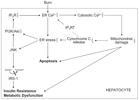
Studies now focus on the identification of which molecular mechanism in a thermal injury leads to hepatocyte apoptosis and dysfunction (Figure 3) (46) and have found that burn injury leads to endoplasmic reticulum (ER) stress and gross alterations in ER calcium with increased cytosolic calcium concentrations. Increased cytosolic calcium induces mitochondrial damage which releases cytochrome c. Cytochrome c binds to the inositol triphosphate receptor (IP3R) augmenting the depletion of ER calcium stores. ER stress/unfolded protein response (UPR) leads to cell apoptosis and activation of Jun-N-terminal kinase (JNK) which phosphorylates the serine IRS-1, blocking phosphorylation of tyrosine IRS-1. ER stress/UPR also impairs the prosurvival PI3K/Akt signaling, resulting in an increased activation of the IP3R increasing ER stress/UPR. This finding is of therapeutic significance because limiting the unfolded protein burden with “chemical chaperones” may promote hepatocyte survival (47). Furthermore, the ongoing development of pharmacologic agents which limit proapoptotic ER stress signaling pathways may have a therapeutic benefit to improve organ function and patient survival (48).
Figure 3.
Schematic of suggested pathways involved in the hepatic response postburn. Thermal injury leads to gross alterations in ER calcium with increased cytosolic calcium concentration. Increased cytosolic calcium induces mitochondrial damage which releases cytochrome c. Cytochrome c increases the existing ER stress/UPR but also binds to the IP3R augmenting the depletion of ER calcium stores. ER stress/UPR leads to cell apoptosis and activation of JNK which phosphorylates the 612 serine of IRS-1, which blocks phosphorylation of tyrosine IRS-1. ER stress/UPR also impairs the prosurvival PI3K/Akt signaling which results in an increased activation of the IP3R increasing ER stress/UPR.
Effects on Biliary System
In trauma and sepsis, intrahepatic cholestasis occurs frequently and appears to be an important pathophysiologic factor, and occurs without demonstrable extrahepatic obstruction. This phenomenon has been described in association with a number of processes, such as hypoxia, drug toxicity, or total parenteral nutrition (49). The mechanisms of intrahepatic cholestasis seem to be associated with an impairment of basolateral and cunicular hepatocyte transport of bile acids and organic anions (50,51). This is most likely due to decreased transporter protein and RNA expression, thus leading to increased bile. Intrahepatic cholestasis, which is one of the prime manifestations of hepatocellular injury, was present in 26% of the cases in a clinical study (28).
Reticuloendothelial System (RES)
Severe burn is associated with a compromised immune system that can lead to infections and sepsis (52–54). Burn has furthermore been shown to depress RES phagocytic activity (55,56). The exact mechanism by which a burn causes RES dysfunction is not known. Trop et al. (55,56) investigated some possible mechanisms by which a burn causes RES dysfunction, and found that hemolysis may play a role in the alterations in RES phagocytic activity. We (15,16,22,43,57), along with others, have shown that the liver also contributes to the inflammatory and acute phase response by expressing proinflammatory cytokines and acute phase proteins, indicating the central role of the liver in the inflammatory, acute phase, and immune response.
METABOLIC SYSTEM
Glucose, Protein, and Lipid Metabolism
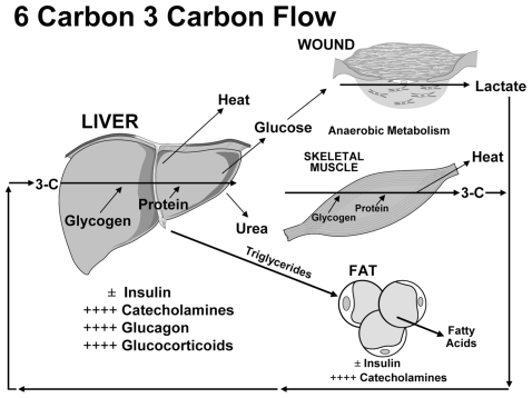
Severe burns covering more than 40% of total body surface area (TBSA) typically are followed by a period of stress, inflammation, and hypermetabolism, characterized by a hyperdynamic circulatory response with increased body temperature, glycolysis, proteolysis, glycogenolysis, gluconeogenesis lipolysis, and futile substrate cycling (58–60). These responses are present in all trauma, surgical, or critically ill patients, but the severity, length, and magnitude is unique for burn patients (7). Marked and sustained increases in catecholamine, glucocorticoid, glucagon, and dopamine secretion are thought to initiate the cascade of events leading to the acute hypermetabolic response with its ensuing catabolic state (7,8,26,61–63) (Figure 4). The cause of this complex response is not well understood. However, interleukin (IL)-1 and IL-6, platelet-activating factor, TNF, endotoxin, neutrophil-adherence complexes, reactive oxygen species, nitric oxide, and coagulation, as well as complement cascades also have been implicated in regulating this response to thermal injury (64). Once these cascades are initiated, their mediators and byproducts appear to stimulate the persistent and increased metabolic rate associated with altered glucose metabolism seen after severe thermal injury (65).
Figure 4.
Metabolic changes postburn with the liver playing an essential role.
Several studies have indicated that these metabolic phenomena postburn occur in a timely manner, suggesting two distinct patterns of metabolic regulation following injury (26,66). The first phase occurs immediately post severe thermal injury and lasts for about 12 to 14 hours and has classically been called the “ebb phase” (66–68). This first phase is characterized by decreases in cardiac output, oxygen consumption, and metabolic rate, as well as by impaired glucose tolerance associated with its hyperglycemic state. These metabolic variables gradually increase within the first 5 days postinjury to a plateau phase (called the “flow” phase), characteristically associated with hyperdynamic circulation and the above-mentioned hypermetabolic state (66). Insulin release, during this time period, was found to be twice that of controls in response to glucose load (26,69), and plasma glucose levels are elevated markedly, indicating the development of an insulin resistance (26,69). Current understanding has been that these metabolic alterations resolve soon after complete wound closure. However, recent studies found that the hypermetabolic response to thermal injury may last for at least 12–36 months after the initial event (16,58,70,71).
Glucose metabolism in healthy subjects is tightly regulated. Under normal circumstances, a postprandial increase in blood glucose concentration stimulates release of insulin from pancreatic β cells. Insulin mediates peripheral glucose uptake into skeletal muscle and adipose tissue and suppresses hepatic gluconeogenesis, thereby maintaining blood glucose homeostasis (72,73). In critical illness, however, metabolic alterations can cause significant changes in energy substrate metabolism. To provide glucose, a major fuel source to vital organs, release of the above-mentioned stress mediators oppose the anabolic actions of insulin (74). By enhancing adipose tissue lipolysis (75) and skeletal muscle proteolysis (76), they increase gluconeogenic substrates, including glycerol, alanine, and lactate, thus augmenting hepatic glucose production in burn patients (72,73,77). Hyperglycemia fails to suppress hepatic glucose release during this time (23), and the suppressive effect of insulin on hepatic glucose release is attenuated, significantly contributing to posttrauma hyperglycemia (78). Catecholamine-mediated enhancement of hepatic glycogenolysis, as well as direct sympathetic stimulation of glycogen breakdown, can further aggravate the hyperglycemia in response to stress (73). Catecholamines also have been shown to impair glucose disposal via alterations of the insulin signaling pathway and GLUT-4 translocation muscle and adipose tissue, resulting in peripheral insulin resistance (72,79). Cree et al. (69) showed an impaired activation of Insulin Receptor Substrate-1 at its tyrosine binding site and an inhibition of Akt in muscle biopsies of children at 7 days postburn. The work of Wolfe et al. (23,75, 78, 80) indicates links between impaired liver and muscle mitochondrial oxidative function, altered rates of lipolysis, and impaired insulin signaling postburn, attenuating both the suppressive actions of insulin on hepatic glucose production and on the stimulation of muscle glucose uptake. Another counterregulatory hormone of interest during stress of the critically ill is glucagon. Glucagon, like epinephrine, leads to increased glucose production through both gluconeogenesis and glycogenolysis (81). The action of glucagon alone is not maintained over time; however, its action on gluconeogenesis is sustained in an additive manner with the presence of epinephrine, cortisol, and GH (74,81). Likewise, epinephrine and glucagon have an additive effect on glycogenolysis (81). Recent studies found that proinflammatory cytokines contribute indirectly to post-burn hyperglycemia via enhancing the release of the above mentioned stress hormones (82–85). Other groups showed that inflammatory cytokines, including TNF, IL-6, and monocyte chemoattractant protein-1 (MCP-1) also act via direct effects on the insulin signal transduction pathway through modification of the signaling properties of insulin receptor substrates, contributing to postburn hyperglycemia via liver and skeletal muscle insulin resistance (86–88). Alterations in metabolic pathways as well as proinflammatory cytokines, such as TNF, also have been implicated in contributing significantly to lean muscle protein breakdown, both during the acute and convalescent phases in response to thermal injury (11,12,89,90). In contrast to starvation, in which lipolysis and ketosis provide energy and protect muscle reserves, thermal injury considerably reduces the ability of the body to utilize fat as an energy source. Counterregulatory hormones such as glucagon, cortisone, and so on, post severe thermal injury, vary in their concentration. While glucagon appears to be decreased, Norbury et al. (63) and Gauglitz et al. (91) have shown recently that cortisone levels are increased markedly up to 3 years post severe thermal injury. The major mechanisms by which thermal injury causes hyperglycemia is probably not due to the hormonal system, but rather is associated on a molecular level with impaired insulin receptor signaling (92).
Skeletal muscle is thus the major source of fuel in the burned patient, which leads to marked wasting of lean body mass (LBM) within days after injury (7,93). This muscle breakdown has been demonstrated with whole body and cross leg nitrogen balance studies in which pronounced negative nitrogen balances persisted for 6 and 9 months after injury (58). Since skeletal muscle has been shown to be responsible for 70% to 80% of whole-body insulin-stimulated glucose uptake, decreases in muscle mass may contribute significantly to this persistent insulin resistance postburn (94). The correlation between hyperglycemia and muscle protein catabolism also has been supported by Flakoll et al. (95) in which an isotopic tracer of leucine was utilized to index whole-body protein flux in normal volunteers. The group showed a significant increase in proteolysis rates occurring without any alteration in either leucine oxidation or non-oxidative disposal (an estimate of protein synthesis), suggesting a hyperglycemia-induced increase in protein breakdown (95). Flakoll et al. (95) further demonstrated that elevations of plasma glucose levels resulted in a marked stimulation of whole body proteolysis during hyperinsulinemia. A 10% to 15% loss in LBM has been shown to be associated with significant increases in infection rate and marked delays in wound healing (96). The resultant muscle weakness was further shown to prolong mechanical ventilatory requirements, inhibit sufficient cough reflexes and delay mobilization in protein-malnourished patients, thus contributing markedly to the incidence of mortality in these patients. Persistent protein catabolism also may account for delay in growth frequently observed in our pediatric patient population for up to 3 years postburn (61,96–99).
Of great interest is the change in fat metabolism, serum triglycerides, and free fatty acids (FFA), all of which are altered significantly throughout almost the entire acute hospital stay. Fat transporter proteins are decreased postburn while triglycerides and FFA are increased, which could explain the fatty infiltration of the liver and other organs postburn. Barret et al. (29) have shown that hepatomegaly with fatty infiltration is associated with increased incidence of sepsis and mortality, implying the importance of organ integrity and function. The occurrence of triglyceride accumulation in the liver has been documented by direct liver biopsy in critically ill patients (100) and in autopsies of patients who died from severe thermal injury (29,101,102). Several investigators (29, 101–103) have shown in clinical studies that the size of the liver may be increased by four-fold or more in burn patients. The rate of gain of liver triglycerides in severely burned patients far exceeds the rate of gain in any other pathological circumstance in which hepatic steatosis develops. The mechanisms leading to hepatic triglyceride accumulation after thermal injury are an excessive delivery of fatty acids to the liver as a consequence of β-adrenergic mediated stimulation of lipolysis (104,105) and a diminished effectiveness of insulin (from carbohydrate intake) in suppressing lipolysis (101). Uptake of fatty acids across the liver was found to be proportionate to delivery in both pigs (106) and human patients (25), so that accelerated delivery of fatty acids due to lipolysis resulted in hepatic uptake at a rate well in excess of the requirement for oxidation. Thus, fatty acids taken up by the liver were channeled to triglyceride synthesis at a markedly accelerated rate in both pigs and humans (105,106). The process of hepatic triglyceride deposition is accelerated when a high proportion of the diet is fat (107). However, substitution of dietary fat with carbohydrate results in the stimulation of de novo fatty acid synthesis, which also contributes to hepatic triglycerides (25). Thus, it appears production of liver triglycerides is a result of the metabolic response to thermal injury, rather than a simple consequence of nutritional support. Nonetheless, accelerated hepatic triglyceride synthesis does not necessarily translate to increased liver triglycerides. In normal individuals, accelerated hepatic triglyceride synthesis is accompanied by increased VLDL-triglyceride secretion, thereby minimizing hepatic accumulation of triglycerides. In contrast, VLDL-triglyceride secretion is low in burn patients and unresponsive to increased hepatic triglyceride synthesis (105). Consequently, it can be predicted that reduction of delivery of FFA to the liver would be the most effective approach to minimizing hepatic triglyceride accumulation. Interestingly, excessive glucose intake and uncontrolled hyperglycemia may also lead to fat deposition in the liver augmenting fatty infiltration of the liver post severe thermal injury (108).
In summary, the liver clearly plays a central role in orchestrating and modulating metabolic processes postburn. The liver is responsible for glucose, fat, and protein metabolism postburn, and alterations in glucose, fat, and protein metabolism have been detrimental outcomes. We, therefore, propose that the liver, with its metabolic function, is one of the essential organs and can determine outcomes of severely burned patients.
Acute Phase Response
After major trauma, such as a severe burn, hepatic protein synthesis shifts from hepatic constitutive proteins, such as albumin, prealbumin, transferrin, and retinol-binding protein to acute phase proteins (13,16,22,26). Previously, acute phase proteins were divided into type I acute phase proteins, such as haptoglobin and α1-acidglycoprotein, mediated by IL-1-like cytokines (IL-1 and TNF), and type II acute phase proteins, such as α2-macroglobulin and fibrinogen, which are mediated by IL-6-like cytokines (IL-6, IL-11) (13). It is unclear whether this division holds true with the discovery of new cytokines and signal transcription factors. Furthermore, it appears that there is a lot of cross-communication between the two responses, indicating that this strict division is not functional, and reflecting the ongoing responses. However, the upregulation of acute phase proteins represents a redirection of the liver to fulfill immune functions, metabolic responses, coagulation, and wound healing processes (13,16,22,26).
In contrast to acute phase proteins, constitutive hepatic proteins are down-regulated (109–112). After a thermal injury, albumin and transferrin decrease by 50% to 70% below normal levels (109–112). Studies have shown that two mechanisms are responsible for the decrease of constitutive hepatic proteins: one is that the liver reprioritizes its protein synthesis from constitutive hepatic proteins to acute phase proteins. This could be shown in many studies in which the mRNA synthesis for constitutive hepatic proteins is decreased. The other mechanism for decreased constitutive hepatic protein concentration is the capillary leakage and the loss of constitutive hepatic proteins into the massive extravascular space and burn wound. Albumin and transferrin, however, have important physiologic functions as they serve as transporter proteins and contribute to osmotic pressure and plasma pH (109–112). Their downregulation after trauma has been described as potentially harmful and the synthesis of these proteins has been used as a predictor of mortality and indicators of recovery (11,12,112–115).
Mediators of the acute phase response are cytokines. In several studies, the biphasic time course of proinflammatory cytokines has been demonstrated. Immediately after burn, IL-1, IL-6, IL-8, and TNF increase two- to ten-fold above normal levels, decrease slightly after approximately 12 hours, increase again, and then start to decrease. Animal and human studies demonstrated that cytokines can either approach normal levels within 2 days after trauma or can be elevated up to 2 weeks after thermal injury (10,16,26,44, 70, 116). The signal transcription cascade includes various pro- and antiinflammatory signal transcription factors such as c-jun/c-fos, nuclear factor-kappa B (NF-κB), CCAAT/enhancer-binding-proteins (C/EBPs), tyrosine phosphorylation and activation of intracellular tyrosine kinases (JAKs), latent cytoplasmic transcription factors, STAT1, STAT3, and STAT5 (signal transducer and activator of transcription), or mitogen-activated protein (57,109,110, 117–121). These signals activate transcription, translation, and expression of acute phase proteins. Particularly IL-6 has been speculated to be the main mediating cytokine. IL-6 activates glycoprotein 130 (gp 130) and the JAK-kinases (JAK-1) leading to activation of STAT 1 and 3 translocating to the nucleus. Intranuclear, the genes for acute phase proteins are turned on. The aim of the acute phase response is to protect the body from further damage, and the aim will be achieved when all elements of the acute phase response coalesce in a balanced fashion. However, a prolonged increase in proinflammatory cytokines and acute phase proteins has been shown to be indicative of a hypercatabolic state, associated with an increased risk of sepsis, multiorgan failure, morbidity and mortality (11,12,112–114).
Vitamin Metabolism
Vitamins play an important role in wound healing, energy, metabolism, inflammation, and antioxidants. Postburn hypermetabolism causes a vitamin deficiency requiring substitution (122–124). There are decreased vitamin A levels in burn patients. This is perhaps due to a reduction of retinol binding protein, which is a vitamin A transporter. Vitamin A has been shown to enhance wound healing, thus its substitution is important for dermal wound repair. Vitamin E encompasses antioxidant properties, and in animal experiments, its administration has been shown to reduce lung injury. After a thermal injury, its concentration is decreased, thus its substitution is suggested. Vitamin D, with its crucial function in the ossear cascade, also is decreased after thermal injury; however, its substitution has not been proven. Thiamin and Riboflavin are vitamins that affect energy and protein metabolism and wound healing. Both factors are decreased after trauma. Thiamin serves as a cofactor in the Krebs cycle and for the oxidation of glucose, thus its need is related to the energy intake. Thiamin is further necessary for the lysyl oxidase function to form collagen. Riboflavin is involved as a coenzyme in oxidation-reduction reactions. Riboflavin is decreased after burn and an increased need for burn victims has been shown (122–124). Folic acid is depleted after a burn and can lead to impaired synthesis of DNA and RNA. Inadequate supplies of vitamin B12 and the indispensable amino acid, methionine, impair folate utilization. Thus, a deficiency of either of these nutrients can produce signs of folate deficiency. Vitamin B complex with B6 and B12 serve as coenzymes in the energy and protein metabolic process. Vitamin B6 is involved in amino acid metabolism, and vitamin B12 in the catabolism of odd-chain-long fatty acids. Both should be substituted in the form of a multivitamin complex, as both are decreased in the postburn hypermetabolic response. Vitamin C plays an important role during the postburn hypermetabolic response. During that period, oxygen-free radicals such as superoxide, peroxide, and hydroxyl may cause increased postburn vascular permeability. Administration of vitamin C, a free radical scavenger, has thus been suggested to be beneficial in terms of reducing microvascular permeability and thus required fluid volume (125).
Coagulation and Clotting Factors
Homeostasis of clotting is complex and has been investigated recently in thermally injured patients (126). Thrombotic and fibrinolytic mechanisms are activated after burn and the extent of activation is increased with the severity of the thermal injury. Most homeostatic markers fell during the early shock phase of burns because of dilutional effects, loss, and degradation of plasma proteins. Clotting factors return to normal levels after the aggressive resuscitation period. Later in the postburn course, thrombogenicity has been suggested to be increased because of a decrease in antithrombin III, protein C, and protein S levels while fibrinolysis activation occurs via increases in tissue plasminogen activation factor, thus leading to an increased risk of thrombosis. The hypercoagulable state places many thermally injured patients at risk for disseminated intravascular coagulation (DIC). DIC has been described post mortem in 30% of the examined cases. Alterations in clotting factors indicate liver damage and should raise concern in terms of poor outcomes.
Hormones
Several hormones are produced by or interact with the liver. Hepatocyte growth factor (HGF) has been shown to accelerate hepatic regeneration, improve hepatic function, and modulate the acute phase response in vivo and in vitro (127–129). Within 30 to 60 minutes of injury, plasma HGF is elevated, presumably sending a strong mitogenic signal to the hepatocytes (127–129). The cause of the increase in plasma HGF is currently unknown; however, it has been postulated to be due either to increased production of HGF in extrahepatic organs such as lung, spleen, kidney, or gut, or to a decrease in hepatic HGF excretion (127–129). The rapid increase in HGF stimulates hepatocyte mitogenesis, motogenesis, and DNA synthesis (27). HGF administered to normal rats only stimulates a small number of hepatocytes to enter the DNA synthesis cycle, indicating that hepatocytes in normal livers are not ready to respond to mitogenic signals without the priming events that switch them into a responsive mode (27).
Our group has shown that rhHGF administration restores homeostasis of the acute phase response postburn (128) by increasing constitutive hepatic proteins and decreasing acute phase proteins. The exact mechanisms by which HGF stimulates constitutive hepatic proteins are unknown; however, HGF is capable of regulating the synthesis of several transcription factors, one of which is C/EBPs (130). Activation of C/EBPα during the acute phase response leads to stimulation of constitutive hepatic proteins (130) while blockade of the C/EBPα leads to decreased synthesis of acute phase proteins (130). HGF, furthermore, exerted beneficial effects after thermal injury by increasing liver weight, liver weight per 100 g body weight, and higher total hepatic protein content compared with rats receiving saline. HGF, furthermore, demonstrated no increase in liver triglyceride content, a phenomenon associated with thermal injury (15, 16).
Another hormone that is synthesized in the liver is IGF-I. IGF-I is a 7.7-kDa single-chain polypeptide of 70 amino acids with sequence homology to proinsulin (131). In the system, 95% to 99% of IGF-I is bound and transported with one of its six binding proteins IGFBP 1–6 (131,132). The IGF-I system is synthesized in the liver and its production is stimulated by GH (131–133). IGF-I has been shown to improve cell recovery, muscle protein synthesis, gut and immune function, and exert promitogenic functions not only after burn, but also in various other pathological states (7,134–137). Recent evidence suggests that IGF-I is instrumental in the early phases of liver regeneration after trauma and modulates the hepatic acute phase response in burned rats restoring hepatic homeostasis and improving hepatic morphology (136,137). Hormones, therefore, play an important function in hepatic recovery and regeneration.
Importance of the Liver for Postburn Outcomes
In the sections above, we have described that the liver has multiple essential functions, but the question remains, is liver function and integrity essential for burn outcomes? The significance of the liver postburn is essentially unknown. There is evidence based on the retrospective study from Price et al. (138) that liver function and integrity affect outcomes of burn victims. Prospective studies examining this do not exist; we therefore conducted several studies to explore whether hepatic function can affect burn outcomes. In our first study, Barret et al. (29) reviewed autopsies of pediatric burn patients and determined the incidence of hepatomegaly, fatty liver, and sepsis. All patients had hepatomegaly at autopsy, while 80% had fatty infiltration of the liver. Barret et al. (29) further found that patients with severe hepatic fatty infiltration had a higher incidence of sepsis indicating that fatty infiltration of the liver is associated with an increased incidence of sepsis. We then asked whether hepatomegaly is specific for nonsurvivors or does it occur in burn patients that recover. In a large prospective study, we (16) found that postburn liver size significantly increased starting the first week postburn (+185 ± 5%), peaked at 2 weeks postburn (+226 ± 19%), and was increased by +189 ± 10% at discharge. At 6, 9, and 12 months postburn, liver weight was increased by 140% to 150% compared with predicted liver weight, indicating prolonged alterations in liver structure. Hepatic protein synthesis was affected up to 12 months after burn (16). We, therefore, concluded that the liver demonstrates a significant enlargement accompanied by impairment in hepatic protein synthesis.
To determine whether hepatomegaly and hepatic steatosis play a role in burn outcomes, Mittendorfer et al. (107) designed an experiment in rodents. Fatty liver was induced by nutritional manipulation and mortality after a 60% TBSA burn was determined. Our data showed that a high-fat diet caused hepatomegaly and induced fatty liver. In rats with hepatomegaly and hepatic steatosis, mortality was 40% while there were no deaths in the control group. We concluded that hepatomegaly and hepatic steatosis and dysfunction in severely burned rats are associated with increased mortality after thermal injury. Data indicated that liver integrity and function are crucial for survival post-burn (107). The liver’s importance for survival was also demonstrated in other diseases (139). Deutschman et al. (139) demonstrated in a mouse sepsis model that IL-6 knockout mice demonstrated a tremendously increased mortality when compared with normal littermates (IL-6 competent). When the authors evaluated hepatic changes, they found that CLP induced cholestasis, steatosis, and hepatocellular injury in IL-6 −/− but not IL-6 +/+, mice. Regeneration was absent following CLP in IL-6 −/− animals but occurred in IL-6 +/+ mice. The authors concluded that the absence of IL-6 is an important determinant of hepatic dysfunction and mortality in sepsis, but more interesting is the fact that hepatic damage and dysfunction was associated with a three- to four-fold increase in mortality (139). Others and we (140,141) hypothesize that the Fas/Fas Ligand is involved in hepatic failure and hepatic apoptosis. Song et al. (141) elegantly demonstrated that RNA interference targeting Fas protected mice from fulminant hepatitis. The authors further showed that Fas silencing prevents death from fulminant hepatitis and concluded that hepatic dysfunction contributes to mortality and restoring hepatic function will improve mortality (141). The function of Fas/FasL in burns is not investigated fully, but there is strong evidence that burn induces Fas/FasL (142), representing one possible pathway by which burn induces hepatic apoptosis and dysfunction.
As the association between organ impairment and mortality is difficult to make in animal experiments, we conducted prospective clinical studies to determine whether liver function affects postburn outcomes. The first study we conducted was in collaboration with the Inflammation and the Host Response to Injury group (Glue grant) (Finnerty, submitted to Nature Medicine). From a cohort of 75 patients with burns over =20% of the TBSA requiring surgical treatment, 16 nonsurvivors were identified and matched to 16 survivors. We analyzed plasma proteins by high throughput proteomic analysis to screen for specific protein expression and to identify a proteomic signature that can predict survival or nonsurvival in severely burned patients (Finnerty, submitted to Nature Medicine). The profiling of the 32 samples led to a total of 4,163 different peptides corresponding to 602 different plasma proteins. Analysis identified 39 proteins that were statistically different between the two groups. Twenty-three of these proteins are produced in the liver and are either part of the coagulation cascade, the complement response, hepatic acute phase response signaling, or hepatic fibrosis and stellate cell activation. These data indicate that the proteomic signature predicts outcomes. Furthermore, data indicate that the majority of the 39 proteins differentially expressed in nonsurvivors after burn are produced in the liver. This strongly supports the hypothesis in a randomized multicenter trial that liver function and integrity orchestrates and determines postburn outcomes and survival. As mortality represents a very strong outcome, we then asked whether liver protein could further predict the incidence of multiorgan failure. Twenty-two patients with similar burn size and injury severity were divided into burn with multiorgan failure (MOF) (DENVER2 > 3) and no MOF. We determined the proteomic signature at admit and at peak DENVER2. We found that 20 proteins were expressed differently in burn patients with MOF and burn patients without MOF. Of these 20 proteins, 15 are produced in the liver and can be categorized into acute phase proteins, coagulation, inflammation, and liver metabolism (unpublished data). This finding indicates that liver function and protein production not only determine survival, but also the incidence of MOF, delineating the importance of the liver postburn.
Lastly, we conducted a large follow-up study and divided 100 control patients (receiving no anabolic or experimental drug) into survivors and nonsurvivors. To screen for possible serum biomarker predictors, we studied data sets comprising hormones, proteins, cytokines, and electrolytes. Different supervised and unsupervised feature extraction techniques and quadratic discriminant analysis (QDA) were applied to classify data using all the features. We used the plot for the empirical cumulative distribution (CDF) of the P values to decide the number of features needed for our study for the purpose of data reduction. Patient demographic and injury severity was similar between the two groups. We found using 38 serum biomarkers that eight “features” have strong discrimination. Biomarkers free fatty acid, retinol binding protein, prealbumin/albumin, α1-acid glycoprotein, alanine amino transferase, apolipoprotein A1, and haptoglobin are the most significant and important proteins indicating if the patient is at a higher risk to die in the early stage postburn. We calculated the probability of survival based on the fitted model, and the performance was > 0.97. These data from severely burned pediatric patients confirm the data from burned adults and again emphasize the importance of hepatic proteins on survival and postburn outcomes.
CONCLUSION
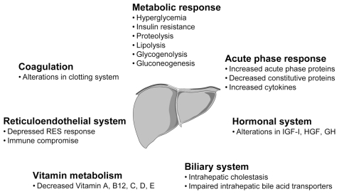
In summary, a severe burn represents a devastating injury affecting nearly every organ system and leading to significant morbidity and mortality (7). In this review, we delineated the central role of the liver during the postburn response. The liver has not one, but myriad functions that are each essential for survival (Figure 5). All of these hepatic functions are affected by a thermal injury, and we have strong evidence that hepatic biomarkers predict and determine morbidity and mortality in severely burned patients. We, therefore, believe that the liver is central for postburn outcome and we propose that attenuation of liver damage and restoration of liver function will improve morbidity and mortality of severely burned patients. Our hypothesis is being supported by a recent book on the liver in which Will Self writes, “Our livers are more valuable than we are, more able, more alive” (143).
Figure 5.
Overview of hepatic functions and responses.
The aim of this review article was to delineate for the first time the role of the liver during the post thermal injury response. We believe the liver is important and determines survival. Currently there are no treatments for liver failure in burns, but based on this review, perhaps research will drive in the direction to develop novel treatments.
ACKNOWLEDGMENTS
This review paper was supported by the American Surgical Association Foundation, Shriners Hospitals for Children 8660, 8760, and 9145, NIH R01-GM56687, T32 GM008256, and P50 GM60338, and NIDRR H133A020102.
Footnotes
Online address: http://www.molmed.org
DISCLOSURE
The authors declare that they have no competing interests as defined by Molecular Medicine, or other interests that might be perceived to influence the results and discussion reported in this paper.
REFERENCES
- 1.Bringham PA, McLoughlin E. Burn incidence and medical care use in the United States: estimates, trends and data sources. J Burn Care Rehabil. 1996;17:95–107. doi: 10.1097/00004630-199603000-00003. [DOI] [PubMed] [Google Scholar]
- 2.World Health Organization. Facts about injuries: Burns. Geneva: WHO, Injuries & Violence Prevention, Non-Communicable Diseases and Mental Health; [place unknown]: International Society for Burn Injuries; (WHO), Injuries & Violence Prevention, Non-Communicable Diseases and Mental Health; p. 5. No date. Listed as a 2004 fact sheet at: http://www.who.int/violence_injury_prevention/publications/other_injury/en/index.html. [Google Scholar]
- 3.Palmieri TL, Greenhalgh DG. Topical treatment of pediatric patients with burns: a practical guide. Am J Clin Dermatol. 2002;3:529–34. doi: 10.2165/00128071-200203080-00003. [DOI] [PubMed] [Google Scholar]
- 4.Nelson KJ, Beierle EA. Exhaust system burn injuries in children. J Pediatr Surg. 2005;40:E43–6. doi: 10.1016/j.jpedsurg.2005.01.030. [DOI] [PubMed] [Google Scholar]
- 5.Foglia RP, Moushey R, Meadows L, Seigel J, Smith M. Evolving treatment in a decade of pediatric burn care. J Pediatr Surg. 2004;39:957–60. doi: 10.1016/j.jpedsurg.2004.04.001. [DOI] [PubMed] [Google Scholar]
- 6.Miller SF, et al. National Burn Repository 2005: a ten-year review. J Burn Care Res. 2006;27:411–36. doi: 10.1097/01.BCR.0000226260.17523.22. [DOI] [PubMed] [Google Scholar]
- 7.Herndon DN, Tompkins RG. Support of the metabolic response to burn injury. Lancet. 2004;363:1895–902. doi: 10.1016/S0140-6736(04)16360-5. [DOI] [PubMed] [Google Scholar]
- 8.Herndon DN, Hart DW, Wolf SE, Chinkes DL, Wolfe RR. Reversal of catabolism by beta-blockade after severe burns. N Engl J Med. 2001;345:1223–9. doi: 10.1056/NEJMoa010342. [DOI] [PubMed] [Google Scholar]
- 9.Van den Berge G, et al. Intensive insulin therapy in critically ill patients. New Engl J Med. 2001;345:1359–67. doi: 10.1056/NEJMoa011300. [DOI] [PubMed] [Google Scholar]
- 10.Finnerty CC, et al. Cytokine expression profile over time in severely burned pediatric patients. Shock. 2006;26:13–9. doi: 10.1097/01.shk.0000223120.26394.7d. [DOI] [PubMed] [Google Scholar]
- 11.Tracey KJ, et al. Anti-cachectin/TNF monoclonal antibodies prevent septic shock during lethal bacteraemia. Nature. 1987;330:662–4. doi: 10.1038/330662a0. [DOI] [PubMed] [Google Scholar]
- 12.Tracey KJ, et al. Cachectin/tumor necrosis factor induces lethal shock and stress hormone responses in the dog. Surg Gynecol Obstet. 1987;164:415–22. [PubMed] [Google Scholar]
- 13.Moshage H. Cytokines and the hepatic acute phase response. J Pathol. 1997;181:257–66. doi: 10.1002/(SICI)1096-9896(199703)181:3<257::AID-PATH756>3.0.CO;2-U. [DOI] [PubMed] [Google Scholar]
- 14.Jeschke MG, Klein D, Herndon DN. Insulin treatment improves the systemic inflammatory reaction to severe trauma. Ann Surg. 2004;239:553–60. doi: 10.1097/01.sla.0000118569.10289.ad. [DOI] [PMC free article] [PubMed] [Google Scholar]
- 15.Jeschke MG, et al. Cell proliferation, apoptosis, NF-kappaB expression, enzyme, protein, and weight changes in livers of burned rats. Am J Physiol Gastrointest Liver Physiol. 2001;280:G1314–20. doi: 10.1152/ajpgi.2001.280.6.G1314. [DOI] [PubMed] [Google Scholar]
- 16.Jeschke MG, Micak RP, Finnerty CC, Herndon DN. Changes in liver function and size after a severe thermal injury. Shock. 2007;28:172–7. doi: 10.1097/shk.0b013e318047b9e2. [DOI] [PubMed] [Google Scholar]
- 17.Jeschke MG, et al. Insulin prevents liver damage and preserves liver function in lipopolysaccharide-induced endotoxemic rats. J Hepatol. 2005;42:870–9. doi: 10.1016/j.jhep.2004.12.036. [DOI] [PubMed] [Google Scholar]
- 18.Moll KJ, Moll M. Anatomie: Kurzlehrbuch zum Gegenstandskatalog. 18th ed. München: Elsevier (Urban & Fischer); 2006. p. 767. [Google Scholar]
- 19.Winkeltau G, Kraas E. Leber. In: Schumpelick V, Bleese NM, Mommsen U, editors. Chirurgie. Enke Verlag; Stuttgart: 2006. pp. 658–75. [Google Scholar]
- 20.Schwartz SI. Liver. In: Schwartz SI, Shires GT, Spencer FC, Daly JM, Fischer JE, Galloway AC, editors. Principles of Surgery. McGraw-Hill; New York: 1999. pp. 1395–436. [Google Scholar]
- 21.Küttler T. Biochemie. In: Küttler T, editor. Biochemie. Jungjohann Verlag; München: 2004. pp. 240–65. [Google Scholar]
- 22.Jeschke MG, Barrow RE, Herndon DN. Extended hypermetabolic response of the liver in severely burned pediatric patients. Arch Surg. 2004;139:641–7. doi: 10.1001/archsurg.139.6.641. [DOI] [PubMed] [Google Scholar]
- 23.Wolfe RR, Durkot MJ, Allsop JR, Burke JF. Glucose metabolism in severely burned patients. Metabolism. 1979;28:1031–9. doi: 10.1016/0026-0495(79)90007-6. [DOI] [PubMed] [Google Scholar]
- 24.Wolfe RR, Herndon DN, Jahoor F, Miyoshi H, Wolfe M. Effect of severe burn injury on substrate cycling by glucose and fatty acids. N Engl J Med. 1987;317:403–8. doi: 10.1056/NEJM198708133170702. [DOI] [PubMed] [Google Scholar]
- 25.Aarsland A, Chinkes D, Wolfe RR. Contributions of de novo synthesis of fatty acids to total VLDL-triglyceride secretion during prolonged hyperglycemia/hyperinsulinemia in normal man. J Clin Invest. 1996;98:2008–17. doi: 10.1172/JCI119005. [DOI] [PMC free article] [PubMed] [Google Scholar]
- 26.Jeschke MG, et al. Pathophysiologic response to severe burn injury. Ann Surg. 2008;248:387–401. doi: 10.1097/SLA.0b013e3181856241. [DOI] [PMC free article] [PubMed] [Google Scholar]
- 27.Gomez-Lechon MJ, et al. Effects of hepatocyte growth factor on the growth and metabolism of human hepatocytes in primary culture. Hepatology. 1995;21:1248–54. [PubMed] [Google Scholar]
- 28.Linares H. Autopsy finding in burned children. In: Carvajal H, Parks D, editors. Burns in Children. Year Book Medical; Chicago: 1991. pp. 1–25. [Google Scholar]
- 29.Barret JP, Jeschke MG, Herndon DN. Fatty infiltration of the liver in severely burned pediatric patients: autopsy findings and clinical implications. J Trauma. 2001;51:736–9. doi: 10.1097/00005373-200110000-00019. [DOI] [PubMed] [Google Scholar]
- 30.Steller H. Mechanisms and genes of cellular suicide. Science. 1995;267:1445–9. doi: 10.1126/science.7878463. [DOI] [PubMed] [Google Scholar]
- 31.Ramzy PI, et al. Gut epithelial apoptosis after severe burn: effects of gut hypoperfusion. J Am Coll Surg. 2000;190:281–7. doi: 10.1016/s1072-7515(99)00269-0. [DOI] [PubMed] [Google Scholar]
- 32.Wolf SE, et al. Cutaneous burn increases apoptosis in the gut epithelium of mice. J Am Coll Surg. 1999;188:10–6. doi: 10.1016/s1072-7515(98)00260-9. [DOI] [PubMed] [Google Scholar]
- 33.Horton JW. Left ventricular contractile dysfunction as a complication of thermal injury. Shock. 2004;22:495–507. doi: 10.1097/01.shk.0000145205.51682.c3. [DOI] [PubMed] [Google Scholar]
- 34.Horton JW, et al. Calcium antagonists improve cardiac mechanical performance after thermal trauma. J Surg Res. 1999;87:39–50. doi: 10.1006/jsre.1999.5726. [DOI] [PubMed] [Google Scholar]
- 35.Baron P, et al. Gut failure and translocation following burn and sepsis. J Surg Res. 1994;57:197–204. doi: 10.1006/jsre.1994.1131. [DOI] [PubMed] [Google Scholar]
- 36.Baron PW, Barrow RE, Pierre EJ, Herndon DN. Prolonged use of propranolol safely decreases cardiac work in burned children. J Burn Care Rehabil. 1997;18:223–7. doi: 10.1097/00004630-199705000-00008. [DOI] [PubMed] [Google Scholar]
- 37.Ikeda H, et al. Apoptosis is a major mode of cell death caused by ischaemia and ischaemia/reperfusion injury to the rat intestinal epithelium. Gut. 1998;42:530–7. doi: 10.1136/gut.42.4.530. [DOI] [PMC free article] [PubMed] [Google Scholar]
- 38.Noda T, Iwakiri R, Fujimoto K, Matsuo S, Aw TY. Programmed cell death induced by ischemia-reperfusion in rat intestinal mucosa. Am J Physiol. 1998;274:G270–6. doi: 10.1152/ajpgi.1998.274.2.G270. [DOI] [PubMed] [Google Scholar]
- 39.Bellas RE, FitzGerald MJ, Fausto N, Sonenshein GE. Inhibition of NF-kappa B activity induces apoptosis in murine hepatocytes. Am J Pathol. 1997;151:891–6. [PMC free article] [PubMed] [Google Scholar]
- 40.Boehning D, et al. Cytochrome c binds to inositol (1,4,5) trisphosphate receptors, amplifying calcium-dependent apoptosis. Nat Cell Biol. 2003;5:1051–61. doi: 10.1038/ncb1063. [DOI] [PubMed] [Google Scholar]
- 41.Strasser A, O’Connor L, Dixit VM. Apoptosis signaling. Annu Rev Biochem. 2000;69:217–45. doi: 10.1146/annurev.biochem.69.1.217. \. [DOI] [PubMed] [Google Scholar]
- 42.Yoneda T, et al. Activation of caspase-12, an endoplastic reticulum (ER) resident caspase, through tumor necrosis factor receptor-associated factor 2-dependent mechanism in response to the ER stress. J Biol Chem. 2001;276:13935–40. doi: 10.1074/jbc.M010677200. [DOI] [PubMed] [Google Scholar]
- 43.Klein D, Schubert T, Horch RE, Jauch KW, Jeschke MG. Insulin treatment improves hepatic morphology and function through modulation of hepatic signals after severe trauma. Ann Surg. 2004;240:340–9. doi: 10.1097/01.sla.0000133353.57674.cd. [DOI] [PMC free article] [PubMed] [Google Scholar]
- 44.Gauglitz GG, et al. Characterization of the inflammatory response during acute and post-acute phases after severe burn. Shock. 2008;30:503–507. doi: 10.1097/SHK.0b013e31816e3373. [DOI] [PMC free article] [PubMed] [Google Scholar]
- 45.Jeschke MG, Boehning DF, Finnerty CC, Herndon DN. Effect of insulin on the inflammatory and acute phase response after burn injury. Crit Care Med. 2007;35:S519–23. doi: 10.1097/01.CCM.0000282027.10288.10. [DOI] [PubMed] [Google Scholar]
- 46.Jeschke MG, et al. Calcium and ER stress mediate hepatic apoptosis after burn injury. J Cell Mol Med. 2009 2009 Jan 14; doi: 10.1111/j.1582-4934.2008.00644.x. [Epub ahead of print] [DOI] [PMC free article] [PubMed] [Google Scholar]
- 47.Ozcan U, et al. Chemical chaperones reduce ER stress and restore glucose homeostasis in a mouse model of type 2 diabetes. Science. 2006;313:1137–40. doi: 10.1126/science.1128294. [DOI] [PMC free article] [PubMed] [Google Scholar]
- 48.Wiseman RL, Balch WE. A new pharmacology—drugging stressed folding pathways. Trends Mol Med. 2005;11:347–50. doi: 10.1016/j.molmed.2005.06.011. [DOI] [PubMed] [Google Scholar]
- 49.Cano N, Gerolami A. Intrahepatic cholestasis during total parenteral nutrition. Lancet. 1983;1:985. doi: 10.1016/s0140-6736(83)92104-9. [DOI] [PubMed] [Google Scholar]
- 50.Bolder U, et al. Heat stress enhances recovery of hepatocyte bile acid and organic anion transporters in endotoxemic rats by multiple mechanisms. Cell Stress Chaperones. 2006;11:89–100. doi: 10.1379/CSC-143R.1. [DOI] [PMC free article] [PubMed] [Google Scholar]
- 51.Bolder U, Ton-Nu HT, Schteingart CD, Frick E, Hofmann AF. Hepatocyte transport of bile acids and organic anions in endotoxemic rats: impaired uptake and secretion. Gastroenterology. 1997;112:214–225. doi: 10.1016/s0016-5085(97)70238-5. [DOI] [PubMed] [Google Scholar]
- 52.Knox J, Demling R, Wilmore D, Sarraf P, Santos A. Increased survival after major thermal injury: the effect of growth hormone therapy in adults. J Trauma. 1995;39:526–32. doi: 10.1097/00005373-199509000-00021. [DOI] [PubMed] [Google Scholar]
- 53.Kobayashi M, et al. An increase in the susceptibility of burned patients to infectious complications due to impaired production of macrophage inflammatory protein 1 alpha. J Immunol. 2002;169:4460–6. doi: 10.4049/jimmunol.169.8.4460. [DOI] [PubMed] [Google Scholar]
- 54.Kobayashi M, et al. Bacterial sepsis and chemokines. Curr Drug Targets. 2006;7:119–34. doi: 10.2174/138945006775270169. [DOI] [PubMed] [Google Scholar]
- 55.Trop M, Schiffrin EJ, Callahan RJ, Strauss HW, Carter EA. Effect of acute burn trauma on reticuloendothelial system phagocytic activity in rats. II: Comparison of uptake of radiolabelled colloid and bacteria. Burns. 1990;16:278–80. doi: 10.1016/0305-4179(90)90139-n. [DOI] [PubMed] [Google Scholar]
- 56.Trop M, Schiffrin EJ, Carter EA. Role of skin in the burn-induced reduction of reticuloendothelial phagocytic activity in rats. Burns. 1990;16:57–9. doi: 10.1016/0305-4179(90)90207-d. [DOI] [PubMed] [Google Scholar]
- 57.Klein D, Einspanier R, Bolder U, Jeschke MG. Differences in the hepatic signal transcription pathway and cytokine expression between thermal injury and sepsis. Shock. 2003;20:536–43. doi: 10.1097/01.shk.0000093345.68755.98. [DOI] [PubMed] [Google Scholar]
- 58.Hart DW, et al. Determinants of skeletal muscle catabolism after severe burn. Ann Surg. 2000;232:455–65. doi: 10.1097/00000658-200010000-00001. [DOI] [PMC free article] [PubMed] [Google Scholar]
- 59.Reiss E, Pearson E, Artz CP. The metabolic response to burns. J Clin Invest. 1956;35:62–77. doi: 10.1172/JCI103253. [DOI] [PMC free article] [PubMed] [Google Scholar]
- 60.Haze K, Yoshida H, Yanagi H, Yura T, Mori K. Mammalian transcription factor ATF6 is synthesized as a transmembrane protein and activated by proteolysis in response to endoplasmic reticulum stress. Mol Biol Cell. 1999;10:3787–99. doi: 10.1091/mbc.10.11.3787. [DOI] [PMC free article] [PubMed] [Google Scholar]
- 61.Hart DW, et al. Persistence of muscle catabolism after severe burn. Surgery. 2000;128:312–9. doi: 10.1067/msy.2000.108059. [DOI] [PubMed] [Google Scholar]
- 62.Norbury WB, Herndon DN. Modulation of the hypermetabolic response after burn injury. In: Herndon DN, editor. Total Burn Care. Saunders Elsevier; New York: 2007. pp. 420–33. [Google Scholar]
- 63.Norbury WB, Herndon DN, Branski LK, Chinkes DL, Jeschke MG. Urinary cortisol and catecholamine excretion following burn injury in children. J Clin Endocrinol Metab. 2008;93:1270–5. doi: 10.1210/jc.2006-2158. [DOI] [PMC free article] [PubMed] [Google Scholar]
- 64.Sherwood ER, Toliver-Kinsky T. Mechanisms of the inflammatory response. Best Pract Res Clin Anaesthesiol. 2004;18:385–405. doi: 10.1016/j.bpa.2003.12.002. [DOI] [PubMed] [Google Scholar]
- 65.Pereira CT, Herndon DN. The pharmacologic modulation of the hypermetabolic response to burns. Adv Surg. 2005;39:245–61. doi: 10.1016/j.yasu.2005.05.005. [DOI] [PubMed] [Google Scholar]
- 66.Wolfe RR. Review: acute versus chronic response to burn injury. Circ Shock. 1981;8:105–15. [PubMed] [Google Scholar]
- 67.Cuthbertson DP, Angeles Valero Zanuy MA, Leon Sanz ML. Post-shock metabolic response. 1942. Nutr Hosp. 2001;16:176–82. [PubMed] [Google Scholar]
- 68.Tredget EE, Yu YM. The metabolic effects of thermal injury. World J Surg. 1992;16:68–79. doi: 10.1007/BF02067117. [DOI] [PubMed] [Google Scholar]
- 69.Cree MG, et al. Insulin sensitivity and mitochondrial function are improved in children with burn injury during a randomized controlled trial of fenofibrate. Ann Surg. 2007;245:214–21. doi: 10.1097/01.sla.0000250409.51289.ca. [DOI] [PMC free article] [PubMed] [Google Scholar]
- 70.Jeschke MG, et al. Burn size determines the inflammatory and hypermetabolic response. Crit Care. 2007;11:R90. doi: 10.1186/cc6102. [DOI] [PMC free article] [PubMed] [Google Scholar]
- 71.Mlcak RP, Jeschke MG, Barrow RE, Herndon DN. The influence of age and gender on resting energy expenditure in severely burned children. Ann Surg. 2006;244:121–30. doi: 10.1097/01.sla.0000217678.78472.d3. [DOI] [PMC free article] [PubMed] [Google Scholar]
- 72.Gearhart MM, Parbhoo SK. Hyperglycemia in the critically ill patient. AACN Clin Issues. 2006;17:50–5. doi: 10.1097/00044067-200601000-00007. [DOI] [PubMed] [Google Scholar]
- 73.Robinson LE, van Soeren MH. Insulin resistance and hyperglycemia in critical illness: role of insulin in glycemic control. AACN Clin Issues. 2004;15:45–62. doi: 10.1097/00044067-200401000-00004. [DOI] [PubMed] [Google Scholar]
- 74.Khani S, Tayek JA. Cortisol increases gluconeogenesis in humans: its role in the metabolic syndrome. Clin Sci (Lond) 2001;101:739–47. doi: 10.1042/cs1010739. [DOI] [PubMed] [Google Scholar]
- 75.Wolfe RR, et al. Regulation of lipolysis in severely burned children. Ann Surg. 1987;206:214–21. doi: 10.1097/00000658-198708000-00016. [DOI] [PMC free article] [PubMed] [Google Scholar]
- 76.Gore DC, Jahoor F, Wolfe RR, Herndon DN. Acute response of human muscle protein to catabolic hormones. Ann Surg. 1993;218:679–84. doi: 10.1097/00000658-199321850-00015. [DOI] [PMC free article] [PubMed] [Google Scholar]
- 77.Carlson GL. Insulin resistance and glucose-induced thermogenesis in critical illness. Proc Nutr Soc. 2001;60:381–8. doi: 10.1079/pns200193. [DOI] [PubMed] [Google Scholar]
- 78.Cree MG, et al. Human mitochondrial oxidative capacity is acutely impaired after burn trauma. Am J Surg. 2008;196:234–9. doi: 10.1016/j.amjsurg.2007.09.048. [DOI] [PMC free article] [PubMed] [Google Scholar]
- 79.Hunt DG, Ivy JL. Epinephrine inhibits insulin-stimulated muscle glucose transport. J Appl Physiol. 2002;93:1638–43. doi: 10.1152/japplphysiol.00445.2002. [DOI] [PubMed] [Google Scholar]
- 80.Cree MG, et al. PPAR-alpha agonism improves whole body and muscle mitochondrial fat oxidation, but does not alter intracellular fat concentrations in burn trauma children in a randomized controlled trial. Nutr Metab (Lond) 2007;4:9. doi: 10.1186/1743-7075-4-9. [DOI] [PMC free article] [PubMed] [Google Scholar]
- 81.Gustavson SM, et al. Glucagon’s actions are modified by the combination of epinephrine and gluconeogenic precursor infusion. Am J Physiol Endocrinol Metab. 2003;285:E534–44. doi: 10.1152/ajpendo.00059.2003. [DOI] [PubMed] [Google Scholar]
- 82.Akita S, et al. Elevated circulating leukemia inhibitory factor in patients with extensive burns. J Burn Care Res. 2006;27:221–5. doi: 10.1097/01.BCR.0000197679.08671.A5. [DOI] [PubMed] [Google Scholar]
- 83.Lang CH. Sepsis-induced insulin resistance in rats is mediated by a beta-adrenergic mechanism. Am J Physiol. 1992;263:E703–11. doi: 10.1152/ajpendo.1992.263.4.E703. [DOI] [PubMed] [Google Scholar]
- 84.Lang CH, Dobrescu C, Bagby GJ. Tumor necrosis factor impairs insulin action on peripheral glucose disposal and hepatic glucose output. Endocrinology. 1992;130:43–52. doi: 10.1210/endo.130.1.1727716. [DOI] [PubMed] [Google Scholar]
- 85.Lang CH, Frost RA, Vary TC. Regulation of muscle protein synthesis during sepsis and inflammation. Am J Physiol Endocrinol Metab. 2007;293:E453–9. doi: 10.1152/ajpendo.00204.2007. [DOI] [PubMed] [Google Scholar]
- 86.Fan J, Li YH, Wojnar MM, Lang CH. Endotoxin-induced alterations in insulin-stimulated phosphorylation of insulin receptor, IRS-1, and MAP kinase in skeletal muscle. Shock. 1996;6:164–70. [PubMed] [Google Scholar]
- 87.del Aguila LF, Claffey KP, Kirwan JP. TNF-alpha impairs insulin signaling and insulin stimulation of glucose uptake in C2C12 muscle cells. Am J Physiol. 1999;276:E849–55. doi: 10.1152/ajpendo.1999.276.5.E849. [DOI] [PubMed] [Google Scholar]
- 88.Sell H, Dietze-Schroeder D, Kaiser U, Eckel J. Monocyte chemotactic protein-1 is a potential player in the negative cross-talk between adipose tissue and skeletal muscle. Endocrinology. 2006;147:2458–67. doi: 10.1210/en.2005-0969. [DOI] [PubMed] [Google Scholar]
- 89.Jahoor F, Desai M, Herndon DN, Wolfe RR. Dynamics of the protein metabolic response to burn injury. Metabolism. 1988;37:330–7. doi: 10.1016/0026-0495(88)90132-1. [DOI] [PubMed] [Google Scholar]
- 90.Jahoor F, Herndon DN, Wolfe RR. Role of insulin and glucagon in the response of glucose and alanine kinetics in burn-injured patients. J Clin Invest. 1986;78:807–14. doi: 10.1172/JCI112644. [DOI] [PMC free article] [PubMed] [Google Scholar]
- 91.Gauglitz GG, Herndon DN, Kulp GA, Meyer WJ, 3rd, Jeschke MG. Abnormal insulin sensitivity persists up to three years in pediatric patients post-burn. J Clin Endocrinol Metab. 2009 2009 Feb 24; doi: 10.1210/jc.2008-1947. [Epub ahead of print] [DOI] [PMC free article] [PubMed] [Google Scholar]
- 92.Ikezu T, Okamoto T, Yonezawa K, Tompkins RG, Martyn JA. Analysis of thermal injury-induced insulin resistance in rodents. Implication of postreceptor mechanisms. J Biol Chem. 1997;272:25289–95. doi: 10.1074/jbc.272.40.25289. [DOI] [PubMed] [Google Scholar]
- 93.Saffle JR, Graves C. Nutritional support of the burned patient. In: Herndon DN, editor. Total Burn Care. Saunders Elsevier; London: 2007. pp. 398–419. [Google Scholar]
- 94.DeFronzo RA, et al. The effect of insulin on the disposal of intravenous glucose. Results from indirect calorimetry and hepatic and femoral venous catheterization. Diabetes. 1981;30:1000–7. doi: 10.2337/diab.30.12.1000. [DOI] [PubMed] [Google Scholar]
- 95.Flakoll PJ, Hill JO, Abumrad NN. Acute hyperglycemia enhances proteolysis in normal man. Am J Physiol. 1993;265:E715–21. doi: 10.1152/ajpendo.1993.265.5.E715. [DOI] [PubMed] [Google Scholar]
- 96.Chang DW, DeSanti L, Demling RH. Anticatabolic and anabolic strategies in critical illness: a review of current treatment modalities. Shock. 1998;10:155–60. doi: 10.1097/00024382-199809000-00001. [DOI] [PubMed] [Google Scholar]
- 97.Hart DW, et al. Determinants of skeletal muscle catabolism after severe burn. Ann Surg. 2000;232:455–65. doi: 10.1097/00000658-200010000-00001. [DOI] [PMC free article] [PubMed] [Google Scholar]
- 98.Przkora R, et al. Body composition changes with time in pediatric burn patients. J Trauma. 2006;60:968–71. doi: 10.1097/01.ta.0000214580.27501.19. [DOI] [PubMed] [Google Scholar]
- 99.Przkora R, et al. Metabolic and hormonal changes of severely burned children receiving long-term oxandrolone treatment. Ann Surg. 2005;242:384–91. doi: 10.1097/01.sla.0000180398.70103.24. [DOI] [PMC free article] [PubMed] [Google Scholar]
- 100.Wolfe BM, Walker BK, Shaul DB, Wong L, Ruebner BH. Effect of total parenteral nutrition on hepatic histology. Arch Surg. 1988;123:1084–90. doi: 10.1001/archsurg.1988.01400330060009. [DOI] [PubMed] [Google Scholar]
- 101.Aarsland A, et al. Insulin therapy in burn patients does not contribute to hepatic triglyceride production. J Clin Invest. 1998;101:2233–9. doi: 10.1172/JCI200. [DOI] [PMC free article] [PubMed] [Google Scholar]
- 102.Barrow RE, Mlcak R, Barrow LN, Hawkins HK. Increased liver weights in severely burned children: comparison of ultrasound and autopsy measurements. Burns. 2004;30:565–8. doi: 10.1016/j.burns.2004.01.027. [DOI] [PubMed] [Google Scholar]
- 103.Barrow RE, Wolfe RR, Dasu MR, Barrow LN, Herndon DN. The use of beta-adrenergic blockade in preventing trauma-induced hepatomegaly. Ann Surg. 2006;243:115–20. doi: 10.1097/01.sla.0000193834.07413.91. [DOI] [PMC free article] [PubMed] [Google Scholar]
- 104.Aarsland A, et al. Beta-blockade lowers peripheral lipolysis in burn patients receiving growth hormone. Rate of hepatic very low density lipoprotein triglyceride secretion remains unchanged. Ann Surg. 1996;223:777–89. doi: 10.1097/00000658-199606000-00016. [DOI] [PMC free article] [PubMed] [Google Scholar]
- 105.Morio B, Irtun O, Herndon DN, Wolfe RR. Propranolol decreases splanchnic triacyl-glycerol storage in burn patients receiving a high-carbohydrate diet. Ann Surg. 2002;236:218–25. doi: 10.1097/00000658-200208000-00010. [DOI] [PMC free article] [PubMed] [Google Scholar]
- 106.Martini WZ, et al. Alteration of hepatic fatty acid metabolism after burn injury in pigs. JPEN J Parenter Enteral Nutr. 2001;25:310–6. doi: 10.1177/0148607101025006310. [DOI] [PubMed] [Google Scholar]
- 107.Mittendorfer B, Jeschke MG, Wolf SE, Sidossis LS. Nutritional hepatic steatosis and mortality after burn injury in rats. Clin Nutr. 1998;17:293–9. doi: 10.1016/s0261-5614(98)80322-1. [DOI] [PubMed] [Google Scholar]
- 108.Deng QG, et al. Steatohepatitis induced by intragastric overfeeding in mice. Hepatology. 2005;42:905–14. doi: 10.1002/hep.20877. [DOI] [PubMed] [Google Scholar]
- 109.Gilpin DA, Hsieh CC, Kuninger DT, Herndon DN, Papaconstantinou J. Effect of thermal injury on the expression of transcription factors that regulate acute phase response genes: the response of C/EBP alpha, C/EBP beta, and C/EBP delta to thermal injury. Surgery. 1996;119:674–83. doi: 10.1016/s0039-6060(96)80192-9. [DOI] [PubMed] [Google Scholar]
- 110.Gilpin DA, Hsieh CC, Kuninger DT, Herndon DN, Papaconstantinou J. Regulation of the acute phase response genes alpha 1-acid glycoprotein and alpha 1-antitrypsin correlates with sensitivity to thermal injury. Surgery. 1996;119:664–73. doi: 10.1016/s0039-6060(96)80191-7. [DOI] [PubMed] [Google Scholar]
- 111.Hiyama DT, et al. Synthesis of albumin and acute-phase proteins in perfused liver after burn injury in rats. J Burn Care Rehabil. 1991;12:1–6. doi: 10.1097/00004630-199101000-00002. [DOI] [PubMed] [Google Scholar]
- 112.Livingston DH, Mosenthal AC, Deitch EA. Sepsis and multiple organ dysfunction syndrome: a clinical-mechanistic overview. New Horiz. 1995;3:257–66. [PubMed] [Google Scholar]
- 113.De Maio A, Mooney ML, Matesic LE, Paidas CN, Reeves RH. Genetic component in the inflammatory response induced by bacterial lipopolysaccharide. Shock. 1998;10:319–23. doi: 10.1097/00024382-199811000-00002. [DOI] [PubMed] [Google Scholar]
- 114.De Maio A, Torres MB, Reeves RH. Genetic determinants influencing the response to injury, inflammation, and sepsis. Shock. 2005;23:11–7. doi: 10.1097/01.shk.0000144134.03598.c5. [DOI] [PubMed] [Google Scholar]
- 115.Selzman CH, et al. Therapeutic implications of interleukin-10 in surgical disease. Shock. 1998;10:309–18. doi: 10.1097/00024382-199811000-00001. [DOI] [PubMed] [Google Scholar]
- 116.Finnerty CC, Herndon DN, Chinkes DL, Jeschke MG. Serum cytokine differences in severely burned children with and without sepsis. Shock. 2007;27:4–9. doi: 10.1097/01.shk.0000235138.20775.36. [DOI] [PubMed] [Google Scholar]
- 117.Siebenlist U, Franzoso G, Brown K. Structure, regulation and function of NF-kappa B. Annu. Rev Cell Biol. 1994;10:405–55. doi: 10.1146/annurev.cb.10.110194.002201. [DOI] [PubMed] [Google Scholar]
- 118.Kishimoto T, Taga T, Akira S. Cytokine signal transduction. Cell. 1994;76:253–62. doi: 10.1016/0092-8674(94)90333-6. [DOI] [PubMed] [Google Scholar]
- 119.Niehof M, et al. Interleukin-6-induced tethering of STAT3 to the LAP/C/EBPbeta promoter suggests a new mechanism of transcriptional regulation by STAT3. J Biol Chem. 2001;276:9016–27. doi: 10.1074/jbc.M009284200. [DOI] [PubMed] [Google Scholar]
- 120.Janes KA, et al. A systems model of signaling identifies a molecular basis set for cytokine-induced apoptosis. Science. 2005;310:1646–53. doi: 10.1126/science.1116598. [DOI] [PubMed] [Google Scholar]
- 121.Mori K, Ma W, Gething MJ, Sambrook J. A transmembrane protein with a cdc2+/CDC28− related kinase activity is required for signaling from the ER to the nucleus. Cell. 1993;74:743–56. doi: 10.1016/0092-8674(93)90521-q. [DOI] [PubMed] [Google Scholar]
- 122.Chan MM, Chan GM. Nutritional therapy for burns in children and adults. Nutrition. 2009;25:261–9. doi: 10.1016/j.nut.2008.10.011. [DOI] [PubMed] [Google Scholar]
- 123.Nguyen TT, et al. Free radical activity and loss of plasma antioxidants, vitamin E, and sulfhydryl groups in patients with burns: the 1993 Moyer Award. J Burn Care Rehabil. 1993;14:602–9. doi: 10.1097/00004630-199311000-00004. [DOI] [PubMed] [Google Scholar]
- 124.Manning AJ, Meyer N, Klein GL. Vitamin and trace element homeostasis following burn injury. In: Herndon DN, editor. Total Burn Care. WB Saunders; Philadelphia: 2007. pp. 251–5. [Google Scholar]
- 125.Matsuda T, et al. Reduced fluid volume requirement for resuscitation of third-degree burns with high-dose vitamin C. J Burn Care Rehabil. 1991;12:525–32. doi: 10.1097/00004630-199111000-00007. [DOI] [PubMed] [Google Scholar]
- 126.Sherwood ER, Traber DL. The systemic inflammatory response syndrome. In: Herndon DN, editor. Total Burn Care. WB Saunders; Philadelphia: 2007. pp. 292–309. [Google Scholar]
- 127.Guillen MI, Gomez-Lechon MJ, Nakamura T, Castell JV. The hepatocyte growth factor regulates the synthesis of acute-phase proteins in human hepatocytes: divergent effect on interleukin-6-stimulated genes. Hepatology. 1996;23:1345–52. doi: 10.1002/hep.510230609. [DOI] [PubMed] [Google Scholar]
- 128.Jeschke MG, et al. Hepatocyte growth factor modulates the hepatic acute-phase response in thermally injured rats. Crit Care Med. 2000;28:504–10. doi: 10.1097/00003246-200002000-00036. [DOI] [PubMed] [Google Scholar]
- 129.Yamashita Y, Jeschke MG, Wolf SE. Differential expression of hepatocyte growth factor in liver, kidney, lung, and spleen following burn in rats. Cytokine. 2000;12:1293–8. doi: 10.1006/cyto.2000.0719. [DOI] [PubMed] [Google Scholar]
- 130.Cho MK, Kim SG. Hepatocyte growth factor activates CCAAT enhancer binding protein and cell replication via PI3-kinase pathway. Hepatology. 2003;37:686–95. doi: 10.1053/jhep.2003.50078. [DOI] [PubMed] [Google Scholar]
- 131.Humbel RE. Insulin-like growth factors I and II. Eur J Biochem. 1990;190:445–62. doi: 10.1111/j.1432-1033.1990.tb15595.x. [DOI] [PubMed] [Google Scholar]
- 132.Baxter RC. Circulating levels and molecular distribution of the acid-labile (alpha) subunit of the high molecular weight insulin-like growth factor-binding protein complex. J Clin Endocrinol Metab. 1990;70:1347–53. doi: 10.1210/jcem-70-5-1347. [DOI] [PubMed] [Google Scholar]
- 133.Jeschke MG, Chrysopoulo MT, Herndon DN, Wolf SE. Increased expression of insulin-like growth factor-I in serum and liver after recombinant human growth hormone administration in thermally injured rats. J Surg Res. 1999;85:171–77. doi: 10.1006/jsre.1999.5623. [DOI] [PubMed] [Google Scholar]
- 134.Herndon DN, et al. Muscle protein catabolism after severe burn, effects of IGF-1/IGFBP3 treatment. Ann Surg. 1999;229:713–20. doi: 10.1097/00000658-199905000-00014. [DOI] [PMC free article] [PubMed] [Google Scholar]
- 135.Jeschke MG, et al. Combination of recombinant human growth hormone and propranolol decreases hypermetabolism and inflammation in severely burned children. Pediatr Crit Care Med. 2008;9:209–16. doi: 10.1097/PCC.0b013e318166d414. [DOI] [PubMed] [Google Scholar]
- 136.Jeschke MG, Herndon DN, Barrow RE. Insulin-like growth factor I in combination with insulin-like growth factor binding protein 3 affects the hepatic acute phase response and hepatic morphology in thermally injured rats. Ann Surg. 2000;231:408–16. doi: 10.1097/00000658-200003000-00014. [DOI] [PMC free article] [PubMed] [Google Scholar]
- 137.Jeschke MG, et al. IGF-I/BP-3 administration preserves hepatic homeostasis after thermal injury which is associated with increases in no and hepatic NF-kappa B. Shock. 2001;16:373–9. doi: 10.1097/00024382-200116050-00009. [DOI] [PubMed] [Google Scholar]
- 138.Price LA, Thombs B, Chen CL, Milner SM. Liver disease in burn injury: evidence from a national sample of 31,338 adult patients. J Burns Wounds. 2007;7:e1. [PMC free article] [PubMed] [Google Scholar]
- 139.Deutschman CS, Cereda M, Ochroch EA, Raj NR. Sepsis-induced cholestasis, steatosis, hepatocellular injury, and impaired hepatocellular regeneration are enhanced in interleukin-6−/− mice. Crit Care Med. 2006;34:2613–20. doi: 10.1097/01.CCM.0000240229.98275.07. [DOI] [PubMed] [Google Scholar]
- 140.Cheng J, et al. Protection from Fas-mediated apoptosis by a soluble form of the Fas molecule. Science. 1994;263:1759–62. doi: 10.1126/science.7510905. [DOI] [PubMed] [Google Scholar]
- 141.Song E, et al. RNA interference targeting Fas protects mice from fulminant hepatitis. Nat Med. 2003;9:347–51. doi: 10.1038/nm828. [DOI] [PubMed] [Google Scholar]
- 142.Jeschke MG, et al. Gut mucosal homeostasis and cellular mediators after severe thermal trauma and the effect of insulin-like growth factor-I in combination with insulin-like growth factor binding protein-3. Endocrinology. 2007;148:354–62. doi: 10.1210/en.2006-0883. [DOI] [PubMed] [Google Scholar]
- 143.Self W. Liver: A Fictional Organ with a Surface Anatomy of Four Lobes. New York: Viking Books; 2008. p. 276. [Google Scholar]


