Abstract
We describe an 87-year-old woman with a large ileal gastrointestinal stromal tumor (GIST) causing hemoperitoneum. A CT scan demonstrated a large heterogeneous mass measuring about 13 cm × 11 cm in the pelvis and hemoperitoneum, with a non-uniform enhancement pattern. The mass was diagnosed as a GIST originating from the gastrointestinal tract. She underwent an urgent laparotomy and an ileal GIST with a rupture was found 130 cm from the anal to the Treitz’s ligament. Hemoperitoneum caused by ileal GIST rupture is a rare condition. Bleeding in the large tumor leading to rupture of the capsule might cause hemoperitoneum in the present case.
Keywords: Intestinal neoplasm, Small intestine, Extraluminal growth, Laparotomy
INTRODUCTION
Gastrointestinal stromal tumor (GIST) is the designation for a major subset of mesenchymal tumors of the gastrointestinal (GI) tract[1–5]. GIST arising in the digestive tract is most commonly located in the stomach and small intestine[6,7]. GIST originating from the small intestine rarely causes hemoperitoneum[8]. Herein, we describe a relatively rare case of extraluminal ileal GIST causing hemoperitoneum.
CASE REPORT
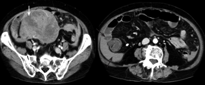
An 87-year-old woman presented with the symptom of a short loss of consciousness. She was in good health with no specific family or past medical history. Her body temperature was 36.7°C, blood pressure was 148/82 mmHg, radial pulse rate was 72 beats/min and regular. She had slight anemia, but no jaundice. Neurological examination revealed no abnormal findings and lymphadenopathy. Abdominal palpation revealed tenderness in the right lower quadrant. Laboratory tests showed a red blood cell count of 315 × 104/μL [normal range (NR), 380-500 × 104/μL], a white blood cell count of 10 500/μL (NR, 4000-9000/μL), a platelet count of 29.8 × 10⁴/μL, and a hemoglobin concentration of 10.1 g/dL (NR, 12-16 g/dL). The levels of hepatic and biliary enzymes, such as aspartate aminotransferase (AST), alanine aminotransferase (ALT), alkaline phosphatase (ALP), leucin aminopeptidase (LAP), and γ-glutamyltranspeptidase (γ-GTP) were normal except for lactate dehydrogenase (LDH) which was 336 IU/L (NR, 106-211 IU/L). A test for C reactive protein revealed a level of 30.3 mg/dL (NR, < 0.5 mg/dL). Renal function tests showed that the blood urea nitrogen level was 38.0 mg/dL (NR, 8-20 mg/dL) and the creatinine level was normal. Serological studies for hepatitis B and C viruses were negative. Urinary protein and sugar were negative. A computed tomography (CT) scan demonstrated a large heterogeneous mass measuring about 13 cm × 11 cm in the pelvis and hemoperitoneum, with a non-uniform enhancement pattern (Figure 1). Based on the imaging examination, this tumor was diagnosed as a GIST originating from the GI tract or omentum, although it should be distinguished from an ovarian tumor or an adenocarcinoma of the small bowel. The patient underwent an urgent laparotomy.
Figure 1.
CT scan demonstrating a large heterogeneous mass with a non-uniform enhancement pattern (white arrow) in the pelvis and hemoperitoneum (black arrows).
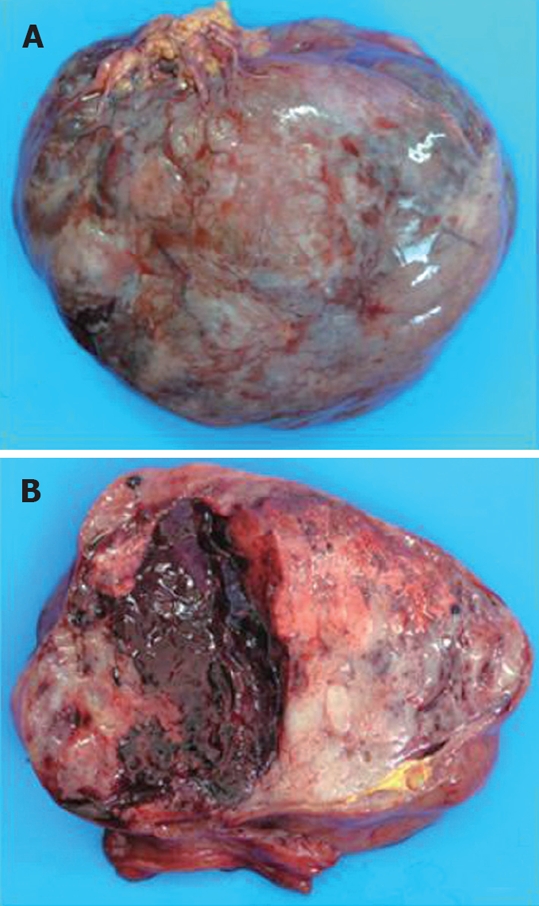
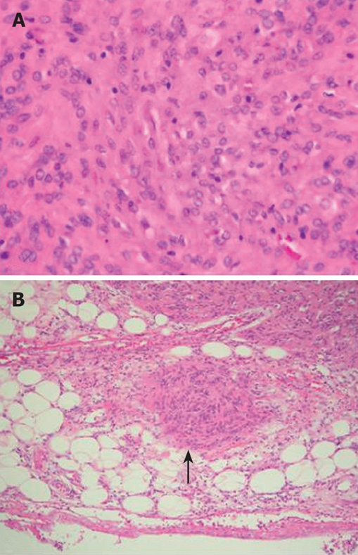
At laparotomy, a 13 cm × 11 cm semipedunculated solid tumor that was 130 cm from the anal to the Treitz’s ligament, showed extraluminal growth (Figure 2A). The tumor was ruptured with no peritoneal metastasis, and partial resection of the ileum was carried out. The resected tumor was brown-red in color, and had bleeding blood clots (Figure 2B). Histological examination of the resected specimen revealed interlaced bundles of large Bizarre spindle-like tumor cells without mitotic figures (Figure 3A). No fission images were evident. Tumor cells were present in the subserosa (Figure 3B). Immunohistological findings were negative for CD34, α-smooth muscle actin (SMA), desmin and S-100 protein, but positive for CD117. Based on the above findings, this tumor was diagnosed as a malignant GIST. The postoperative course was uneventful. The patient has been followed up for 16 mo with no evidence of recurrence.
Figure 2.

Macroscopic finding of the tumor. A: A large tumor (measuring 13 cm × 11 cm) arising from the ileum with extraluminal growth; B: The cut surface showing bleeding blood clots in the tumor.
Figure 3.

Microscopic findings of the tumor. A: Histological examination demonstrating interlaced bundles of large Bizarre spindle cells without mitotic figures (HE, × 100); B: Tumor cells present in the subserosa (black arrow) (HE, × 20).
DISCUSSION
GIST is the most common mesenchymal tumor of the GI tract and expresses c-kit protein, also known as CD117, which is considered a highly specific marker differentiating GIST from other mesenchymal tumors, such as leiomyomas[9–11]. The majority of GISTs occur in the stomach (60%-70%) and small intestine (20%-30%)[10]. Approximately, 10%-30% of patients with GIST may be asymptomatic[11]. Gastric and small intestinal stromal tumors are usually associated with abdominal pain, GI bleeding or palpable mass[12]. However, GIST in the small intestine rarely causes hemoperitoneum. A MEDLINE search of the literature has revealed only 10 cases of GIST in the small intestine with hemoperitoneum since 2000, including the present case in Japan[8,13] (Table 1). The tumor size was over 5 cm in 9 of the 10 cases. However, Hisaoka et al[13] have reported a case of small intestinal GIST, measuring about 1 cm in size, with intraperitoneal hemorrhage. Thus, a small GIST does not necessarily have a low risk of bleeding. We found that 7 of the 10 patients were in their sixties or over and GIST was predominant in older patients. The male to female ratio was 6:4. The major symptom was abdominal pain. Two patients had consciousness loss, including our case, which might be related to bleeding.
Table 1.
Summary of 10 cases of small bowel GIST causing hemoperitoneum in Japan (since 2000)
| AuthorRef | Age | Sex | Year | Tumor location | Tumor size (cm) | Symptoms | |
| 1 | Ri | 45 | Male | 2000 | Ileum | 5 | Abdominal pain |
| 2 | Yanaginuma | 58 | Male | 2001 | Ileum | 13 × 10 × 8 | Abdominal pain |
| 3 | Sugawara[8] | 37 | Male | 2001 | Ileum | 4 × 4 × 3 | Abdominal pain |
| 4 | Okita | 79 | Male | 2003 | Ileum | 18.5 × 15.7 × 6.5 | Abdominal pain |
| 5 | Hirose | 71 | Female | 2003 | Ileum | 7 | Vomiting |
| 6 | Kinoshita | 70 | Male | 2005 | Jejunum | 11 × 7 | Abdominal pain |
| 7 | Saito | 62 | Male | 2006 | Ileum | 5.0 × 4.5 × 3 | Abdominal pain |
| 8 | Goto | 71 | Female | 2006 | Ileum | 9 × 4 × 5 | Consciousness loss, vomiting |
| 9 | Hisaoka[13] | 73 | Female | 2007 | Ileum | 1 | Abdominal pain |
| 10 | Present case | 87 | Female | 2007 | Ileum | 13 × 11 | Consciousness loss, abdominal pain |
The mechanism underlying hemoperitoneum might be due to bleeding in the tumor leading to hematoma and rupture of the capsule, or transudation of blood components from the tumor. In the present case, bleeding in the tumor leading to rupture of the capsule might have caused hemoperitoneum. Currently, there is no single prognostic factor that can be used alone to predict tumor behavior. The biological behavior of tumors depends on the location (GIST arising from the small bowel is generally associated with a less favorable outcome than that arising in the stomach)[14]. Radiological and surgical factors that have been used to determine malignancy include invasion to adjacent organs, omental or peritoneal seeding, tumor recurrence after surgical resection, and distant metastasis[9,15]. Pathological factors that determine malignancy are tumor size, mitotic activity, pleomorphism of nuclei, degree of cellularity, nucleus/cytoplasm ratio, and mucosal invasion[16]. The present patient was diagnosed with malignant GIST because of the tumor size and rupture. Although peritoneal metastasis was not seen in the present patient, we should pay attention to tumor recurrence because the tumor was ruptured. This patient remains alive without disease 16 mo after surgery. We should carefully follow up with CT or MRI images and this patient is going to undergo imatinib mesilate therapy if tumor recurrence is identified.
Computed tomography (CT) and MR imaging are useful for the diagnosis of GIST and demonstration of the tumor extent[17–19]. Because of the high soft-tissue contrast, MR imaging shows a tendency of GIST toward necrosis and hemorrhage[20]. In particular, hemorrhage observed in large tumors is associated with large necrosis. Because GIST can rupture and result in hemoperitoneum, as in the present case, the presence of hemorrhage inside and outside the tumor should be detected. It is impossible to differentiate benign from malignant GIST confidently based on imaging findings alone. It was recently reported that a markedly enhanced GIST on MRI might demonstrate a higher mitosis index even if it is relatively small[20]. To clarify the relationship between MRI and pathological findings, we should accumulate and analyze many cases of GIST. Because of hemoperitoneum, our patient underwent urgent laparotomy but not MRI. In addition, previous reports of contrast-enhanced CT indicate that tumor density and heterogenetity after contrast enhancement may reflect the poor prognosis and tumor response to chemotherapy, respectively[21,22]. Further studies on the correlation between imaging diagnosis and pathological findings of GIST are certainly required.
In conclusion, we reported the case of a woman with a large ileal GIST causing hemoperitoneum. It is necessary to know that a large GIST may cause hemoperitoneum, although GIST in the small intestine rarely causes hemo-peritoneum.
Peer reviewers: Ton Lisman, PhD, Thrombosis and Haemostasis Laboratory, Department of Haematology G.03.550, University Medical Centre, Heidelberglaan 100, 3584 CX Utrecht, The Netherlands; Vincent W Yang, Professor and Director, 201 Whitehead Research Building, 615 Michael Street, Atlanta, GA 30322, United States; Jayaram Menon, Head, Department of Medicine, Queen Elizabeth Hospital, Department of Medicine, Queen Elizabeth Hospital, Kota Kinabalu, Sabah, Malaysia
S- Editor Zhong XY L- Editor Wang XL E- Editor Liu Y
References
- 1.Miettinen M, Lasota J. Gastrointestinal stromal tumors--definition, clinical, histological, immunohistochemical, and molecular genetic features and differential diagnosis. Virchows Arch. 2001;438:1–12. doi: 10.1007/s004280000338. [DOI] [PubMed] [Google Scholar]
- 2.Fletcher CD, Berman JJ, Corless C, Gorstein F, Lasota J, Longley BJ, Miettinen M, O'Leary TJ, Remotti H, Rubin BP, et al. Diagnosis of gastrointestinal stromal tumors: A consensus approach. Hum Pathol. 2002;33:459–465. doi: 10.1053/hupa.2002.123545. [DOI] [PubMed] [Google Scholar]
- 3.Joensuu H, Fletcher C, Dimitrijevic S, Silberman S, Roberts P, Demetri G. Management of malignant gastrointestinal stromal tumours. Lancet Oncol. 2002;3:655–664. doi: 10.1016/s1470-2045(02)00899-9. [DOI] [PubMed] [Google Scholar]
- 4.Suzuki K, Kaneko G, Kubota K, Horigome N, Hikita H, Senga O, Miyakawa M, Shimojo H, Uehara T, Itoh N. Malignant tumor, of the gastrointestinal stromal tumor type, in the greater omentum. J Gastroenterol. 2003;38:985–988. doi: 10.1007/s00535-003-1182-z. [DOI] [PubMed] [Google Scholar]
- 5.Miettinen M, Lasota J. Gastrointestinal stromal tumors (GISTs): definition, occurrence, pathology, differential diagnosis and molecular genetics. Pol J Pathol. 2003;54:3–24. [PubMed] [Google Scholar]
- 6.DeMatteo RP, Lewis JJ, Leung D, Mudan SS, Woodruff JM, Brennan MF. Two hundred gastrointestinal stromal tumors: recurrence patterns and prognostic factors for survival. Ann Surg. 2000;231:51–58. doi: 10.1097/00000658-200001000-00008. [DOI] [PMC free article] [PubMed] [Google Scholar]
- 7.Grassi N, Cipolla C, Torcivia A, Mandala S, Graceffa G, Bottino A, Latteri F. Gastrointestinal stromal tumour of the rectum: Report of a case and review of literature. World J Gastroenterol. 2008;14:1302–1304. doi: 10.3748/wjg.14.1302. [DOI] [PMC free article] [PubMed] [Google Scholar]
- 8.Sugawara G, Yamaguchi A, Isogai M, Kaneoka Y, Suzuki M. A case of gastrointestinal stromal tumor of the ileum with intraabdominal hemorrhage (in Japanese) J Jpn Soc Clin Surg. 2003;64:3092–3096. [Google Scholar]
- 9.Miettinen M, Sarlomo-Rikala M, Lasota J. Gastrointestinal stromal tumors: recent advances in understanding of their biology. Hum Pathol. 1999;30:1213–1220. doi: 10.1016/s0046-8177(99)90040-0. [DOI] [PubMed] [Google Scholar]
- 10.Fletcher CD, Berman JJ, Corless C, Gorstein F, Lasota J, Longley BJ, Miettinen M, O'Leary TJ, Remotti H, Rubin BP, et al. Diagnosis of gastrointestinal stromal tumors: A consensus approach. Hum Pathol. 2002;33:459–465. doi: 10.1053/hupa.2002.123545. [DOI] [PubMed] [Google Scholar]
- 11.Miettinen M, Sobin LH, Sarlomo-Rikala M. Immunohisto-chemical spectrum of GISTs at different sites and their differential diagnosis with a reference to CD117 (KIT) Mod Pathol. 2000;13:1134–1142. doi: 10.1038/modpathol.3880210. [DOI] [PubMed] [Google Scholar]
- 12.Mehta RM, Sudheer VO, John AK, Nandakumar RR, Dhar PS, Sudhindran S, Balakrishnan V. Spontaneous rupture of giant gastric stromal tumor into gastric lumen. World J Surg Oncol. 2005;3:11. doi: 10.1186/1477-7819-3-11. [DOI] [PMC free article] [PubMed] [Google Scholar]
- 13.Hisaoka S, Wakata T, Hirose C, Kakehisa M, kajikawa A, Yoneda A, Hirokawa M. A case of small intestinal GIST with intraperitoneal hemorrhage (in Japanese) Rinsho Hosyasen. 2007;52:820–824. [Google Scholar]
- 14.Emory TS, Sobin LH, Lukes L, Lee DH, O'Leary TJ. Prognosis of gastrointestinal smooth-muscle (stromal) tumors: dependence on anatomic site. Am J Surg Pathol. 1999;23:82–87. doi: 10.1097/00000478-199901000-00009. [DOI] [PubMed] [Google Scholar]
- 15.Miettinen M, Sarlomo-Rikala M, Lasota J. Gastrointestinal stromal tumours. Ann Chir Gynaecol. 1998;87:278–281. [PubMed] [Google Scholar]
- 16.Franquemont DW. Differentiation and risk assessment of gastrointestinal stromal tumors. Am J Clin Pathol. 1995;103:41–47. doi: 10.1093/ajcp/103.1.41. [DOI] [PubMed] [Google Scholar]
- 17.Levy AD, Remotti HE, Thompson WM, Sobin LH, Miettinen M. Anorectal gastrointestinal stromal tumors: CT and MR imaging features with clinical and pathologic correlation. AJR Am J Roentgenol. 2003;180:1607–1612. doi: 10.2214/ajr.180.6.1801607. [DOI] [PubMed] [Google Scholar]
- 18.Kim HC, Lee JM, Kim SH, Kim KW, Lee M, Kim YJ, Han JK, Choi BI. Primary gastrointestinal stromal tumors in the omentum and mesentery: CT findings and pathologic correlations. AJR Am J Roentgenol. 2004;182:1463–1467. doi: 10.2214/ajr.182.6.1821463. [DOI] [PubMed] [Google Scholar]
- 19.Lau S, Tam KF, Kam CK, Lui CY, Siu CW, Lam HS, Mak KL. Imaging of gastrointestinal stromal tumour (GIST) Clin Radiol. 2004;59:487–498. doi: 10.1016/j.crad.2003.10.018. [DOI] [PubMed] [Google Scholar]
- 20.Amano M, Okuda T, Amano Y, Tajiri T, Kumazaki T. Magnetic resonance imaging of gastrointestinal stromal tumor in the abdomen and pelvis. Clin Imaging. 2006;30:127–131. doi: 10.1016/j.clinimag.2005.09.025. [DOI] [PubMed] [Google Scholar]
- 21.Tateishi U, Hasegawa T, Satake M, Moriyama N. Gastrointestinal stromal tumor. Correlation of computed tomography findings with tumor grade and mortality. J Comput Assist Tomogr. 2003;27:792–798. doi: 10.1097/00004728-200309000-00018. [DOI] [PubMed] [Google Scholar]
- 22.Choi H, Charnsangavej C, de Castro Faria S, Tamm EP, Benjamin RS, Johnson MM, Macapinlac HA, Podoloff DA. CT evaluation of the response of gastrointestinal stromal tumors after imatinib mesylate treatment: a quantitative analysis correlated with FDG PET findings. AJR Am J Roentgenol. 2004;183:1619–1628. doi: 10.2214/ajr.183.6.01831619. [DOI] [PubMed] [Google Scholar]


