Abstract
A cavernous hemangioma of the cecum is a rare vascular malformation but is clinically important because of the possibility of massive bleeding. We report a case of a large cavernous hemangioma with pericolic infiltration in the cecum which was removed successfully using minimally invasive surgery.
Keywords: Cavernous hemangioma, Cecum, Laparoscopic surgery
INTRODUCTION
Hemangiomas of the large intestine are relatively uncommon benign tumors which can occur anywhere in the gastrointestinal tract[1]. These are clinically important vascular malformations because of the possibility of massive bleeding. Although a variety of histological and clinical types of hemangioma exist, the cavernous and capillary subtypes are most commonly encountered. Recent advances in colonoscopic techniques have led to successful endoscopic resection in selected cases[2–4], but most large lesions have been treated surgically[5,6]. We report the case of a large cavernous hemangioma in the cecum with pericolic infiltration which was removed successfully using minimally invasive surgery.
CASE REPORT
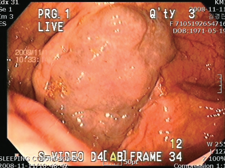
A 37-year-old woman was referred to her local hospital because of intermittent rectal bleeding. Colonoscopy revealed a blue polypoid lesion with a nodular surface in the cecum (Figure 1). A hemangioma was suspected. She was transferred to our hospital for further evaluation and surgical treatment.
Figure 1.

Colonoscopy of the cecum showing a bluish nodular lesion.
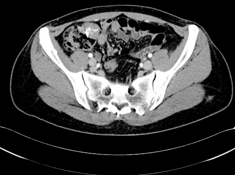
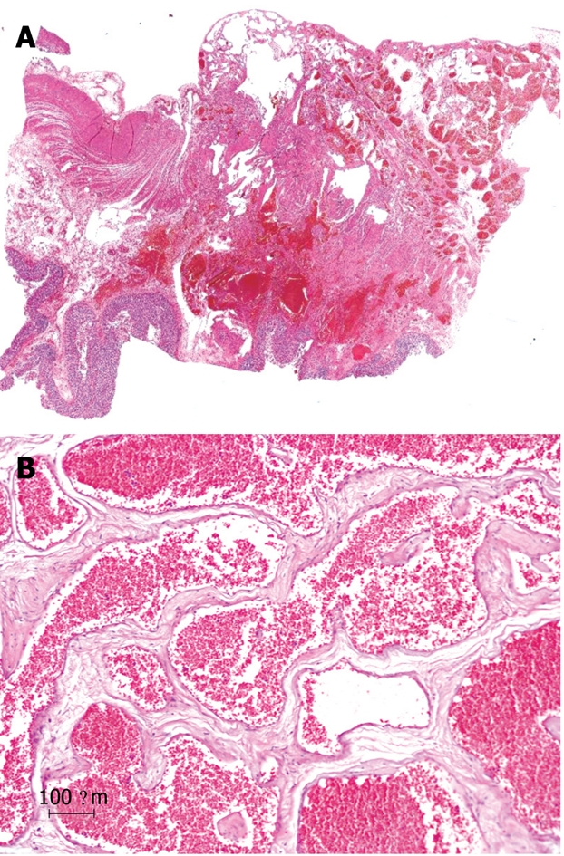
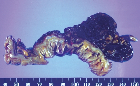
The physical examination revealed no specific signs. Abdominopelvic computed tomography (CT) showed a lesion in the cecum protruding extraluminally with heterogenous enhancement (Figure 2). The operative findings suggested a tumor involving the cecum and extending into the pericolic fat. Laparoscopic-assisted ileocecal resection and side-to-end ileo-ascending colon anastomosis were carried out. Macroscopically, the surgical specimen was a 5.8 cm × 4.2 cm cavernous hemangioma involving the entire wall of the colon and extending into the pericolic fat (Figure 3). Microscopically, the tumor consisted of large, dilated, blood-filled vessels lined by flattened endothelium. The vascular walls were thickened focally by adventitial fibrosis (Figure 4). Postoperatively, the patient has recovered well with no recurrence of symptoms.
Figure 2.

Abdominopelvic computed tomography showing a lesion in the cecum protruding extraluminally with heterogenous enhancement.
Figure 3.
Macroscopically, the surgical specimen was a cavernous hemangioma.
Figure 4.

HE staining of the hemangioma. A: Histological section showing large, dilated, blood-filled vessels lined by flattened endothelium (HE, × 1); B: The vascular walls were thickened focally by adventitial fibrosis (HE, × 100).
DISCUSSION
Hemangiomas are rare lesions of the colon, but vascular malformations of the gastrointestinal tract have been reported since 1839[7]. According to the literature, these lesions originate from embryonic sequestrations of mesodermal tissue[8]. Histologically, hemangiomas are distinct from telangiectasias and angiodysplasias, and most colonic hemangiomas are the capillary or cavernous subtype. Capillary hemangiomas consist of a proliferation of small capillaries composed of thin-walled spaces lined by endothelial cells, while cavernous hemangiomas consist of large spaces lined by single or multiple layers of endothelial cells[9]. The capillary subtype is usually solitary and causes no symptoms, while the cavernous subtype presents with bleeding (60%-90%), anemia (43%), obstruction (17%), and, rarely, with platelet sequestration, although approximately 10% of patients remain asymptomatic[10]. The endoscopic findings of hemangiomas of the colon vary. Grossly, hemangiomas appear as soft, compressible bluish or deep red submucosal lesions, with dilated, engorged veins in the rectal wall[5]. However, they can be difficult to diagnose in some cases, especially if the hemangioma has an unusual color or is covered. A histological diagnosis before treatment may be difficult because of the risk of uncontrollable bleeding following a biopsy[1,4]. Hence, these lesions should not be biopsied. Abdominal CT can provide useful information about the size of the lesion and its extension to adjacent organs. Visceral angiography may also be useful in excluding coexisting hemangiomas at other sites in the gastrointestinal tract[5].
Complete surgical resection is the best treatment for large or diffuse lesions[5], although endoscopic resection has sometimes been recommended for hemangiomas that are pedunculated, polypoid, small, and limited to the submucosal layer according to endoscopic ultrasonography[4]. Polypoid hemangiomas sometimes involve the entire wall of the colon, extending through the mesocolon and mesentery, as in our case[5]. In summary, we performed laparoscopic resection of a large cavernous hemangioma in the cecum. Although these lesions are rare, a better understanding of these lesions should help to obtain a definite diagnosis and devise an appropriate treatment plan.
Footnotes
Peer reviewer: Zvi Fireman, MD, Associate Professor of Medicine, Head, Gastroenterology Department, Hillel Yaffe Medical Center, POB 169, 38100, Hadera, Israel; Rafiq A Sheikh, MBBS, MD, MRCP, FACP, FACG, Department of Gastroenterology, Kaiser Permanente Medical Center, 6600 Bruceville Road, Sacramento, CA 95823, United States
S- Editor Li LF L- Editor Cant MR E- Editor Ma WH
References
- 1.Pontecorvo C, Lombardi S, Mottola L, Donisi M, DiTuoro A. Hemangiomas of the large bowel. Report of a case. Dis Colon Rectum. 1983;26:818–820. doi: 10.1007/BF02554759. [DOI] [PubMed] [Google Scholar]
- 2.van Deursen CT, Buijs J, Nap M. An uncommon polyp in the colon: a pedunculated cavernous hemangioma. Endoscopy. 2008;40 Suppl 2:E127. doi: 10.1055/s-2007-995697. [DOI] [PubMed] [Google Scholar]
- 3.Kimura S, Tanaka S, Kusunoki H, Kitadai Y, Sumii M, Tazuma S, Yoshihara M, Haruma K, Chayama K. Cavernous hemangioma in the ascending colon treated by endoscopic mucosal resection. J Gastroenterol Hepatol. 2007;22:280–281. doi: 10.1111/j.1440-1746.2006.03302.x. [DOI] [PubMed] [Google Scholar]
- 4.Hasegawa K, Lee WY, Noguchi T, Yaguchi T, Sasaki H, Nagasako K. Colonoscopic removal of hemangiomas. Dis Colon Rectum. 1981;24:85–89. doi: 10.1007/BF02604292. [DOI] [PubMed] [Google Scholar]
- 5.Sylla P, Deutsch G, Luo J, Recavarren C, Kim S, Heimann TM, Steinhagen RM. Cavernous, arteriovenous, and mixed hemangioma-lymphangioma of the rectosigmoid: rare causes of rectal bleeding--case series and review of the literature. Int J Colorectal Dis. 2008;23:653–658. doi: 10.1007/s00384-008-0466-4. [DOI] [PubMed] [Google Scholar]
- 6.Marinis A, Kairi E, Theodosopoulos T, Kondi-Pafiti A, Smyrniotis V. Right colon and liver hemangiomatosis: a case report and a review of the literature. World J Gastroenterol. 2006;12:6405–6407. doi: 10.3748/wjg.v12.i39.6405. [DOI] [PMC free article] [PubMed] [Google Scholar]
- 7.Head HD, Baker JQ, Muir RW. Hemangioma of the colon. Am J Surg. 1973;126:691–694. doi: 10.1016/s0002-9610(73)80024-8. [DOI] [PubMed] [Google Scholar]
- 8.Lyon DT, Mantia AG. Large-bowel hemangiomas. Dis Colon Rectum. 1984;27:404–414. doi: 10.1007/BF02553013. [DOI] [PubMed] [Google Scholar]
- 9.Levy AD, Abbott RM, Rohrmann CA Jr, Frazier AA, Kende A. Gastrointestinal hemangiomas: imaging findings with pathologic correlation in pediatric and adult patients. AJR Am J Roentgenol. 2001;177:1073–1081. doi: 10.2214/ajr.177.5.1771073. [DOI] [PubMed] [Google Scholar]
- 10.Hsu RM, Horton KM, Fishman EK. Diffuse cavernous hemangiomatosis of the colon: findings on three-dimensional CT colonography. AJR Am J Roentgenol. 2002;179:1042–1044. doi: 10.2214/ajr.179.4.1791042. [DOI] [PubMed] [Google Scholar]


