Abstract
A single exercise bout can increase insulin-independent glucose transport immediately postexercise and insulin-dependent glucose transport (GT) for several hours postexercise. Akt substrate of 160 kDa (AS160) and TBC1D1 are paralog Rab GTPase-activating proteins that have been proposed to contribute to these exercise effects. Previous research demonstrated greater AS160 and Akt threonine phosphorylation in rat skeletal muscle at 3–4 h postexercise concomitant with enhanced insulin-stimulated GT. To further probe whether these signaling events or TBC1D1 phosphorylation were important for the enhanced postexercise insulin-stimulated GT, male Wistar rats were studied using four experimental protocols (2-h swim exercise, differing with regard to timing of muscle sampling and whether food was provided postexercise) that were known to vary in their influence of insulin-independent and insulin-dependent GT postexercise. The results indicated that, in isolated rat epitrochlearis muscle, 1) elevated phosphorylation of AS160 (measured using anti-phospho-Akt substrate, PAS-AS160, and phosphospecific anti-Thr642-AS160, pThr642-AS160) consistently tracked with elevated insulin-stimulated GT; 2) PAS-TBC1D1 was not different from sedentary values at 3 or 27 h postexercise, when insulin sensitivity was increased; 3) insulin-stimulated Akt activity was not increased postexercise in muscles with increased insulin sensitivity; 4) PAS-TBC1D1 was increased immediately postexercise, when insulin-independent GT was elevated, and reversed at 3 and 27 h postexercise, when insulin-independent GT was also reversed; and 5) there was no significant effect of exercise or insulin on total abundance of AS160, TBC1D1, Akt, or GLUT4 protein with any of the protocols. The results are consistent with increased AS160 phosphorylation (PAS-AS160 or pThr642-AS160) but not increased PAS-TBC1D1 or Akt activity, which is important for increased postexercise insulin-stimulated GT in rat skeletal muscle. They also support the idea that increased TBC1D1 phosphorylation may play a role in the insulin-independent increase in GT postexercise.
Keywords: Akt substrate of 160 kDa, glucose transporter 4, Akt, glucose transport
a single bout of exercise leads to a subsequent increase in insulin-stimulated glucose transport (7, 9, 22, 40). The postexercise increase in insulin sensitivity is attributable to increased insulin-stimulated GLUT4 cell surface localization (24). However, many studies have found that prior exercise has no effect on proximal insulin-signaling steps, including insulin receptor binding (4, 5, 55), insulin receptor tyrosine phosphorylation (24, 26, 47, 53), insulin receptor tyrosine kinase activity (46, 47, 52), insulin receptor substrate tyrosine phosphorylation (24, 26, 54), insulin receptor substrate-associated phosphatidylinositol-3-kinase activity (46, 53), and Akt serine phosphorylation (1, 16, 23, 46, 52).
The Rab GTPase-activating protein (Rab GAP) Akt substrate of 160 kDa (AS160; also known as TBC1D4) is the most distal signaling protein that has been implicated in insulin-mediated glucose transporter 4 (GLUT4) translocation (8, 32, 42, 43). Arias et al. (1) found that phosphorylation of AS160 is greater in insulin-stimulated muscles from exercised (3–4 h postexercise) rats compared with sedentary controls. However, this increase was not because of greater insulin sensitivity for AS160 phosphorylation. Rather, it was because the baseline phosphorylation of AS160 in the absence of insulin remained increased 3–4 h postexercise. In other words, basal AS160 phosphorylation remained higher at 3–4 h postexercise compared with resting, and this elevated baseline accounted for the greater AS160 phosphorylation found in muscles that were subsequently stimulated with insulin. This observation is also supported by results in humans that indicated that skeletal muscle AS160 phosphorylation can remain elevated for 2–14 h postexercise without elevated insulin (17, 44, 49). Arias et al. (1) proposed that the persistent increase in insulin-stimulated glucose transport is attributable, at least in part, to the sustained increase in AS160 phosphorylation after acute exercise.
Phosphorylation of TBC1D1, a paralog Rab GAP of AS160, may also regulate GLUT4 translocation (10, 28, 41). In skeletal muscle, TBC1D1 phosphorylation is increased in response to insulin (19, 45), contraction (3, 18, 19, 45), or incubation with the AMP-activated protein kinase (AMPK) activator 5-aminoimidazole-4-carboxamide-1-β-d-ribofuranoside (AICAR) (45). However, it is unknown whether TBC1D1 phosphorylation is increased in response to in vivo exercise. The relationship between TBC1D1 phosphorylation and the postexercise increase in insulin-stimulated glucose transport has also not been investigated. Experiments using 3T3-L1 (10) or L6 (28) cells suggest that, although insulin can induce TBC1D1 phosphorylation, TBC1D1's inhibitory effects on GLUT4 translocation may not be subject to regulation by insulin. It remains possible that TBC1D1 participates in GLUT4 trafficking induced by insulin-independent stimuli, e.g., in vivo exercise.
Consistent with previous reports (12, 16, 23, 46, 52), Arias et al. (1) found that insulin-stimulated Akt serine phosphorylation (pSerAkt) was not enhanced at 3–4 h postexercise. However, in the same muscles, insulin-stimulated Akt threonine phosphorylation (pThrAkt) was greater at 3–4 h postexercise compared with sedentary controls. The insulin-stimulated phosphorylation of two Akt substrates (GSK-3 and AS160) was not greater at 3–4 h postexercise, suggesting that greater insulin-stimulated pThrAkt after exercise did not result in greater Akt activity. Nonetheless, it would be important to determine whether this prediction is true, because enhanced insulin-stimulated Akt activity could potentially contribute to increase insulin sensitivity.
To further probe the possible functional importance of AS160, TBC1D1, and Akt for the postexercise increase in insulin-stimulated glucose transport, we used four experimental protocols (differing with regard to the timing of muscle sampling and whether food was provided postexercise) that were known to vary in their influence on insulin-independent and insulin-dependent glucose transport after exercise (male Wistar rats, 2-h swim exercise). We hypothesized that the protocols with enhanced insulin-dependent glucose transport after exercise would be accompanied by increased AS160 phosphorylation and that protocols without enhanced insulin-dependent glucose transport after exercise would not be characterized by elevated AS160 phosphorylation. We also hypothesized that increased TBC1D1 phosphorylation for exercised vs. sedentary groups would be found in protocols with elevated insulin-independent glucose transport but not in protocols with an exercise effect on insulin-dependent glucose transport. We further hypothesized that the increased pThrAkt found in insulin-stimulated skeletal muscle at 3 h postexercise would not be accompanied by an increase in Akt activity compared with sedentary controls.
METHODS
Materials.
Human recombinant insulin was obtained from Eli Lilly (Indianapolis, IN). Reagents and apparatus for SDS-PAGE and immunoblotting were purchased from Bio-Rad (Hercules, CA). Bicinchoninic acid protein assay reagent (no. 23227), T-PER tissue protein extraction reagent (no. 78510), and West Dura Extended Duration Substrate (no. 34075) were from Pierce Biotechnology (Rockford, IL). Anti-Akt (no. 9272), anti-phospho-Thr308Akt (pThrAkt, no. 9275), anti-phospho-(Ser/Thr) Akt substrate (PAS; no. 9611), anti-GLUT4 (no. 2299), and goat anti-rabbit IgG horseradish peroxidase conjugate (no. 7074) were from Cell Signaling Technology (Danvers, MA). PAS was designed to recognize Akt phosphorylation motif peptide sequences (RXRXXpT/S). TBC1D1 polyclonal antibody was provided by Dr. Makoto Kanzaki at Tohoku University (35). Anti-AS160 (no. 07-741), anti-phospho-Thr642AS160 (pThr-AS160; no. 07-802), protein G-agarose beads (no. 16-266), and Akt immunoprecipitation kinase assay kit (no. 17-188) were from Upstate USA (Charlottesville, VA). 3-O-methyl-[3H]glucose ([3H]3-MG) was from Sigma-Aldrich. [14C]mannitol and [γ-32P]ATP were from PerkinElmer (Waltham, MA). Other reagents were from Sigma-Aldrich and Fisher Scientific (Pittsburgh, PA).
Animal treatment.
Procedures for animal care were approved by the University of Michigan Committee on Use and Care of Animals. Male Wistar rats (∼140–200 g; Harlan, Indianapolis, IN) were studied using four experimental protocols. In each protocol, rats were provided with rodent chow (Lab Diet; PMI Nutritional International, Brentwood, MO) and water ad libitum. At 1700 on the night before the experiments, rats were housed individually and provided ad libitum water and 4 g of chow each.
On the following day, rats were randomly assigned to a postexercise (PEX) or sedentary (SED) treatment. Beginning at ∼0900, PEX rats swam in a barrel filled with water (35°C) to a depth of ∼60 cm (6 or 7 rats/barrel) for 4 × 30 min bouts, with a 5-min rest period between each bout. The four experimental protocols differed for the PEX groups and their respective SED controls only with regard to 1) the time after completion of exercise when muscles were dissected out from anesthetized rats and 2) whether rats had access to chow after exercise. The eight groups (a PEX and a SED group for each protocol) were 1) anesthetized immediately postexercise without access to food after exercise (0-h PEX and 0-h SED), 2) anesthetized 3 h postexercise without access to food after exercise (3-h PEX and 3-h SED), 3) anesthetized 3 h postexercise with unlimited access to food after exercise (3-h PEX-chow and 3-h SED-chow), and 4) anesthetized 27 h postexercise without access to food after exercise (27-h PEX and 27-h SED). While the rats were under deep anesthesia, both epitrochlearis muscles were rapidly dissected out and either freeze-clamped immediately or transferred to vials for subsequent incubation.
Muscle incubations.
For all incubation steps, flasks were continuously gassed from above with 95% O2-5% CO2 and shaken in a heated water bath. Isolated epitrochlearis muscles were incubated in Krebs-Henseleit buffer (KHB) + 0.1% bovine serum albumin (BSA) + 8 mM glucose + 2 mM mannitol (solution 1) for 30 min in a water bath at 35°C. During this step, one muscle from each rat was incubated in solution 1 supplemented with 50 μU/ml of insulin, and the contralateral muscle was incubated in solution 1 without insulin. Insulin remained present at the same concentration throughout all subsequent incubations. After the initial incubation, muscles were transferred to vials containing KHB + BSA + 2 mM pyruvate + 6 mM mannitol (solution 2) at 30°C for 10 min. Finally, muscles were transferred to flasks containing KHB, 0.1% BSA with 8 mM 3-MG (including 0.25 mCi/mmol [3H]3-MG), and 2 mM mannitol (including 0.1 mCi/mmol [14C]mannitol). After incubation with 3-MG for 15 min, the muscles were rapidly blotted on filter paper dampened with incubation medium, trimmed, freeze-clamped, and stored at −80°C until being processed as described below.
3-MG transport and protein phosphorylation.
The procedures for 3-MG transport, immunoprecipitation, and immunoblotting were conducted as described previously (19). For PAS-AS160, muscles were immunoprecipitated using anti-PAS, followed by immunoblotting by anti-AS160. For PAS-TBC1D1, muscles were immunoprecipitated using anti-TBC1D1, followed by immunoblotting by anti-PAS. The mean values for basal muscles (sedentary without insulin) on each blot were normalized to equal 1.0, and then all samples on the blot were expressed relative to the normalized basal value.
Akt activity measurement.
Akt activity was determined according to the manufacturer's instructions (Upstate USA). Briefly, protein G-agarose beads were rotated overnight with anti-Akt/PH domain clone SKB1 (no. 05-591). The antibody/protein G-agarose mixture was combined with 300 μg of protein from each sample and rotated for 2 h at 4°C. The antigen/antibody/protein G-agarose complex was combined with Akt substrate peptide (no. 12-340) and [γ-32P]ATP (final concentration of 1 μCi/μl) and shaken at room temperature for 60 min. Next, the complex was centrifuged (4,000 g for 1 min), and 40 μl of supernatant was collected and transferred to phosphocellulose paper. After washes (3 times with 1.5% phosphoric acid and once with acetone), the phosphocellulose paper was transferred to a vial containing scintillation cocktail for scintillation counting. The mean values for basal muscles (sedentary without insulin) on each experiment day were normalized to equal 1.0, and then all samples were expressed relative to the normalized basal value.
Muscle glycogen concentration.
Muscles used for measurement of glycogen were frozen immediately after dissection, weighed, and homogenized in ice-cold 0.3 M perchloric acid. An aliquot of the homogenate was stored at −80°C for later determination of glycogen concentration by the amyloglucosidase method (37).
Statistical analysis.
Statistical analyses were performed using Sigma Stat (San Rafael, CA) version 2.0. Data are expressed as means ± SE. P ≤ 0.05 was considered statistically significant. Data from the 3-h PEX and 27-h PEX experiments were analyzed with two-way ANOVA and the Student-Newman-Keuls post hoc test. When data failed the Levene Median test for equal variance, the Kruskal-Wallis nonparametric ANOVA on ranks was used with Dunn's post hoc test. The insulin-stimulated increases (Δinsulin) in glucose transport, protein phosphorylation, and Akt activity were calculated by subtracting the values for muscles incubated without insulin from the respective values of paired muscles incubated with insulin. Student's t-test was used for the analysis of glucose transport and protein phosphorylation for 0-h PEX experiments and Δinsulin values for 3-h PEX and 27-h PEX experiments.
RESULTS
3-MG transport.
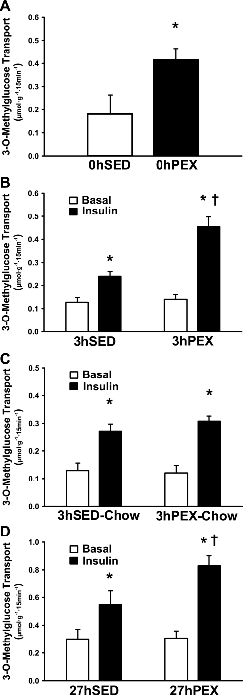
Insulin-independent glucose transport was ∼2.3-fold greater (P < 0.05) for 0-h PEX rats compared with sedentary (0-h SED) controls (Fig. 1A), and this increase was completely reversed at both 3 and 27 h postexercise (Fig. 1, B–D). Insulin-treated muscles had a greater glucose transport than paired muscles incubated without insulin for all groups (Fig. 1, B–D). PEX vs. SED groups that were not refed chow after exercise (Fig. 1, B and D) had significantly greater glucose transport for insulin-stimulated muscles at both 3-h PEX (P < 0.001) and 27-h PEX (P < 0.01). Significant (P < 0.01) effects of prior exercise were also found for Δinsulin glucose transport for the 3-h PEX (0.322 ± 0.052 μmol·g−1·15 min−1) vs. 3-h SED (0.125 ± 0.024 μmol·g−1·15 min−1) and 27-h PEX (0.523 ± 0.075 μmol·g−1·15 min−1) vs. 27-h SED (0.240 ± 0.068 μmol·g−1·15 min−1) groups. The exercise effects on glucose transport by insulin-stimulated muscles and Δinsulin glucose transport were lost with chow refeeding (Fig. 1C); i.e., there were no significant differences between the 3-h SED-chow (0.144 ± 0.039 μmol·g−1·15 min−1) and 3-h PEX-Chow (0.187 ± 0.037 μmol·g−1·15 min−1) groups.
Fig. 1.
Rate of 3-O-methylglucose (3-MG) transport in isolated rat epitrochlearis muscles. A: rats anesthetized immediately postexercise without access to food after exercise (0-h PEX); B: rats anesthetized 3 h postexercise without access to food after exercise (3-h PEX); C: rats anesthetized 3 h postexercise with unlimited access to food after exercise (3-h PEX-chow); D: rats anesthetized 27 h postexercise without access to food after exercise (27-h PEX). For rats in the 0-h PEX and 0-h SED groups, all muscles that were used to measure 3-MG transport were incubated without insulin to determine the insulin-independent effect of exercise. For rats in the 3-h PEX, 3-h PEX-chow, and 27-h PEX groups and their respective sedentary controls (3-h SED, 3-h SED-chow, and 27-h SED, respectively), 1 of the paired muscles was incubated without insulin, and the contralateral muscle was incubated with insulin. A: data are meanss ± SE; n = 6/group. *P < 0.05 (exercise effect, t-test). Open bar, 0-h SED; filled bar, 0-h PEX. B–D: data are means ± SE; n = 6–13/group. *P < 0.05 (insulin effect, post hoc test); †P < 0.05 (exercise effect, post hoc test). Open bars, without insulin; filled bars, with insulin.
Total protein abundance.
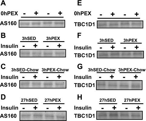
The total protein abundance of AS160 (Fig. 2, A–D), TBC1D1 (Fig. 2, E–H), GLUT4 (data not shown), and Akt (data not shown) was unaltered by the experimental treatments (insulin or exercise; n = 4 for each protein within every exercise and insulin treatment group) in every protocol studied (0-h PEX, 3-h PEX, 3-h PEX-chow, or 27-h PEX).
Fig. 2.
Akt substrate of 160 kDa (AS160) and TBC1D1 total protein abundance in rat epitrochlearis muscles. There were no significant effects of exercise or insulin with any of the protocols (n = 4 for each exercise and insulin treatment group). A: AS160 of 0-h PEX; B: AS160 of 3-h PEX; C: AS160 of 3-h PEX-chow; D: AS160 of 27-h PEX; E: TBC1D1 of 0-h PEX; F: TBC1D1 of 3-h PEX; G: TBC1D1 of 3-h PEX-chow; H: TBC1D1 of 27-h PEX.
AS160 phosphorylation.
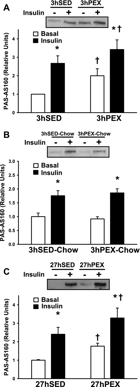
Insulin treatment compared with no insulin resulted in significantly greater PAS-phosphorylation of AS160 (PAS-AS160) in all groups (Fig. 3, A–C). Consistent with our previous study (1), PAS-AS160 in muscles incubated without insulin was significantly greater (P < 0.05) in the 3-h PEX compared with the 3-h SED group (Fig. 3A). Also consistent with our previous study (1), PAS-AS160 in muscles incubated with insulin was significantly greater (P < 0.05) in the 3-h PEX compared with the 3-h SED group (Fig. 3A). In contrast, PAS-AS160 was not different between 3-h SED-chow and 3-h PEX-chow groups with or without insulin during the incubations (Fig. 3B). PAS-AS160 in muscles incubated without insulin was significantly greater (P < 0.05) in the 27-h PEX compared with the 27-h SED group (Fig. 3C). PAS-AS160 in muscles incubated with insulin was also significantly greater (P < 0.05) in 27-h PEX compared with 27-h PEX group (Fig. 3C). The Δinsulin values were not significantly different between SED and PEX rats for any of the groups (data not shown).
Fig. 3.
AS160 phospho-(Ser/Thr) Akt substrate (PAS)-phosphorylation in rat epitrochlearis muscles. A: 3-h PEX; B: 3-h PEX-chow; C: 27-h PEX. One of the paired muscles from each rat was incubated without insulin, and the contralateral muscle was incubated with insulin. Data are means ± SE; n = 6–13/group. *P < 0.05 (insulin effect, post hoc test); †P < 0.05 (exercise effect, post hoc test). Open bars, without insulin; filled bars, with insulin. The SE value for the basal 3-h SED group in Fig. 2A is too small to be visible.
AS160 Thr642 phosphorylation.
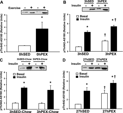
AS160 Thr642 phosphorylation was significantly elevated (P < 0.05) for the 0-h PEX compared with the 0-h SED group (Fig. 4A). Insulin treatment compared with no insulin resulted in significantly greater pThr642-AS160 in all groups (Fig. 4, B–D). Consistent with the results for PAS-AS160, pThr642-AS160 in muscles incubated without insulin was significantly greater (P < 0.001) in the 3-h PEX compared with the 3-h SED group (Fig. 4B). Also consistent with the results on PAS-AS160, pThr642-AS160 in muscles incubated with insulin was significantly greater (P < 0.001) in the 3-h PEX compared with the 3-h SED group (Fig. 4B). As was found with PAS-AS160, pThr642-AS160 was not different between 3-h SED-chow and 3-h PEX-chow groups (Fig. 4C). The 27-h PEX group compared with the 27-h SED group had greater pThr642-AS160 with (P < 0.05) or without (P < 0.005) insulin (Fig. 4D). The Δinsulin values were not significantly different between SED and PEX rats for any of the protocols (data not shown).
Fig. 4.
AS160 Thr642 phosphorylation in rat epitrochlearis muscles. A: 0-h PEX; B: 3-h PEX; C: 3-h PEX-chow; D: 27-h PEX. For rats in the 0-h PEX group, muscles were frozen immediately after dissection and used to determine the insulin-independent effect of exercise. For rats in the 3-h PEX, 3-h PEX-chow, and 27-h PEX groups and their respective SED controls (3-h SED, 3-h SED-chow, and 27-h SED, respectively), 1 paired muscle from each rat was incubated without insulin, and the contralateral muscle was incubated with insulin. A: data are means ± SE; n = 4/group. *P < 0.05 (exercise effect, t-test). Open bar, 0-h SED; filled bar, 0-h PEX. B–D: data are means ± SE; n = 4/group. *P < 0.05 (insulin effect, post hoc test); †P < 0.05 (exercise effect, post hoc test). Open bars, without insulin; filled bars, with insulin.
TBC1D1 phosphorylation.
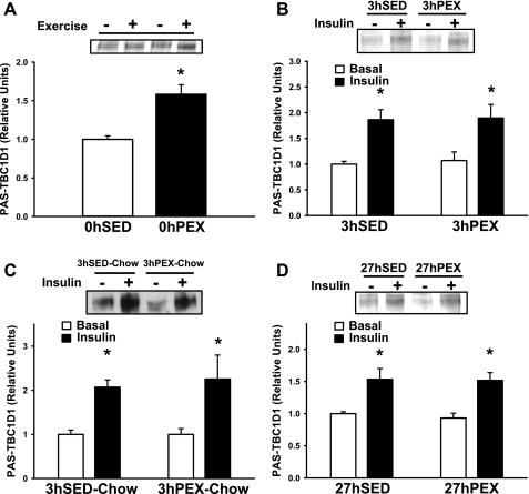
PAS-phosphorylation of TBC1D1 (PAS-TBC1D1) was significantly elevated (P < 0.001) in 0-h PEX compared with 0-h SED rats (Fig. 5A). This increase was completely reversed at 3 h postexercise, with or without refeeding, and at 27 h postexercise (Fig. 5, B–D). Insulin treatment compared with no insulin resulted in significantly greater PAS-TBC1D1 in all groups (Fig. 5, B–D). However, in contrast to PAS-AS160, PAS-TBC1D1 was not different for 3-h PEX, 3-h PEX-chow, or 27-h PEX compared with the respective sedentary controls (3-h SED, 3-h SED-chow, and 27-h SED) regardless of insulin concentration. The Δinsulin values were also not significantly different between SED and PEX rats for any of the groups (data not shown).
Fig. 5.
TBC1D1 PAS-phosphorylation in rat epitrochlearis muscles. A: 0-h PEX; B: 3-h PEX; C: 3-h PEX-chow; D: 27-h PEX. For rats in the 0-h PEX group, muscles were frozen immediately after dissection and used to determine the insulin-independent effect of exercise. For rats in the 3-h PEX, 3-h PEX-chow, and 27-h PEX groups and their respective SED controls (3-h SED, 3-h SED-chow, and 27-h SED, respectively), 1 of the paired muscles from each rat was incubated without insulin, and the contralateral muscle was incubated with insulin. A: data are means ± SE; n = 6/group. *P < 0.05 (exercise effect, t-test). Open bar, 0-h SED; filled bar, 0-h PEX. B–D: data are means ± SE; n = 6–10/group. *P < 0.05 (insulin effect, post hoc test). Open bars, without insulin; filled bars, with insulin.
Akt threonine phosphorylation.
Insulin treatment compared with no insulin resulted in significantly greater pThrAkt in all groups (Fig. 6, A–C). Consistent with our previous results (1), pThrAkt in muscles incubated with insulin was greater (P < 0.05) for the 3-h PEX group compared with 3-h SED controls (Fig. 6A). Furthermore, there was also a significant (P < 0.05) increase for the 27-h PEX vs. the 27-h SED group (Fig. 6C). The Δinsulin values were significantly greater for the 3-h PEX vs. 3-h SED and 27-h PEX vs. 27-h SED groups (P < 0.05). In contrast, neither the pThrAkt in muscles incubated with insulin (Fig. 6B) nor the Δinsulin values (data not shown) were significantly different between the 3-h SED-chow and 3-h PEX-chow groups.
Fig. 6.
Akt threonine phosphorylation and Akt activity in rat epitrochlearis muscles. One of the paired muscles from each rat was incubated without insulin, and the contralateral muscle was incubated with insulin. A: phospho-Thr308Akt (pThrAkt) 3-h PEX; B: pThrAkt 3-h PEX-chow; C: pThrAkt 27-h PEX; D: Akt activity 3-h PEX. Data are means ± SE; n = 7–12/group. *P < 0.05 (insulin effect, post hoc test); †P < 0.05 (exercise effect, post hoc test). Open bars, without insulin; filled bars, with insulin. The SE value for the basal 3-h SED group in Fig. 5A is too small to be visible.
Akt activity.
Insulin treatment compared with no insulin resulted in significantly greater Akt activity (Fig. 6D). Unlike pThrAkt, Akt activity for insulin-stimulated muscles was not significantly different between the 3-h SED and 3-h PEX groups. The Δinsulin values were also not different between 3-h SED (0.831 ± 0.173 relative units) and 3-h PEX rats (0.886 ± 0.373 relative units).
Glycogen concentration.
Epitrochlearis glycogen concentration (Table 1) was reduced by ∼60% (P < 0.001) immediately postexercise (0-h PEX vs. 0-h SED). In the exercised groups that were not refed (3-h PEX and 27-h PEX), glycogen did not increase significantly above the 0-h PEX values. Glycogen values were also essentially unchanged for 3-h SED vs. 0-h SED, but there was an ∼30% reduction (P < 0.05) for the 27-h SED group compared with the other sedentary groups that were not refed (0-h SED and 3-h SED). With this decline there was no significant difference for the 27-h SED compared with any of the exercised groups that were not refed (0-h PEX, 3-h PEX, and 27-h PEX). Glycogen, which was increased in the 3-h PEX-chow above all other groups (P < 0.05), was approximately fourfold greater than the 0-h PEX group and ∼50% greater than the 3-h SED-chow rats. Chow refeeding did not significantly increase glycogen for the 3-h SED group compared with the 0-h SED or 3-h SED animals.
Table 1.
Muscle glycogen concentration
| Time, h | SED | PEX | SED-Chow | PEX-Chow |
|---|---|---|---|---|
| 0 | 14.4±1.5* | 5.9±0.6# | ||
| 3 | 13.5±1.9* | 5.8±1.1# | 17.5±2.2* | 25.9±1.8† |
| 27 | 9.4±0.3# | 8.8±1.1# |
Values are means ± SE (μmol/g muscle); n = 6-8/group. SED, sedentary; PEX, postexercise; SED- and PEX-chow, SED and PEX rats, respectively, with unlimited access to food. Time refers to the time at which muscles were sampled relative to the completion of exercise by PEX groups. Values not marked with the same symbol (
) were significantly different (P < 0.05). 0 = immediately PEX; 3 = 3 h PEX; 27 = 27 h PEX.
DISCUSSION
The results support the hypothesis that a sustained increase in AS160 phosphorylation, but not TBC1D1 phosphorylation, consistently occurs with protocols that cause a sustained postexercise increase in insulin-stimulated glucose transport. Specifically, the data 1) indicated an increased PAS-AS160 and pThr642-AS160 in skeletal muscle, with or without insulin, and enhanced insulin-stimulated glucose transport at 3 h postexercise in rats that were not refed; 2) extended the previously published results (1) by demonstrating the postexercise increase in PAS-AS160 and pThr642-AS160 together with increased insulin-stimulated glucose transport at 27 h postexercise in rats not refed; 3) demonstrated the elimination of increased PAS-AS160, pThr642-AS160, and increased insulin-stimulated glucose transport at 3 h postexercise in rats that were refed; and 4) found that neither insulin nor exercise altered the total abundance of AS160, TBC1D1, Akt, or GLUT4 protein in any of the protocols tested. In addition, there was not a persistent increase in PAS-TBC1D1 at 3 or 27 h postexercise, supporting the idea that AS160 phosphorylation, rather than TBC1D1 phosphorylation, is important for improved insulin sensitivity after exercise. Furthermore, the lack of an increase in insulin-stimulated Akt activity after exercise indicates that this mechanism cannot account for the improved insulin sensitivity.
There was a persistent increase in basal (without insulin) AS160 phosphorylation in the 3-h PEX and 27-h PEX groups, and this higher baseline value accounted for the greater AS160 phosphorylation in insulin-stimulated muscles after exercise. What is a possible mechanism that could link the elevated AS160 phosphorylation without insulin with increased insulin-stimulated glucose transport after exercise? It is important to recognize that AS160 does not regulate all of the steps required for GLUT4 to be redistributed from intracellular storage vesicles to the cell surface membranes, where they are able to facilitate glucose uptake (27, 51). Studies in both 3T3-L1 adipocytes (2) and L6 myoblasts (38) suggest that AS160 phosphorylation is required for insulin-stimulated docking of GLUT4 vesicles to cell surface membranes. AS160 apparently does not regulate several of the other steps that are required for complete GLUT4 translocation and increased glucose transport rate, including the recruitment, tethering, and fusion of GLUT4 vesicles with surface membranes and possibly GLUT4 activation (2, 20, 27, 51). We speculate that the persistent increase in AS160 phosphorylation in the absence of insulin results in a change in the localization of a portion of the GLUT4 vesicles so that they are more susceptible to the insulin-stimulated, AS160-independent steps of GLUT4 vesicle traffic. The idea is that the persistent increase in AS160 phosphorylation found several hours after exercise may serve to “prime the pump” so that, when the muscle is subsequently stimulated with insulin, there is a greater pool of GLUT4 that is susceptible to being recruited by a given amount of insulin. This scenario is similar to a mechanism that was previously proposed by Holloszy (25). We propose that AS160-independent regulatory steps that become activated upon insulin stimulation, acting in concert with the persistent effect of exercise on AS160 phosphorylation, culminate in the enhanced postexercise insulin-stimulated GLUT4 translocation and glucose transport. This model does not exclude the possibility that prior exercise also amplifies insulin-signaling steps other than AS160 phosphorylation.
In contrast to PAS-AS160 and pThr642-AS160, PAS-TBC1D1 was not different for the 3-h PEX, 3-h PEX-chow, and 27-h PEX groups compared with their respective sedentary control groups (3-h SED, 3-h SED-chow, and 27-h SED), demonstrating that enhanced PAS-TBC1D1 is not essential for the postexercise increase in insulin-stimulated glucose transport. It remains possible that there is a persistent effect of exercise on TBC1D1 by another mechanism such as phosphorylation on sites not recognized by the PAS antibody or changes in subcellular localization (10, 11, 41). Therefore, we cannot rule out the possibility that TBC1D1 participates in the increased insulin-stimulated glucose transport after exercise. Nevertheless, the results suggest that 1) PAS-phosphorylation of TBC1D1 is not essential for increased insulin-stimulated glucose transport postexercise and 2) postexercise reversal of the increased phosphorylation of TBC1D1 and AS160 is regulated differently.
We confirmed the previous observation indicating that there is an increase in insulin-stimulated pThrAkt at 3 h postexercise in rats not fed after exercise (1) and extended this result to show enhanced insulin-stimulated pThrAkt at 27 h postexercise in rats that remained unfed. Furthermore, insulin-stimulated pThrAkt was not increased 3 h postexercise in rats that were refed. Thus, the effect of exercise on insulin-stimulated pThrAkt tracked with insulin-stimulated glucose transport for each of the protocols. However, because there was not a concomitant increase in insulin-stimulated Akt activity at 3 h postexercise in rats not refed, it seems unlikely that the elevated pThrAkt could account for improved insulin sensitivity. These data suggest that, in postexercise muscles, 1) the postexercise enhancement of insulin-stimulated pThrAkt does not induce greater than usual increase in insulin-stimulated Akt activity and 2) enhanced Akt activity is not responsible for the enhanced insulin-stimulated glucose transport postexercise. The similar Akt activity found in sedentary compared with postexercise groups is also consistent with our previous results indicating that exercise did not alter the insulin-stimulated (Δinsulin) increase in phosphorylation of Akt substrates pGSK-3 and PAS-AS160 (1). The current study also found that prior exercise did not alter the ability of insulin to induce the phosphorylation of Akt substrates (as assessed by PAS-AS160, pThr642-AS160 and PAS-TBC1D1) in skeletal muscle. These results are consistent with a number of studies indicating that acute exercise does not amplify proximal insulin signaling in skeletal muscle.
Although elevated PAS-TBC1D1 could not explain the long-lasting increase in insulin-dependent glucose transport found at 3 or 27 h postexercise, the increased PAS-TBC1D1 tracked closely with the more transient postexercise effect on insulin-independent glucose transport (14, 50). The exercise effects on PAS-TBC1D1 and glucose transport in the absence of insulin were both substantially elevated at 0-h PEX, and the exercise effects in both were also completely reversed in the 3-h PEX, 3-h PEX-chow, and 27-h PEX groups. This consistent association supports the idea that PAS-TBC1D1 may play an important role in contraction-stimulated glucose transport (3, 19). The relationship between PAS-TBC1D1 and insulin-independent glucose transport with contraction or exercise is in striking contrast to the results for PAS-AS160, which remained increased without insulin at 3-h PEX and 27-h PEX compared with respective sedentary controls in the absence of an exercise effect on insulin-independent glucose transport. We previously found that wortmannin completely eliminated the contraction-stimulated increase in PAS-AS160 without altering the contraction-stimulated increases in PAS-TBC1D1 or glucose transport (19). Furthermore, an AMPK inhibitor eliminated the contraction effect on PAS-TBC1D1 and reduced contraction-stimulated glucose transport by 65% without attenuating the contraction-induced increase in PAS-AS160. In conjunction with many other results (1, 6, 15, 19, 48), these data suggested that AS160 phosphorylation is neither necessary nor sufficient for increased insulin-independent glucose transport after exercise or contraction. The results are also consistent with the possibility that TBC1D1 plays a role in a portion of the insulin-independent increase in glucose transport induced by exercise or contraction. Our working hypothesis is that AS160 phosphorylation is more important for insulin-stimulated glucose transport, whereas TBC1D1 phosphorylation is more important for exercise-stimulated glucose transport. Regulation of AS160 by its calmodulin-binding domain has been implicated in a portion of the contraction-stimulated increase in glucose transport (31). TBC1D1 also has a calmodulin-binding domain, but its role in contraction-stimulated glucose transport has not been assessed.
Both AS160 and TBC1D1 can be phosphorylated by multiple kinases (11, 21). Many studies have shown that in vivo exercise can activate AMPK (23, 39, 44, 48), and Arias et al. (1) found increased pAMPK at 0-h PEX using the same protocol as that in the current study. Although increased Akt activity after in vivo exercise has been reported (44, 48), some studies have not detected Akt activation in skeletal muscle with in vivo exercise (17, 33, 53), including Arias et al. (1), who found unaltered pThrAkt and pSerAkt at 0-h PEX. These results suggest AMPK as a candidate for the increased AS160 and TBC1D1 phosphorylation at 0-h PEX, but we cannot rule out the possibility that Akt was transiently activated in the early stages of the exercise or a role for other kinases that were not tested.
Both AS160 and TBC1D1 can be phosphorylated on multiple sites (11, 21, 29). Treebak et al. (49) recently reported that one-legged exercise by humans resulted in an elevation of AS160 phosphorylation on Ser318, Ser341, and Ser751, with a trend for an increase on Ser588 in muscle in the absence of insulin infusion (at 240 min postexercise) and with insulin infusion (at 340 min postexercise). There was no effect of prior exercise on AS160 phosphorylation detected on Thr642 or Ser666 or using anti-PAS. Previous research with humans had found increased PAS-AS160 at 2.5 h postexercise (44). Although the explanation for the different results for PAS-AS160 and pThr642-AS160 after exercise is uncertain, there is a great deal of evidence for a persistent increase in AS160 in skeletal muscle after acute exercise.
As expected, exercise resulted in decreased muscle glycogen content. It has been hypothesized that reduced glycogen concentration is involved in the postexercise increase in insulin-stimulated glucose transport (13, 34, 36). However, activation of AMPK by AICAR can result in subsequently elevated insulin-stimulated glucose transport in the absence of altered glycogen levels (16). Furthermore, a study that compared multiple in situ contraction protocols found that all protocols that resulted in decreased glycogen concentration also resulted in greater insulin sensitivity postexercise, but not all protocols that induced a similar decrement in glycogen levels were also characterized by improved insulin sensitivity (30). These findings suggest that glycogen reduction may be necessary, but not sufficient, for a postexercise-induced increase in insulin-stimulated glucose transport. The reduced muscle glycogen levels at the 3-h PEX and 27-h PEX groups compared with the 0-h SED group are consistent with the idea that glycogen reduction may contribute to the postexercise increase in insulin-stimulated glucose transport, and the higher glycogen content in 3-h PEX-chow group vs. the 3-h SED group is consistent with the idea that glycogen resynthesis may contribute to the reversal of the postexercise effect on insulin sensitivity. However, the lack of difference in glycogen content between 27-h SED and 27-h PEX groups, taken together with earlier results (30), suggests that lower glycogen values do not fully explain the mechanism for the prolonged increase in insulin sensitivity on the day after exercise.
In conclusion, the results of this study support a model in which AS160 and TBC1D1, two Rab GAP paralogs, have different roles in the postexercise effects on skeletal muscle glucose transport. The major new findings indicate that 1) elevated PAS-AS160 or pThr642-AS160 after exercise consistently tracked with elevated insulin-stimulated glucose transport, supporting the idea that AS160 phosphorylation plays a role in the postexercise increase in insulin sensitivity; 2) PAS-TBC1D1 was not different from sedentary control values at 3 or 27 h after exercise, when insulin-stimulated glucose transport was increased, demonstrating that the postexercise dephosphorylation of TBC1D1 is regulated differently from AS160, and PAS-TBC1D1 is not important for the postexercise increase in insulin-stimulated glucose transport; 3) insulin-stimulated Akt activity was not increased at 3 h after exercise, at which time insulin-stimulated glucose transport was enhanced, demonstrating that it cannot account for increased postexercise insulin sensitivity; and 4) the temporal relationship between the postexercise effects on insulin-independent glucose transport and PAS-TBC1D1 (both of which are elevated immediately after in vivo exercise and reversed at 3 and 27 h postexercise) supports the hypothesis that TBC1D1 phosphorylation may play a role in the transient exercise-induced increase in glucose transport in the absence of insulin. It will be important for future research to identify the unique phosphosignatures of AS160 and TBC1D1 in response to exercise and insulin to determine their protein-protein interactions (especially with 14-3-3 proteins) and to characterize how their subcellular localization is influenced by exercise and insulin.
GRANTS
This research was supported by National Institutes of Health Grants AG-010026 and DK-071771 (to G. D. Cartee).
REFERENCES
- 1.Arias EB, Kim J, Funai K, Cartee GD. Prior exercise increases phosphorylation of Akt substrate of 160 kDa (AS160) in rat skeletal muscle. Am J Physiol Endocrinol Metab 292: E1191–E1200, 2007. [DOI] [PubMed] [Google Scholar]
- 2.Bai L, Wang Y, Fan J, Chen Y, Ji W, Qu A, Xu P, James DE, Xu T. Dissecting multiple steps of GLUT4 trafficking and identifying the sites of insulin action. Cell Metab 5: 47–57, 2007. [DOI] [PubMed] [Google Scholar]
- 3.Blair DR, Funai K, Schweitzer GG, Cartee GD. A myosin II ATPase inhibitor reduces force production, glucose transport, and phosphorylation of AMPK and TBC1D1 in electrically stimulated rat skeletal muscle. Am J Physiol Endocrinol Metab 296: E993–E1002, 2009. [DOI] [PMC free article] [PubMed] [Google Scholar]
- 4.Bonen A, Tan MH, Clune P, Kirby RL. Effects of exercise on insulin binding to human muscle. Am J Physiol Endocrinol Metab 248: E403–E408, 1985. [DOI] [PubMed] [Google Scholar]
- 5.Bonen A, Tan MH, Watson-Wright WM. Effects of exercise on insulin binding and glucose metabolism in muscle. Can J Physiol Pharmacol 62: 1500–1504, 1984. [DOI] [PubMed] [Google Scholar]
- 6.Bruss MD, Arias EB, Lienhard GE, Cartee GD. Increased phosphorylation of Akt substrate of 160 kDa (AS160) in rat skeletal muscle in response to insulin or contractile activity. Diabetes 54: 41–50, 2005. [DOI] [PubMed] [Google Scholar]
- 7.Cartee GD, Holloszy JO. Exercise increases susceptibility of muscle glucose transport to activation by various stimuli. Am J Physiol Endocrinol Metab 258: E390–E393, 1990. [DOI] [PubMed] [Google Scholar]
- 8.Cartee GD, Wojtaszewski JF. Role of Akt substrate of 160 kDa in insulin-stimulated and contraction-stimulated glucose transport. Appl Physiol Nutr Metab 32: 557–566, 2007. [DOI] [PubMed] [Google Scholar]
- 9.Cartee GD, Young DA, Sleeper MD, Zierath J, Wallberg-Henriksson H, Holloszy JO. Prolonged increase in insulin-stimulated glucose transport in muscle after exercise. Am J Physiol Endocrinol Metab 256: E494–E499, 1989. [DOI] [PubMed] [Google Scholar]
- 10.Chavez JA, Roach WG, Keller SR, Lane WS, Lienhard GE. Inhibition of GLUT4 translocation by Tbc1d1, a Rab GTPase-activating protein abundant in skeletal muscle, is partially relieved by AMP-activated protein kinase activation. J Biol Chem 283: 9187–9195, 2008. [DOI] [PMC free article] [PubMed] [Google Scholar]
- 11.Chen S, Murphy J, Toth R, Campbell DG, Morrice NA, Mackintosh C. Complementary regulation of TBC1D1 and AS160 by growth factors, insulin and AMPK activators. Biochem J 409: 449–459, 2008. [DOI] [PubMed] [Google Scholar]
- 12.Chibalin AV, Yu M, Ryder JW, Song XM, Galuska D, Krook A, Wallberg-Henriksson H, Zierath JR. Exercise-induced changes in expression and activity of proteins involved in insulin signal transduction in skeletal muscle: differential effects on insulin-receptor substrates 1 and 2. Proc Natl Acad Sci USA 97: 38–43, 2000. [DOI] [PMC free article] [PubMed] [Google Scholar]
- 13.Coderre L, Kandror KV, Vallega G, Pilch PF. Identification and characterization of an exercise-sensitive pool of glucose transporters in skeletal muscle. J Biol Chem 270: 27584–27588, 1995. [DOI] [PubMed] [Google Scholar]
- 14.Constable SH, Favier RJ, Cartee GD, Young DA, Holloszy JO. Muscle glucose transport: interactions of in vitro contractions, insulin, and exercise. J Appl Physiol 64: 2329–2332, 1988. [DOI] [PubMed] [Google Scholar]
- 15.Dreyer HC, Drummond MJ, Glynn EL, Fujita S, Chinkes DL, Volpi E, Rasmussen BB. Resistance exercise increases human skeletal muscle AS160/TBC1D4 phosphorylation in association with enhanced leg glucose uptake during postexercise recovery. J Appl Physiol 105: 1967–1974, 2008. [DOI] [PMC free article] [PubMed] [Google Scholar]
- 16.Fisher JS, Gao J, Han DH, Holloszy JO, Nolte LA. Activation of AMP kinase enhances sensitivity of muscle glucose transport to insulin. Am J Physiol Endocrinol Metab 282: E18–E23, 2002. [DOI] [PubMed] [Google Scholar]
- 17.Frosig C, Rose AJ, Treebak JT, Kiens B, Richter EA, Wojtaszewski JF. Effects of endurance exercise training on insulin signaling in human skeletal muscle: interactions at the level of phosphatidylinositol 3-kinase, Akt, and AS160. Diabetes 56: 2093–2102, 2007. [DOI] [PubMed] [Google Scholar]
- 18.Funai K, Cartee GD. Contraction-stimulated glucose transport in rat skeletal muscle is sustained despite reversal of increased PAS-phosphorylation of AS160 and TBC1D1. J Appl Physiol 105: 1788–1795, 2008. [DOI] [PMC free article] [PubMed] [Google Scholar]
- 19.Funai K, Cartee GD. Inhibition of contraction-stimulated AMP-activated protein kinase inhibits contraction-stimulated increases in PAS-TBC1D1 and glucose transport without altering PAS-AS160 in rat skeletal muscle. Diabetes 58: 1096–1104, 2009. [DOI] [PMC free article] [PubMed] [Google Scholar]
- 20.Furtado ML, Poon V, Klip A. GLUT4 activation: thoughts on possible mechanisms. Acta Physiol Scand 178: 287–296, 2003. [DOI] [PubMed] [Google Scholar]
- 21.Geraghty KM, Chen S, Harthill JE, Ibrahim AF, Toth R, Morrice NA, Vandermoere F, Moorhead GB, Hardie DG, MacKintosh C. Regulation of multisite phosphorylation and 14-3-3 binding of AS160 in response to IGF-1, EGF, PMA and AICAR. Biochem J 407: 231–241, 2007. [DOI] [PMC free article] [PubMed] [Google Scholar]
- 22.Gulve EA, Cartee GD, Zierath JR, Corpus VM, Holloszy JO. Reversal of enhanced muscle glucose transport after exercise: roles of insulin and glucose. Am J Physiol Endocrinol Metab 259: E685–E691, 1990. [DOI] [PubMed] [Google Scholar]
- 23.Hamada T, Arias EB, Cartee GD. Increased submaximal insulin-stimulated glucose uptake in mouse skeletal muscle after treadmill exercise. J Appl Physiol 101: 1368–1376, 2006. [DOI] [PubMed] [Google Scholar]
- 24.Hansen PA, Nolte LA, Chen MM, Holloszy JO. Increased GLUT-4 translocation mediates enhanced insulin sensitivity of muscle glucose transport after exercise. J Appl Physiol 85: 1218–1222, 1998. [DOI] [PubMed] [Google Scholar]
- 25.Holloszy JO Exercise-induced increase in muscle insulin sensitivity. J Appl Physiol 99: 338–343, 2005. [DOI] [PubMed] [Google Scholar]
- 26.Howlett KF, Sakamoto K, Hirshman MF, Aschenbach WG, Dow M, White MF, Goodyear LJ. Insulin signaling after exercise in insulin receptor substrate-2-deficient mice. Diabetes 51: 479–483, 2002. [DOI] [PubMed] [Google Scholar]
- 27.Huang S, Czech MP. The GLUT4 glucose transporter. Cell Metab 5: 237–252, 2007. [DOI] [PubMed] [Google Scholar]
- 28.Ishikura S, Klip A. Muscle cells engage Rab8A and myosin Vb in insulin-dependent GLUT4 translocation. Am J Physiol Cell Physiol 295: C1016–C1025, 2008. [DOI] [PubMed] [Google Scholar]
- 29.Kane S, Sano H, Liu SC, Asara JM, Lane WS, Garner CC, Lienhard GE. A method to identify serine kinase substrates. Akt phosphorylates a novel adipocyte protein with a Rab GTPase-activating protein (GAP) domain. J Biol Chem 277: 22115–22118, 2002. [DOI] [PubMed] [Google Scholar]
- 30.Kim J, Solis RS, Arias EB, Cartee GD. Postcontraction insulin sensitivity: relationship with contraction protocol, glycogen concentration, and 5′ AMP-activated protein kinase phosphorylation. J Appl Physiol 96: 575–583, 2004. [DOI] [PubMed] [Google Scholar]
- 31.Kramer HF, Taylor EB, Witczak CA, Fujii N, Hirshman MF, Goodyear LJ. Calmodulin-binding domain of AS160 regulates contraction- but not insulin-stimulated glucose uptake in skeletal muscle. Diabetes 56: 2854–2862, 2007. [DOI] [PubMed] [Google Scholar]
- 32.Kramer HF, Witczak CA, Taylor EB, Fujii N, Hirshman MF, Goodyear LJ. AS160 regulates insulin- and contraction-stimulated glucose uptake in mouse skeletal muscle. J Biol Chem 281: 31478–31485, 2006. [DOI] [PubMed] [Google Scholar]
- 33.Markuns JF, Wojtaszewski JF, Goodyear LJ. Insulin and exercise decrease glycogen synthase kinase-3 activity by different mechanisms in rat skeletal muscle. J Biol Chem 274: 24896–24900, 1999. [DOI] [PubMed] [Google Scholar]
- 34.McBride A, Ghilagaber S, Nikolaev A, Hardie DG. The glycogen-binding domain on the AMPK beta subunit allows the kinase to act as a glycogen sensor. Cell Metab 9: 23–34, 2009. [DOI] [PMC free article] [PubMed] [Google Scholar]
- 35.Nedachi T, Fujita H, Kanzaki M. Contractile C2C12 myotube model for studying exercise-inducible responses in skeletal muscle. Am J Physiol Endocrinol Metab 295: E1191–E1204, 2008. [DOI] [PubMed] [Google Scholar]
- 36.Nolte LA, Gulve EA, Holloszy JO. Epinephrine-induced in vivo muscle glycogen depletion enhances insulin sensitivity of glucose transport. J Appl Physiol 76: 2054–2058, 1994. [DOI] [PubMed] [Google Scholar]
- 37.Passoneau JV, Lauderdale VR. A comparison of three methods of glycogen measurement. Anal Biochem 60: 405–412, 1974. [DOI] [PubMed] [Google Scholar]
- 38.Randhawa VK, Ishikura S, Talior-Volodarsky I, Cheng AW, Patel N, Hartwig JH, Klip A. GLUT4 vesicle recruitment and fusion are differentially regulated by Rac, AS160, and Rab8A in muscle cells. J Biol Chem 283: 27208–27219, 2008. [DOI] [PubMed] [Google Scholar]
- 39.Rasmussen BB, Winder WW. Effect of exercise intensity on skeletal muscle malonyl-CoA and acetyl-CoA carboxylase. J Appl Physiol 83: 1104–1109, 1997. [DOI] [PubMed] [Google Scholar]
- 40.Richter EA, Garetto LP, Goodman MN, Ruderman NB. Muscle glucose metabolism following exercise in the rat: increased sensitivity to insulin. J Clin Invest 69: 785–793, 1982. [DOI] [PMC free article] [PubMed] [Google Scholar]
- 41.Roach WG, Chavez JA, Miinea CP, Lienhard GE. Substrate specificity and effect on GLUT4 translocation of the Rab GTPase-activating protein Tbc1d1. Biochem J 403: 353–358, 2007. [DOI] [PMC free article] [PubMed] [Google Scholar]
- 42.Sakamoto K, Holman GD. Emerging role for AS160/TBC1D4 and TBC1D1 in the regulation of GLUT4 traffic. Am J Physiol Endocrinol Metab 295: E29–E37, 2008. [DOI] [PMC free article] [PubMed] [Google Scholar]
- 43.Sano H, Kane S, Sano E, Miinea CP, Asara JM, Lane WS, Garner CW, Lienhard GE. Insulin-stimulated phosphorylation of a Rab GTPase-activating protein regulates GLUT4 translocation. J Biol Chem 278: 14599–14602, 2003. [DOI] [PubMed] [Google Scholar]
- 44.Sriwijitkamol A, Coletta DK, Wajcberg E, Balbontin GB, Reyna SM, Barrientes J, Eagan PA, Jenkinson CP, Cersosimo E, DeFronzo RA, Sakamoto K, Musi N. Effect of acute exercise on AMPK signaling in skeletal muscle of subjects with type 2 diabetes: a time-course and dose-response study. Diabetes 56: 836–848, 2007. [DOI] [PMC free article] [PubMed] [Google Scholar]
- 45.Taylor EB, An D, Kramer HF, Yu H, Fujii NL, Roeckl KS, Bowles N, Hirshman MF, Xie J, Feener EP, Goodyear LJ. Discovery of TBC1D1 as an insulin-, AICAR-, and contraction-stimulated signaling nexus in mouse skeletal muscle. J Biol Chem 283: 9787–9796, 2008. [DOI] [PMC free article] [PubMed] [Google Scholar]
- 46.Thong FS, Derave W, Kiens B, Graham TE, Urso B, Wojtaszewski JF, Hansen BF, Richter EA. Caffeine-induced impairment of insulin action but not insulin signaling in human skeletal muscle is reduced by exercise. Diabetes 51: 583–590, 2002. [DOI] [PubMed] [Google Scholar]
- 47.Treadway JL, James DE, Burcel E, Ruderman NB. Effect of exercise on insulin receptor binding and kinase activity in skeletal muscle. Am J Physiol Endocrinol Metab 256: E138–E144, 1989. [DOI] [PubMed] [Google Scholar]
- 48.Treebak JT, Birk JB, Rose AJ, Kiens B, Richter EA, Wojtaszewski JF. AS160 phosphorylation is associated with activation of α2β2γ1- but not α2β2γ3-AMPK trimeric complex in skeletal muscle during exercise in humans. Am J Physiol Endocrinol Metab 292: E715–E722, 2007. [DOI] [PubMed] [Google Scholar]
- 49.Treebak JT, Frøsig C, Pehmøller C, Chen S, Maarbjerg SJ, Brandt N, MacKintosh C, Zierath JR, Hardie DG, Kiens B, Richter EA, Pilegaard H, Wojtaszewski JF. Potential role of TBC1D4 in enhanced post-exercise insulin action in human skeletal muscle. Diabetologia 52: 891–900, 2009. [DOI] [PMC free article] [PubMed] [Google Scholar]
- 50.Wallberg-Henriksson H, Constable SH, Young DA, Holloszy JO. Glucose transport into rat skeletal muscle: interaction between exercise and insulin. J Appl Physiol 65: 909–913, 1988. [DOI] [PubMed] [Google Scholar]
- 51.Watson RT, Pessin JE. GLUT4 translocation: the last 200 nanometers. Cell Signal 19: 2209–2217, 2007. [DOI] [PubMed] [Google Scholar]
- 52.Wojtaszewski JF, Hansen BF, Gade J, Kiens B, Markuns JF, Goodyear LJ, Richter EA. Insulin signaling and insulin sensitivity after exercise in human skeletal muscle. Diabetes 49: 325–331, 2000. [DOI] [PubMed] [Google Scholar]
- 53.Wojtaszewski JF, Higaki Y, Hirshman MF, Michael MD, Dufresne SD, Kahn CR, Goodyear LJ. Exercise modulates postreceptor insulin signaling and glucose transport in muscle-specific insulin receptor knockout mice. J Clin Invest 104: 1257–1264, 1999. [DOI] [PMC free article] [PubMed] [Google Scholar]
- 54.Zhou Q, Dohm GL. Treadmill running increases phosphatidylinositol 3-kinase activity in rat skeletal muscle. Biochem Biophys Res Commun 236: 647–650, 1997. [DOI] [PubMed] [Google Scholar]
- 55.Zorzano A, Balon TW, Garetto LP, Goodman MN, Ruderman NB. Muscle α-aminoisobutyric acid transport after exercise: enhanced stimulation by insulin. Am J Physiol Endocrinol Metab 248: E546–E552, 1985. [DOI] [PubMed] [Google Scholar]


