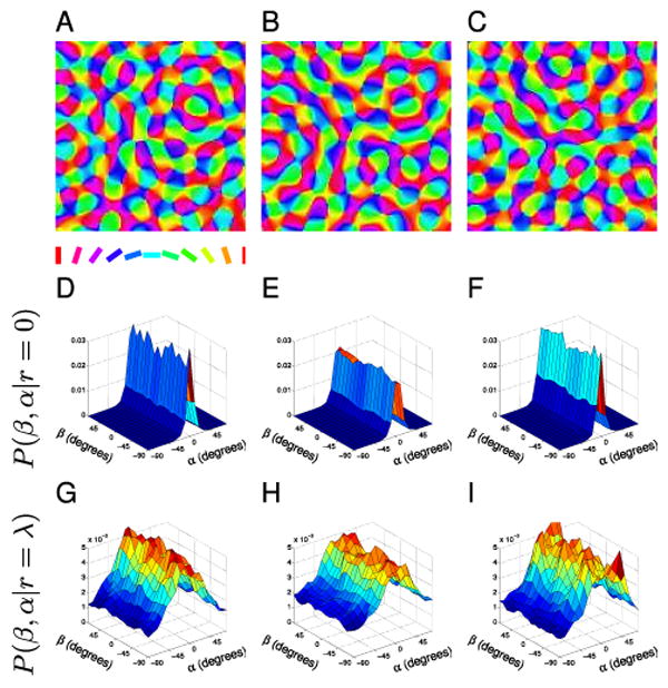
Figure 6. ECP maps.

(A–C) Sample ECP orientation maps. (D–F) The associated P(β, α|r = 0) distributions for cortical points with small separation do not have reduced symmetry since pairs tend to be orientated similarly regardless of direction β. (G–I) The P(β, α|r = λ) distributions for cortical points separated by one wavelength also do not show any clear co-circularity.
