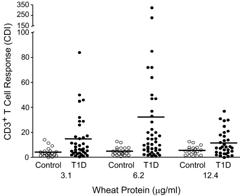
FIG. 1.
Antigen-specific CD3+ T-cell proliferation. 1.2 × 10⁶ CFSE-labeled PBMNCs from patients with type 1 diabetes (T1D) or healthy control subjects were cultured for 8 days in the absence or presence of different concentrations of WPs. On day 8, cells were stained with Cy-chrome conjugated anti-CD3 monoclonal antibody. CDI was calculated based on a fixed number of 5,000 CD3+ CFSEbright cells using the following formula: CDI = number of CD3+, CFSEdim cells with antigen/number of CD3+, CFSEdim cells without antigen (medium). The horizontal line indicates the mean.

