Abstract
This paper briefly reviews the optically stimulated luminescence (OSL) properties of dental enamel and discusses the potential and challenges of OSL for filling the technology gap in biodosimetry required for medical triage following a radiological/nuclear accident or terrorist event. The OSL technique uses light to stimulate a radiation-induced luminescence signal from materials previously exposed to ionizing radiation. This luminescence originates from radiation-induced defects in insulating crystals and is proportional to the absorbed dose of ionizing radiation. In our research conducted to date, we focused on fundamental investigations of the OSL properties of dental enamel using extracted teeth and tabletop OSL readers. The objective was to obtain information to support the development of the necessary instrumentation for retrospective dosimetry using dental enamel in laboratory, or for in situ and non-invasive accident dosimetry using dental enamel in emergency triage. An OSL signal from human dental enamel was detected using blue, green, or IR stimulation. Blue/green stimulation associated with UV emission detection seems to be the most appropriate combination in the sense that there is no signal from un-irradiated samples and the shape of the OSL decay is clear. Improvements in the minimum detection level were achieved by incorporating an ellipsoidal mirror in the OSL system to maximize light collection. Other possibilities to improve the sensitivity and research steps necessary to establish the feasibility of the technique for retrospective assessment of radiation exposure are also discussed.
1. Introduction
Current technologies for retrospective assessment of radiation exposure after a radiological/nuclear (R/N) event were recently the subject of a technology assessment by a Joint Interagency Working Group (JIWG), resulting in a roadmap to improve technology available to retrospectively estimate radiation dose (JIWG, 2006). The JIWG concluded that “presently available methods are not satisfactory for managing the medical casualties from an R/N event and there is urgent need to develop new capabilities to assess radiation dose quickly with at least moderate precision.” The JIWG also stated that there is a need to develop tools that can estimate whole or near whole body radiation doses in the range of 1-8 Gy with a throughput of 1 assay every 5 minutes.
Some clinical signs are available to indicate the degree of exposure to an individual, but in general they have limited quantitative precision. While numerous genetic assays have been developed and are the subject of many studies, complementary techniques are vitally needed. Both Electron Paramagnetic Resonance (EPR) (Chumak et al, 1998; Desrosiers and Schauer, 2001; Swartz et al, 2005) and OSL using dental enamel (Godfrey-Smith and Pass, 1997) offer such a promise and have the advantage of assaying a type of biological material (teeth) which is available in practically all cases of exposure.
In the OSL technique, light is used to stimulate the luminescence from materials previously exposed to ionizing radiation, the total luminescence emitted being proportional to the absorbed dose of radiation to which the material was exposed (Bøtter-Jensen et al., 2003). Some of the advantages offered by the OSL technique are associated with the all-optical readout process, namely: (i) optical stimulation can be controlled more easily and in a more flexible way than heating, since the stimulation intensity can be continuous, pulsed, or ramped and the stimulation wavelength can be tuned to the properties of the material (Bøtter-Jensen et al., 2003); (ii) measurements can be made remotely using optical fibers (Huston et al., 2001; Polf et al., 2002); (iii) the measurements of biological material (teeth) can potentially be made in-situ (Pass et al., 2003), since heating is not required. In addition, further advances in the OSL technique are likely since technological advances in LED and laser light sources, making them brighter, cheaper and more compact, have been rapid and are likely to continue.
The possibility of performing OSL measurements of dental enamel was first proposed by Godfrey-Smith and Pass (1997), who observed dose-dependent IR-stimulated and green-stimulated luminescence signals in deproteinated and non-deproteinated dental enamel. That study demonstrated the feasibility of using OSL from dental enamel for radiation dosimetry using infrared stimulation and concluded that OSL can “become the first non-invasive, simple, reliable, and portable means of retrospective radiation dosimetry in humans” if the sensitivity can be improved by 2-3 orders of magnitude.
In this paper we briefly review the OSL properties of dental enamel and report on our recent and preliminary investigations of the technique. We also discuss further steps to potentially improve the sensitivity of the technique and research steps necessary to establish its usefulness for retrospective assessment of radiation exposure as applied in medical triage and epidemiological studies.
2. Laboratory Investigations into OSL
The work by Godfrey-Smith and Pass (1997) demonstrated the presence of an OSL signal in deproteinated enamel stimulated with green and/or IR light. Green light was found to be more efficient in depleting the OSL signal than IR light. IR stimulated OSL was also observed in natural (undeproteinated) enamel. The samples investigated were aliquots of ∼7.5mg prepared from enamel fragments crushed and sieved to obtain grain sizes smaller than 355 μm. The readout system used was a Risø TL/OSL-DA-12-A reader (Risø National Laboratory, Denmark) equipped with IR diodes (880 ± 80 nm, 40 mW of power incident in the sample) and green light obtained by filtering the light from a 75 W quartz envelope tungsten filament lamp (523 ± 37 nm). To the best of our knowledge, no other investigations on the OSL properties of dental enamel have been reported until now.
We recently extended this initial characterization on the OSL properties of natural (undeproteinated) dental enamel using green, blue, and IR stimulation. The investigations were carried out using samples prepared from teeth provided by the National Institutes of Health. The results reported in this study were obtained with powdered samples prepared by crushing pieces of enamel using an agate mortar and pestle. The OSL measurements were carried out using a Risø TL/OSL reader (TL/OSL-DA-15, Risø National Laboratory). The samples were stimulated with either blue LEDs (470±20nm) or an IR laser diode (830±10nm). The irradiations were performed using a 90Sr/90Y beta source incorporated into the Risø TL/OSL reader. Light detection was performed with a photomultiplier tube (PMT) from Electron Tubes Inc. model 9235 QB fitted with appropriate filters for discrimination between the stimulation light and sample luminescence. Using UV transmitting (Hoya U-340 filters, 7.5 mm total thickness) it is possible to perform blue and IR stimulation. Using broadband visible filters (Corning BG-39, 6 mm total thickness), only IR stimulation is possible.
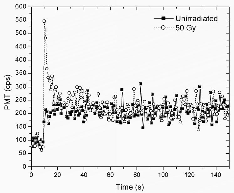
Figure 1 illustrates a typical OSL curve of dental enamel obtained under blue stimulation. This graph shows the PMT signal monitored during a total period of 150 s, the stimulation being switched on at t = 10s. In the unirradiated sample, an increase in the background due to leakage of stimulation light is observed when the LEDs are switched on. For the sample irradiated with 50 Gy, a clear OSL signal that decays to the background level in ∼10-20 s is observed. The OSL curves obtained here for natural (non-deproteinated) enamel are similar to those presented by Godfrey-Smith and Pass (1997) for deproteinated enamel obtained using green light stimulation.
Figure 1.
PMT signal of an enamel piece before and after irradiation with 50 Gy, the stimulation light was switched on at t = 10s. The measurements were performed using a using a Risø TL/OSL reader (TL/OSL-DA-15, Risø National Laboratory) with blue LED stimulation and with Hoya U-340 filters (7.5mm total thickness) in front of the PMT. The irradiation was performed with a 90Sr/90Y beta source, incorporated in the Risø TL/OSL reader.
The OSL signal of dental enamel was observed to be correlated with absorbed dose up to the maximum dose investigated, 1000 Gy. The dose response of dental enamel was linear up to 100 Gy in most cases. Although an OSL signal was observed with IR stimulation, the OSL decay was much slower than the decay observed using blue stimulation. Preliminary investigations on the stability of the OSL signal indicated a decay of approximately 20% in the first 10 min after irradiation and before OSL readout. Stability was investigated only up to a period of 5 h after irradiation, indicating a decay of up to 50% in this period.
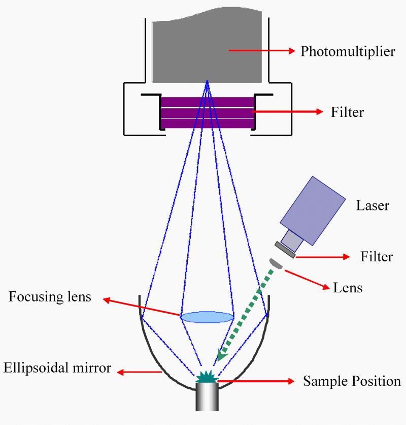
With the experimental conditions of our initial measurements, the minimum detectable doses were of the order of tens of Grays (20-30 Gy). For that reason, tests were performed using a new OSL system designed specifically for this investigation, the High-Sensitivity OSL (HS-OSL) system (Figure 2). The system uses an ellipsoidal mirror to improve the light collection of the sample luminescence, as previously suggested by Markey et al. (1997). Green stimulation (532 nm) is obtained using a compact green laser and blue stimulation (472 nm) is obtained using an argon ion laser from (Spectra-Physics, model 2020). The objective of this instrument is to allow fundamental investigations on the OSL properties of dental enamel.
Figure 2.
Scheme of the high-sensitivity OSL (HS-OSL) system tested to improve the light collection of the sample's luminescence. The system consists of: i) blue or green laser for optical stimulation, properly filtered in each case (GG-420 and OG-515, respectively), and defocusing lens, ii) light collection system composed of an ellipsoidal mirror (rhodium coated) and focusing lens to collect the maximum luminescence from the sample; and iii) detection optics composed of a UV transmitting (Hoya U-340 filter) to cut the stimulation wavelength and a PMT (Electron Tubes, model 9235QB).
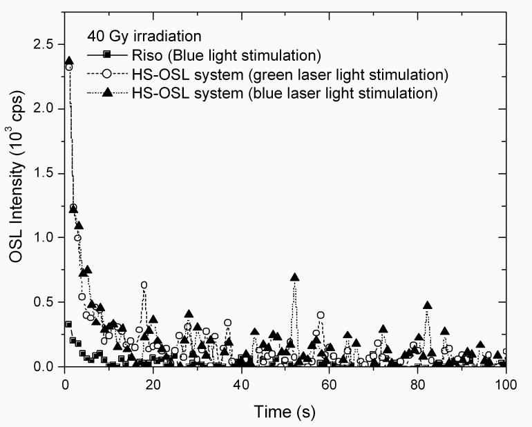
Figure 3 shows a comparison of the OSL curves obtained using the Risø TL/OSL reader and the HS-OSL system using the same sample and same detection filters in both cases (see figure caption for experimental details). The sample was irradiated with 40 Gy prior to the measurements. The data show that the light collection in the HS-OSL system is improved six times. In addition, the graph shows that similar OSL intensity and decay shape was obtained with either green or blue laser stimulation.
Figure 3.
OSL curves of a dental enamel powder sample of 15 mg irradiated with 40 Gy and readout using the Risø TL/OSL reader or the HS-OSL system designed for these experiments. The stimulation power was approximately 27 mW/cm2 and 23 mW/cm2 for blue and green stimulation, respectively.
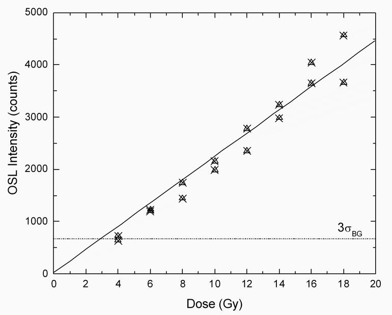
Figure 4 shows the dose response of a sample of dental enamel (15 mg) obtained using blue laser stimulation in the HS-OSL system. The figure also shown the intensity corresponding to three times the standard deviation of the background, estimated by repeating the measurements with an un-irradiated sample. Comparing these values, the estimated minimum detectable dose with this system is of the order of 4-6 Gy. While these improvements have achieved minimum detectable dose levels that approach that required medical triage following accidental exposures, these measurements were obtained under laboratory conditions. We have yet to try and replicate this level of sensitivity in applications outside the laboratory.
Figure 4.
Dose response of a 15 mg dental enamel sample irradiated with various doses of 90Sr beta particles. The standard deviation of the background was estimated from 3 measurements of an un-irradiated sample. The OSL intensity corresponds to the OSL signal integrated for the initial 10 s of stimulation.
3. Discussion
3.1 Advantages of OSL
There are many advantages to the OSL method. First, the OSL signal is easier to interpret than the EPR spectrum, which requires spectrum deconvolution. The total OSL signal (total light emitted) is proportional to the absorbed dose. In addition, as already mentioned, the OSL technique offers a wider variety of stimulation approaches since the optical stimulation can be continuous or modulated in some desired fashion (e.g. pulsed, or ramped). Furthermore, the stimulation wavelength can be selected according to the properties of the material. In addition, the all-optical nature of the OSL technique makes it suitable for non-invasive in situ measurements if readout equipment with appropriate sensitivity is developed.
3.2 Possibilities for Improvement
Despite the improvements in sensitivity described here, compared with earlier measurements, further improvements are likely to be the major challenge in developing OSL of dental enamel as a useful tool for retrospective dosimetry. However, in our opinion further advances are possible by taking advantage of several techniques, including:
Implementation of POSL technique: The pulsed OSL technique, which consists of synchronizing the detection system to the pulsed stimulation source in such a way that detection is performed only when the stimulation source is off. This allows a significant improvement in signal-to-noise ratio (Akselrod and McKeever, 1999). This technique is suitable when the lifetime of the luminescence centers in the material are longer than the period of the stimulation pulses.
Use of cooled PMT to decrease noise: If the limiting factor for the detection of the weak signals is the dark count from the PMT, the background can be reduced by the use of a cooled PMT. This would potentially decrease the minimum detectable dose.
Further investigations on detection filters: There is very little information available on the emission spectrum of the OSL of dental enamel and the UV detection window may not be the most suitable for this material. The use of filters with higher transmittance in the visible region may improve the OSL signal. However, it may also result in an increase in the background due to the stimulation light. For this reason, it is important to investigate the possibility of using the POSL technique in conjunction with different filters in order to eliminate the background from the stimulation light.
Improvement in sample preparation to avoid possible heating: It was reported by Godfrey-Smith and Pass (1997) that heating the enamel resulted in a decrease in sensitivity. The effect of sample preparation on the radiation-induced signal and on the sample sensitivity needs to be investigated. Ultimately, the sample preparation method should preserve the signal (at least for the laboratory characterizations) and its sensitivity to radiation.
3.3. Other investigations
Other areas of investigation are also necessary to establish the OSL technique as a retrospective dosimetry tool, including: (i) establish the thermal stability of the OSL signal; (ii) determine the optical stability of the OSL signal; (iii) establish the influence of UV exposure prior to OSL readout; (iv) study the tooth-to-tooth variability; and (v) study differences by age, gender, race, provenance, etc. The studies of fading of the OSL signal with time have so far been very limited and it will be necessary to determine the stability of the signal on time scales of days, weeks, and months. It will also be important to determine to what extent the OSL from the teeth are stable under natural light exposure while inside the mouth. The influence of UV is likely to be restricted to the incisors, especially the labial surface, but this effect needs to be investigated for the canine and molars as well, in order to establish the reliability of OSL as a dosimetry technique. If the challenges above are overcome and the OSL technique proves to have sufficient sensitivity for retrospective dose assessment, the next step would then be to conduct measurements to determine the tooth-to-tooth variability and differences by age, gender, race, provenance, etc.
The OSL readers described in this paper are suitable for carrying out the fundamental investigations required to establish the feasibility of using the OSL from dental enamel for retrospective and accident dosimetry. For the specific application of this technique in emergency triage, a portable OSL system capable of performing the OSL measurements in a non-invasive way and with a high throughput needs to be developed, as proposed by Pass et al. (2003).
4. Summary and Conclusions
This paper briefly reviews the data on the optically stimulated luminescence (OSL) of dental enamel. OSL of dental enamel was observed using IR, blue, and green stimulation. However, blue or green stimulation combined with UV detection seems to be the most appropriate combination so far, in the sense that there is no signal from un-irradiated samples, and the OSL decay is very clear. The minimum detection dose obtained with a high-sensitivity OSL system is estimated to be about 4-6 Gy in laboratory conditions. Some proposals to improve the viability of this technique, such as the use of the pulsed OSL technique, are discussed.
Making further improvements in the sensitivity and studying the thermal and optical stability of the OSL signal in the conditions in which the technique is to be applied will be the main challenges for the development of the technique as a tool for retrospective dosimetry. If the technical challenges described in this paper can be overcome, the Optically Stimulated Luminescence (OSL) technique using dental enamel may offer a fast and practical new method for retrospective assessment of radiation exposure for use on medical triage or epidemiological studies, depending on the minimum detectable doses that need to be achieved.
Acknowledgments
This work was supported by the Intra-agency agreement between the National Institute of Allergy and Infectious Diseases and the National Cancer Institute, NIAID agreement #Y2-Al-5077 and NCI agreement #Y3-CO-5117.
Footnotes
Publisher's Disclaimer: This is a PDF file of an unedited manuscript that has been accepted for publication. As a service to our customers we are providing this early version of the manuscript. The manuscript will undergo copyediting, typesetting, and review of the resulting proof before it is published in its final citable form. Please note that during the production process errors may be discovered which could affect the content, and all legal disclaimers that apply to the journal pertain.
References
- Akselrod MS, McKeever SWS. A radiation dosimetry methods using pulsed optically stimulated luminescence. Radiat Prot Dosim. 1999;81:167–176. [Google Scholar]
- Bøtter-Jensen L, McKeever SWS, Wintle AG. Optically Stimulated Luminescence Dosimetry. Amsterdam: Elsevier; 2003. [Google Scholar]
- Chumak V, Likhtarev I, Sholom S, Meckbach R, Krjuchkov V. Chernobyl experience in field of retrospective dosimetry: reconstruction of doses to the population and liquidators involved in the accident. Radiat Prot Dosim. 1998;77:91–95. [Google Scholar]
- Desrosiers M, Schauer DA. Electron paramagnetic resonance (EPR) biodosimetry. Nucl Instr Meth Phys Res B. 2001;184:219–228. [Google Scholar]
- Godfrey-Smith DI, Pass B. A new method of retrospective radiation dosimetry: optically stimulated luminescence in dental enamel. Health Physics. 1997;72:744–749. doi: 10.1097/00004032-199705000-00010. [DOI] [PubMed] [Google Scholar]
- Huston AL, Justus BL, Falkenstein PL, Miller RW, Ning H, Altemus R. Remote optical fiber dosimetry. Nucl Instr Meth Phys Res B. 2001;184:55–67. [Google Scholar]
- Joint Interagency Working Group (JIWG) Technology assessment and roadmap for the Emergency Radiation Dose Assessment Program. Department of Homeland Security; 2005. UCRL-TR-215887. [Google Scholar]
- Markey BG, Bøtter-Jensen L, Duller GAT. A new flexible system for measuring thermally and optically stimulated luminescence. Radiat Meas. 1997;27:93–89. [Google Scholar]
- Pass B, Godfrey-Smith DI, Scallion P. Retrospective Radiation Dosimetry Using Optically Stimulated Luminescence in Dental Enamel: Possibilities for Invivo Dosimetry. Proceedings of the 36th Midyear Topical Meeting “Radiation Safety Aspects of Homeland Security and Emergency Response”; San Antonio, Texas. January 27-29, 2003.pp. 210–217. [Google Scholar]
- Polf JC, McKeever SWS, Akselrod MS, Holmstrom S. A real-time, fibre optic dosimetry system using Al2O3 fibres Radiat. Prot Dosim. 2002;100:301–304. doi: 10.1093/oxfordjournals.rpd.a005873. [DOI] [PubMed] [Google Scholar]
- Swartz HM, Iwasaki A, Walczak T, Demidenko E, Salikov I, Lesniewski P, Starewicz P, Schauer D, Romanyukha A. Measurements of clinically significant doses of ionizing radiation using non-invasive in vivo EPR spectroscopy of teeth in situ. Appl Radiat Isot. 2005;62:293–299. doi: 10.1016/j.apradiso.2004.08.016. [DOI] [PubMed] [Google Scholar]


