Abstract
Oxidative injury to the lung is associated with widespread injury to the alveolar epithelium, which can be fatal unless the process is controlled and repaired. Keratinocyte growth factor (KGF), a member of the fibroblast growth factor family, has been shown to protect the lung from a variety of oxidative insults. The mechanism(s) underlying the protective effects of KGF in lung injury is being investigated in many laboratories. Although KGF has potent mitogenic effects on epithelial cells, the proliferative effect of KGF was shown to be abolished in oxygen-breathing animals, but KGF was still able to inhibit alveolar damage. This demonstrates that the protective effect of KGF cannot simply be explained by the ability of KGF to stimulate type II cell proliferation. To identify the mechanisms involved in the protective effects of KGF, we used an inducible lung-specific transgenic approach to overexpress KGF in murine lungs, since constitutive overexpression of KGF in the mouse affects lung development. The transgenic system allowed us to identify the pro-survival Akt pathway as an important mediator of the protective effects of KGF both in vivo and in vitro. In addition, use of a yeast two-hybrid system led to the identification two proteins p90RSK and PAK4 that associate with the KGF receptor and are important for the protective functions of KGF. Experiments are underway to determine whether the different pathways triggered by KGF all converge on the Akt pathway, or whether they independently induce protective mechanisms that along with Akt are crucial for cell survival.
Keywords: Akt, hyperoxia, keratinocyte growth factor, lung, PAK4, p90RSK
The detrimental consequences of hyperoxia on the lung are well recognized; high concentrations of oxygen are toxic for most cellular constituents of the lung and can cause widespread irreversible damage that can be fatal. The damaging effects of oxygen are caused by reactive oxygen species (ROS) such as the superoxide anion (O2−) and through subsequent enzymatic steps, hydroxyl radicals (OH−) (e.g., in the iron-catalyzed Fenton reaction), which are potent oxidants. The capacity of hyperoxia to induce diffuse alveolar damage has been studied in humans as well as several other species including rats (1), primates (2), rabbits (3), and mice (4, 5). The pathologic sequence of injury and repair are similar across species. The changes seen in the alveolar epithelium in the late stage of hyperoxic injury are similar to those seen in many other forms of alveolar injury. Extensive necrosis of type I epithelial cells, and hyperplasia of cuboidal type II cell occurs. Areas of denuded epithelial basement membrane are present, and hyaline membranes consisting of fibrin, other plasma proteins, and debris from necrotic cells fill the alveolar space. As in other forms of diffuse alveolar damage, proliferating type II epithelial cells eventually cover the denuded epithelium. In animals that survive beyond this proliferative phase, either the type II cells differentiate into type I cells (6) and alveolar architecture returns to normal, or progressive alveolar fibrosis occurs (7).
Keratinocyte growth factor (KGF), a member of the fibroblast growth factor (FGF) family (also known as FGF-7), is a potent mitogen for epithelial cells (8, 9). The pattern of expression of KGF and its receptor suggest an important role for KGF in mediating mesenchymal–epithelial interactions (10). The receptor for KGF (also called FGFR2-IIIb), which has intrinsic tyrosine kinase activity (11), is expressed specifically on epithelial cells and is diffusely expressed in the Day 11 lung epithelium (12). The expression of KGF in the developing lung needs to be tightly controlled, because transgenic mice transgenic mice constitutively overexpressing KGF display lethal papillary cystadenoma, with marked enlargement of bronchial air spaces (13). However, the need for KGF signaling during development is essential because transgenic mice expressing a dominant-negative mutant of the KGF receptor (KGFR) under the control of the SP-C promoter exhibit embryonic lethality with grossly abnormal lung development, with only two primordial epithelial tubes and no branching morphogenesis (14).
Administration of KGF has been shown to protect lungs of animals from a variety of insults including oxygen (15), radiation and chemotherapy (bleomycin) (5, 16), and acid (17). Thus, intratracheal administration of KGF before oxygen exposure protects rats from hyperoxic injury (15). Intravenous administration of KGF was found to protect mice from oxygen toxicity when administered either before or after oxygen exposure (18). Since KGF mRNA expression has been shown to be induced by cytokines and growth factors such as interleukin (IL)-1 and transforming growth factor (TGF)-α (19), increased production of these molecules from activated alveolar macrophages and other inflammatory cells during hyperoxic injury could potentially stimulate local KGF production, which, in turn, could act on type II cells triggering repair of alveolar damage.
The precise mechanism(s) underlying the protective effects of KGF in lung injury is being investigated in many laboratories. In vitro studies reported to date suggest a role in type II cell proliferation (8), inhibition of increased oxidant-induced epithelial cell permeability (20), stimulation of transepithelial Na ion transport (21) and stimulation of DNA repair (22). In vivo studies show decreased protein-rich pulmonary edema upon KGF pretreatment both in humans and in animal studies (23, 24). Although KGF induces type II cell proliferation, this proliferative effect of KGF was abolished in oxygen-breathing animals, but KGF was still able to inhibit alveolar damage (18). This demonstrates that the protective effect of KGF within the lung cannot simply be explained by the ability of KGF to stimulate type II cell proliferation. Although in vitro studies have shown increased surfactant protein synthesis in the presence of KGF, only modest effects were noted in in vivo studies (18). Collectively, while all studies clearly demonstrate a protective role of KGF in hyperoxic alveolar damage, the molecular mechanism(s) by which this is achieved has not been adequately addressed. We have used different approaches to investigate the protective effects of KGF in oxidative lung injury. We developed a lung-specific inducible transgenic system to overexpress KGF in lungs of mice in a tightly controlled fashion to study the role of KGF-induced signaling pathways in the protective effects of KGF. In another approach, we have used the yeast two-hybrid system to identify KGFR-associated proteins that are involved in the transduction of pro-survival signals by KGF.
IDENTIFICATION OF THE PRO-SURVIVAL AKT PATHWAY IN KGF-MEDIATED PROTECTION USING A CELL-SPECIFIC INDUCIBLE TRANSGENIC SYSTEM
To study the protective effects of KGF in vivo, we generated transgenic (Tg) mice that express KGF in an inducible, lung-specific fashion. To minimize leaky expression of the transgene, we used a modified dual repressor-activator tet system to tightly control transgene expression (25). These mice were backcrossed for 10 generations onto the C57BL/6 background. Using these Tg mice in a model of hyperoxic lung injury, we have shown that KGF protects lung epithelial cells from hyperoxia-induced cell death (25). By electron microscopy, we demonstrated preservation of the alveolar epithelial surface in the presence of KGF overexpression (25). However, the endothelium and accessory cells lining the capillaries were not protected in the KGF Tg mice. The epithelial-specific protection by KGF was not surprising, since the KGF receptor is expressed on epithelial and not on endothelial cells. KGF expression in these animals was accompanied by activation of the pro-survival Akt pathway in the lungs of the animals (25). In in vitro studies, the protective effect of KGF on oxidant-induced lung epithelial cell death was inhibited by a dominant-negative mutant of Akt (DN Akt), which demonstrated the biological significance of KGF-induced Akt activation (25).
Since KGF-induced Akt activation appeared to be important for the protective effects of KGF, we investigated whether Akt activation in vivo would protect cells from oxidant-induced cell death and prolong survival of mice. Toward this end, we used a constitutively active form of Akt (myristoylated Akt; myr-Akt) in which the Src myristoylation signal was fused to the N-terminus of Akt. Myristoylation causes targeting of the enzyme to the cell membrane, which results in its phosphorylation and consequently activation. By intratracheal instillation of Ad-myr-Akt, we have demonstrated inhibition of oxidant-induced injury to the lung and prolongation of survival of mice (26). The protection from hyperoxia correlated with decreased hemorrhage, inflammation, and edema in the lung, and a dramatic suppression of airway cell death (26). Since the adenoviral gene delivery allowed expression of activated Akt in both epithelial and endothelial cells, which could not be achieved by KGF expression alone (because the KGFR is not expressed on endothelial cells), the animals were more resistant to hyperoxia-induced death. Thus, use of a stringently regulated inducible transgenic system for selective expression of KGF in the adult mouse lung allowed us to study the protective effects of KGF on lung epithelial cells. Collectively, our studies show that Akt activation in vivo promotes resistance to oxidant-induced cell death (26, 25). Additional recent reports have also shown activation of Akt by KGF in lung epithelial cells (27–29). These results suggest that protection of both epithelial and endothelial cells through activation of pro-survival pathways such as Akt using pharmaceutical agents may provide a better survival advantage during oxidative stress.
IDENTIFICATION OF KGFR-ASSOCIATED PROTEINS USING THE YEAST TWO-HYBRID SYSTEM
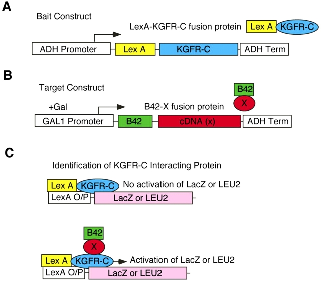
The Akt pathway is induced downstream of multiple growth factor receptors. While dual phosphorylation of Akt on residues Thr308 and Ser473 is known to be important for Akt function, the mechanisms involved in Ser473 phosphorylation and Akt activation are not well understood. Also, although our studies showed Akt activation to be important in KGF-mediated protection, we could not rule out the involvement of additional pathways that may crosstalk with the Akt pathway or may also be important in addition to the Akt pathway in KGF-mediated protection. To identify adaptor proteins that might mediate KGF-induced Akt activation and additional pro-survival mechanisms, we used the yeast two-hybrid system to identify KGFR-interacting proteins. The yeast two-hybrid system has been successfully used to identify interacting proteins for a number of receptor tyrosine kinases (RTKs) including insulin receptor, CSF-1R, Met (HGFR), EGFR, and c-kit (30–32). A key event that triggers interactions between RTKs and other proteins is phosphorylation of the receptor induced by the binding of the cognate ligand. It has been shown that phosphorylation of the receptor is also induced when the receptor is overexpressed in cells. Thus, receptor-interacting proteins have been identified by overexpression of the receptors in yeast cells and many of the interacting proteins identified using this approach have been found to interact in a tyrosine phosphorylation–dependent fashion, demonstrating the validity of this approach. We used the carboxyl terminal domain of FGFR2IIIb (for simplicity called KGFR-C) as bait to identify intracellular proteins that interact with the receptor (Figure 1). We isolated multiple clones that encoded proteins interacting with KGFR-C. We have studied in detail interactions with PAK4 (33) and with p90RSK (34). The following is a brief description of these studies:
Figure 1.
Schematic of the strategy used for screening KGFR-interacting proteins using the yeast two-hybrid assay. (A) The KGFR cytoplasmic domain containing the tyrosine kinase domain was cloned into the yeast expression plasmid pEG202-NLS to generate the bait construct. (B) A cDNA expression library constructed from cDNA derived from a 19-d postcoital mouse embryo was fused to the B42 activation domain–HA-tagged expression vector pJG4-5 to generate the target construct. (C) Reporter gene expression was induced only when the KGFR cytoplasmic domain fusion protein (bait) interacted with a target protein. After selecting clones potentially expressing interacting proteins, the specificity of the interaction was confirmed by a yeast mating assay.
PAK4
PAK4 is a member of the PAK family of proteins. The p21 Rac/Cdc42-activated kinases (PAKs) are serine/threonine kinases that were identified as downstream targets of the GTPases Rac and Cdc42Hs (35). The PAK members have an N-terminal GTPase (Rac/Cdc42)-binding regulatory domain and a C-terminal kinase domain. PAK4 is the most divergent member of the family in which the kinase domain shares 53% identity with the kinase domains of other PAKs (36). A common property of PAK proteins is their ability to regulate cytoskeletal organization. However, the proteins have been shown to have different roles in the apoptotic response of cells. While PAK2 promote apoptosis (37), PAK1 has been shown to protect cells from apoptosis induced by growth factor withdrawal (38). PAK4 was shown to protect cells from apoptosis induced by multiple stimuli including serum withdrawal, TNF-α treatment, or ultraviolet irradiation (39). Although PAK4 has been shown to be a target for Cdc42, extracellular stimuli that regulate endogenous PAK4 activity have not been identified. Our studies show the ability of activated KGFR to interact with and induce phosphorylation of PAK4 (33). We have also shown that DNPAK4 prevents the ability of KGF to protect small airway epithelial (SAE) cells from hyperoxia-induced cell death (33).
p90RSK
The RSK (or p90RSK) family includes three members, RSK1-3, which show 75 to 80% similarity at the amino acid level (40). RSK family members contain two heterologous kinase domains (41). Although originally identified as ribosome S6 protein kinase, S6 protein phosphorylation by RSK family proteins is very restricted and p70S6K is the major physiologic S6 kinase (40). RSKs are activated in response to several growth factors and mitogens including EGF (42, 43), insulin, and IGF-I (44, 45). The activated RSKs phosphorylate a number of proteins containing the consensus sequences (R/L)xRxxS and are involved in a wide range of cellular activities (40). Interestingly, the kinase PDK1 has been shown to be recruited by RSK2, resulting in coordinated phosphorylation and activation of PDK1 and RSK2 (46). PDK1 is critical for the activation of Akt and related enzymes, including RSK, that regulate many physiologic processes such as cell survival, proliferation, and gene expression. Since RSK was found to associate with the KGF receptor, it seemed plausible that RSK might actually be involved in Akt activation. Indeed, it has been shown that ERK may be involved in Akt activation under certain circumstances, and the RSK homolog MSK was shown to phosphorylate and activate Akt (47–49).
To test whether RSK1 is involved in Akt activation, we used a mutant of RSK1, RSK1d1, lacking the carboxyl-terminus of RSK1, which includes the ERK binding site of RSK, and checked the Akt kinase activity in cells overexpressing wt or mutant RSK1. KGF stimulation of cells caused a fivefold increase in Akt kinase activity. Overexpression of wtRSK1 caused a three- to fivefold increase in Akt kinase activity independent of KGF stimulation. Overexpression of the mutant RSK1d1, however, caused inhibition of KGF-induced Akt kinase activation (34). These results suggested that RSK proteins are an important component of KGF-induced Akt activation (34).
RSK1 MUTANT PREVENTS KGF-MEDIATED INHIBITION OF OXIDANT-INDUCED POLY (ADP-RIBOSE) POLYMERASE CLEAVAGE
Since our data suggested an important role for RSK1 in KGF-induced Akt activation, we investigated whether blocking RSK1 function would influence anti-apoptotic functions induced by KGF in epithelial cells. For this purpose, we treated cells with H2O2, which has been shown to induce pro-apoptotic pathways in these cells (50). Since apoptosis is associated with caspase-3 activation, a convergence point of different pro-apoptotic pathways, we investigated the activation of both pro–caspase-3 and cleavage of the substrate of caspase-3, poly (ADP-ribose) polymerase (PARP), in the cells under different conditions of treatment. To inhibit the functions of endogenous RSK1, we overexpressed RSK1d1. Treatment of cells with H2O2 promoted cleavage of both pro–caspase-3 and the substrate of caspase-3, the PARP protein. Cleavage of both proteins was inhibited in the presence of KGF. However, when the RSK1 mutant was expressed in the cells, KGF was unable to inhibit protein cleavage. Addition of the ERK inhibitor PD98059 inhibited the effects of KGF, suggesting ERK-stimulated RSK activity to be important for KGF-induced effects. These results were similar to our previous observations of the ability of DNAkt and DNPAK4 to block inhibition of PARP cleavage by KGF (25, 33).
CONCLUSIONS
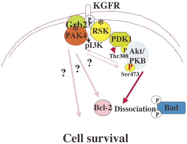
We have shown that inhibition of any of the three pathways, Akt, PAK4, or RSK1, prevents KGF from exerting its protective effects on epithelial cells. How can we then define a common framework for interactions between these pathways in KGF-induced protective effects? The Akt pathway has been shown to crosstalk with the mitogen-activated protein kinase pathway and promote cell survival. While PAK has been shown to be a downstream target of of Ras via PI 3-kinase, one study showed that Akt mediates PAK activation by Ras, and activated PAK (PAK1) promoted cell survival in this study (51). In another study, HIV-1 Nef-associated PI3-kinase and PAK were shown to stimulate Akt-independent Bad phosphorylation to induce cell survival signals (52). Thus, it will be interesting to determine whether the different pathways triggered by PAK4 and RSK1 activation all converge on Akt, or whether they independently induce protective mechanisms that along with Akt are also crucial for cell survival. As illustrated in Figure 2, in the next phase of these studies, we are interested in characterizing the relationships between the different pathways downstream of the KGF receptor that collectively determine the protective functions of KGF.
Figure 2.
A model depicting intracellular signaling pathways that have been identified (solid red arrows) downstream of the KGFR with potential interactions shown by patterned lines. The asterisks indicate KGFR-interacting proteins identified by yeast two-hybrid assay.
Acknowledgments
The author thanks Drs. ZhongZong Pan, Yunbiao Lu, and Yvan Devaux for their contribution to the experiments discussed in this manuscript. The author apologizes for inadvertent omission of relevant citations.
This work was supported by grants HL 69810 and P01-HL70807 from the National Institutes of Health (to P.R.).
Conflict of Interest Statement: P.R. does not have a financial relationship with a commercial entity that has an interest in the subject of this manuscript.
References
- 1.Nogee LM, Wispe JR. Effects of pulmonary oxygen injury on airway content of surfactant- associated protein A. Pediatr Res 1988;24:568–573. [DOI] [PubMed] [Google Scholar]
- 2.Simonson SG, Welty-Wolf KE, Huang YCT, Taylor DE, Kantrow SP, Carraway MS, Crapo JD, Piantadosi CA. Aerosolized manganese SOD decreases hyperoxic pulmonary injury in primates: I. Physiology and biochemistry. J Appl Physiol 1997;83:550–558. [DOI] [PubMed] [Google Scholar]
- 3.Novotny WE, Hudak BB, Matalon S, Holm BA. Hyperoxic lung injury reduces exogenous surfactant clearance in vivo. Am J Respir Crit Care Med 1995;151:1843–1847. [DOI] [PubMed] [Google Scholar]
- 4.Barazzone C, Horowitz S, Donati YR, Rodriguez I, Piguet PF. Oxygen toxicity in mouse lung: pathways to cell death. Am J Respir Cell Mol Biol 1998;19:573–581. [DOI] [PubMed] [Google Scholar]
- 5.Guo J, Yi ES, Havill AM, Sarosi I, Whitcomb L, Yin S, Middleton SC, Piguet P, Ulich TR. Intravenous keratinocyte growth factor protects against experimental pulmonary injury. Am J Physiol 1998;275:L800–L805. [DOI] [PubMed] [Google Scholar]
- 6.Adamson IY, Bowden DH. The type 2 cell as progenitor of alveolar epithelial regeneration. A cytodynamic study in mice after exposure to oxygen. Lab Invest 1974;30:35–42. [PubMed] [Google Scholar]
- 7.Basset F, Ferrans VJ, Soler P, Takemura T, Fukuda Y, Crystal RG. Intraluminal fibrosis in interstitial lung disorders. Am J Pathol 1986;122:443–461. [PMC free article] [PubMed] [Google Scholar]
- 8.Panos RJ, Rubin JS, Csaky KG, Aaronson SA, Mason RJ. Keratinocyte growth factor and hepatocyte growth factor/scatter factor are heparin-binding growth factors for alveolar type II cells in fibroblast-conditioned medium. J Clin Invest 1993;92:969–977. [published erratum appears in J Clin Invest 1994 Mar;93(3):1347]. [DOI] [PMC free article] [PubMed] [Google Scholar]
- 9.Rubin JS, Osada H, Finch PW, Taylor WG, Rudikoff S, Aaronson SA. Purification and characterization of a newly identified growth factor specific for epithelial cells. Proc Natl Acad Sci USA 1989;86:802–806. [DOI] [PMC free article] [PubMed] [Google Scholar]
- 10.Finch PW, Cunha GR, Rubin JS, Wong J, Ron D. Pattern of keratinocyte growth factor and keratinocyte growth factor receptor expression during mouse fetal development suggests a role in mediating morphogenetic mesenchymal-epithelial interactions. Dev Dyn 1995;203:223–240. [DOI] [PubMed] [Google Scholar]
- 11.Miki T, Fleming TP, Bottaro DP, Rubin JS, Ron D, Aaronson SA. Expression cDNA cloning of the KGF receptor by creation of a transforming autocrine loop. Science 1991;251:72–75. [DOI] [PubMed] [Google Scholar]
- 12.Cardoso WV, Itoh A, Nogawa H, Mason I, Brody JS. FGF-1 and FGF-7 induce distinct patterns of growth and differentiation in embryonic lung epithelium. Dev Dyn 1997;208:398–405. [DOI] [PubMed] [Google Scholar]
- 13.Simonet WS, DeRose ML, Bucay N, Nguyen HQ, Wert SE, Zhou L, Ulich TR, Thomason A, Danilenko DM, Whitsett JA. Pulmonary malformation in transgenic mice expressing human keratinocyte growth factor in the lung. Proc Natl Acad Sci USA 1995;92:12461–12465. [DOI] [PMC free article] [PubMed] [Google Scholar]
- 14.Peters K, Werner S, Liao X, Wert S, Whitsett J, Williams L. Targeted expression of a dominant negative FGF receptor blocks branching morphogenesis and epithelial differentiation of the mouse lung. EMBO J 1994;13:3296–3301. [DOI] [PMC free article] [PubMed] [Google Scholar]
- 15.Panos RJ, Bak PM, Simonet WS, Rubin JS, Smith LJ. Intratracheal instillation of keratinocyte growth factor decreases hyperoxia-induced mortality in rats. J Clin Invest 1995;96:2026–2033. [DOI] [PMC free article] [PubMed] [Google Scholar]
- 16.Yi ES, Williams ST, Lee H, Malicki DM, Chin EM, Yin S, Tarpley J, Ulich TR. Keratinocyte growth factor ameliorates radiation- and bleomycin-induced lung injury and mortality. Am J Pathol 1996;149:1963–1970. [PMC free article] [PubMed] [Google Scholar]
- 17.Yano T, Deterding RR, Simonet WS, Shannon JM, Mason RJ. Keratinocyte growth factor reduces lung damage due to acid instillation in rats. Am J Respir Cell Mol Biol 1996;15:433–442. [DOI] [PubMed] [Google Scholar]
- 18.Barazzone C, Donati YR, Rochat AF, Vesin C, Kan CD, Pache JC, Piguet PF. Keratinocyte growth factor protects alveolar epithelium and endothelium from oxygen-induced injury in mice. Am J Pathol 1999;154:1479–1487. [DOI] [PMC free article] [PubMed] [Google Scholar]
- 19.Brauchle M, Angermeyer K, Hubner G, Werner S. Large induction of keratinocyte growth factor expression by serum growth factors and pro-inflammatory cytokines in cultured fibroblasts. Oncogene 1994;9:3199–3204. [PubMed] [Google Scholar]
- 20.Waters CM, Savla U, Panos RJ. KGF prevents hydrogen peroxide-induced increases in airway epithelial cell permeability. Am J Physiol 1997;272:L681–L689. [DOI] [PubMed] [Google Scholar]
- 21.Borok Z, Danto SI, Dimen LL, Zhang XL, Lubman RL. Na(+)-K(+)-ATPase expression in alveolar epithelial cells: upregulation of active ion transport by KGF. Am J Physiol 1998;274:L149–L158. [DOI] [PubMed] [Google Scholar]
- 22.Wu KI, Pollack N, Panos RJ, Sporn PH, Kamp DW. Keratinocyte growth factor promotes alveolar epithelial cell DNA repair after H2O2 exposure. Am J Physiol 1998;275:L780–L787. [DOI] [PubMed] [Google Scholar]
- 23.Haddad IY, Panoskaltsis-Mortari A, Ingbar DH, Resnik ER, Yang S, Farrell CL, Lacey DL, Cornfield DN, Blazar BR. Interactions of keratinocyte growth factor with a nitrating species after marrow transplantation in mice. Am J Physiol 1999;277:L391–L400. (In Process Citation). [DOI] [PubMed] [Google Scholar]
- 24.Verghese GM, McCormick-Shannon K, Mason RJ, Matthay MA. Hepatocyte growth factor and keratinocyte growth factor in the pulmonary edema fluid of patients with acute lung injury. Biologic and clinical significance. Am J Respir Crit Care Med 1998;158:386–394. [DOI] [PubMed] [Google Scholar]
- 25.Ray P, Devaux Y, Stolz DB, Yarlagadda M, Watkins SC, Lu Y, Chen L, Yang XF, Ray A. Inducible expression of keratinocyte growth factor (KGF) in mice inhibits lung epithelial cell death induced by hyperoxia. Proc Natl Acad Sci USA 2003;100:6098–6103. [DOI] [PMC free article] [PubMed] [Google Scholar]
- 26.Lu YB, Parkyn L, Otterbein L, Kureishi Y, Walsh K, Ray A, Ray P. Activated Akt protects the lung from oxidant-induced injury and delays death of mice. J Exp Med 2001;193:545–549. [DOI] [PMC free article] [PubMed] [Google Scholar]
- 27.Bao S, Wang Y, Sweeney P, Chaudhuri A, Doseff AI, Marsh CB, Knoell DL. Keratinocyte growth factor induces Akt kinase activity and inhibits Fas-mediated apoptosis in A549 lung epithelial cells. Am J Physiol Lung Cell Mol Physiol 2005;288:L36–L42. [DOI] [PubMed] [Google Scholar]
- 28.Portnoy J, Curran-Everett D, Mason RJ. Keratinocyte growth factor stimulates alveolar type II cell proliferation through the extracellular signal-regulated kinase and phosphatidylinositol 3-OH kinase pathways. Am J Respir Cell Mol Biol 2004;30:901–907. [DOI] [PubMed] [Google Scholar]
- 29.Truong SV, Monick MM, Yarovinsky TO, Powers LS, Nyunoya T, Hunninghake GW. Extracellular signal-regulated kinase activation delays hyperoxia-induced epithelial cell death in conditions of Akt downregulation. Am J Respir Cell Mol Biol 2004;31:611–618. [DOI] [PubMed] [Google Scholar]
- 30.Bourette RP, Myles GM, Choi JL, Rohrschneider LR. Sequential activation of phoshatidylinositol 3-kinase and phospholipase C-gamma2 by the M-CSF receptor is necessary for differentiation signaling. EMBO J 1997;16:5880–5893. [DOI] [PMC free article] [PubMed] [Google Scholar]
- 31.Galcheva-Gargova Z, Konstantinov KN, Wu IH, Klier FG, Barrett T, Davis RJ. Binding of zinc finger protein ZPR1 to the epidermal growth factor receptor. Science 1996;272:1797–1802. [DOI] [PubMed] [Google Scholar]
- 32.O'Neill TJ, Craparo A, Gustafson TA. Characterization of an interaction between insulin receptor substrate 1 and the insulin receptor by using the two-hybrid system. Mol Cell Biol 1994;14:6433–6442. [DOI] [PMC free article] [PubMed] [Google Scholar]
- 33.Lu Y, Pan ZZ, Devaux Y, Ray P. p21-activated protein kinase 4 (PAK4) interacts with the keratinocyte growth factor receptor and participates in keratinocyte growth factor-mediated inhibition of oxidant-induced cell death. J Biol Chem 2003;278:10374–10380. [DOI] [PubMed] [Google Scholar]
- 34.Pan ZZ, Devaux Y, Ray P. Ribosomal S6 kinase as a mediator of keratinocyte growth factor-induced activation of akt in epithelial cells. Mol Biol Cell 2004;15:3106–3113. [DOI] [PMC free article] [PubMed] [Google Scholar]
- 35.Knaus UG, Bokoch GM. The p21Rac/Cdc42-activated kinases (PAKs). Int J Biochem Cell Biol 1998;30:857–862. [DOI] [PubMed] [Google Scholar]
- 36.Abo A, Qu J, Cammarano MS, Dan C, Fritsch A, Baud V, Belisle B, Minden A. PAK4, a novel effector for Cdc42Hs, is implicated in the reorganization of the actin cytoskeleton and in the formation of filopodia. EMBO J 1998;17:6527–6540. [DOI] [PMC free article] [PubMed] [Google Scholar]
- 37.Rudel T, Bokoch GM. Membrane and morphological changes in apoptotic cells regulated by caspase-mediated activation of PAK2. Science 1997;276:1571–1574. [DOI] [PubMed] [Google Scholar]
- 38.Schurmann A, Mooney AF, Sanders LC, Sells MA, Wang HG, Reed JC, Bokoch GM. p21-activated kinase 1 phosphorylates the death agonist bad and protects cells from apoptosis. Mol Cell Biol 2000;20:453–461. [DOI] [PMC free article] [PubMed] [Google Scholar]
- 39.Gnesutta N, Qu J, Minden A. The serine/threonine kinase PAK4 prevents caspase activation and protects cells from apoptosis. J Biol Chem 2001;276:14414–14419. [DOI] [PubMed] [Google Scholar]
- 40.Frodin M, Gammeltoft S. Role and regulation of 90 kDa ribosomal S6 kinase (RSK) in signal transduction. Mol Cell Endocrinol 1999;151:65–77. [DOI] [PubMed] [Google Scholar]
- 41.Fisher TL, Blenis J. Evidence for two catalytically active kinase domains in pp90rsk. Mol Cell Biol 1996;16:1212–1219. [DOI] [PMC free article] [PubMed] [Google Scholar]
- 42.Sassone-Corsi P, Mizzen CA, Cheung P, Crosio C, Monaco L, Jacquot S, Hanauer A, Allis CD. Requirement of Rsk-2 for epidermal growth factor-activated phosphorylation of histone H3. Science 1999;285:886–891. [DOI] [PubMed] [Google Scholar]
- 43.Zhao Y, Bjorbaek C, Moller DE. Regulation and interaction of pp90(rsk) isoforms with mitogen-activated protein kinases. J Biol Chem 1996;271:29773–29779. [DOI] [PubMed] [Google Scholar]
- 44.Alessi DR, Cuenda A, Cohen P, Dudley DT, Saltiel AR. PD 098059 is a specific inhibitor of the activation of mitogen-activated protein kinase kinase in vitro and in vivo. J Biol Chem 1995;270:27489–27494. [DOI] [PubMed] [Google Scholar]
- 45.Lazar DF, Wiese RJ, Brady MJ, Mastick CC, Waters SB, Yamauchi K, Pessin JE, Cuatrecasas P, Saltiel AR. Mitogen-activated protein kinase kinase inhibition does not block the stimulation of glucose utilization by insulin. J Biol Chem 1995;270:20801–20807. [DOI] [PubMed] [Google Scholar]
- 46.Frodin M, Jensen CJ, Merienne K, Gammeltoft S. A phosphoserine-regulated docking site in the protein kinase RSK2 that recruits and activates PDK1. EMBO J 2000;19:2924–2934. [DOI] [PMC free article] [PubMed] [Google Scholar]
- 47.Hsu EH, Lochan AC, Cowen DS. Activation of Akt1 by human 5-hydroxytryptamine (serotonin)1B receptors is sensitive to inhibitors of MEK. J Pharmacol Exp Ther 2001;298:825–832. [PubMed] [Google Scholar]
- 48.Nomura M, Kaji A, Ma WY, Zhong S, Liu G, Bowden GT, Miyamoto KI, Dong Z. Mitogen- and stress-activated protein kinase 1 mediates activation of Akt by ultraviolet B irradiation. J Biol Chem 2001;276:25558–25567. [DOI] [PubMed] [Google Scholar]
- 49.Yu CF, Roshan B, Liu ZX, Cantley LG. Erk regulates the hepatocyte growth factor-mediated interaction of gab1 and the phosphatidylinositol 3-kinase. J Biol Chem 2001;276:32552–32558. [DOI] [PubMed] [Google Scholar]
- 50.Gotoh Y, Cooper JA. Reactive oxygen species- and dimerization-induced activation of apoptosis signal-regulating kinase 1 in tumor necrosis factor-alpha signal transduction. J Biol Chem 1998;273:17477–17482. [DOI] [PubMed] [Google Scholar]
- 51.Tang Y, Zhou H, Chen A, Pittman RN, Field J. The Akt proto-oncogene links Ras to Pak and cell survival signals. J Biol Chem 2000;275:9106–9109. [DOI] [PubMed] [Google Scholar]
- 52.Wolf D, Witte V, Laffert B, Blume K, Stromer E, Trapp S, d'Aloja P, Schurmann A, Baur AS. HIV-1 Nef associated PAK and PI3-kinases stimulate Akt-independent Bad-phosphorylation to induce anti-apoptotic signals. Nat Med 2001;7:1217–1224. [DOI] [PubMed] [Google Scholar]


