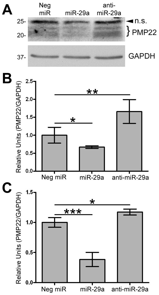
Figure 7. miR-29a regulates endogenous PMP22 levels in Schwann cells.
(A) Schwann cells were transfected with Neg. miR, miR-29a, or anti-miR-29a and processed for Western blotting (40 μg/lane) with the indicated antibodies. Arrowhead corresponds to a non-specific (n.s.) band and the bracket indicates differentially glycosylated forms of PMP22. GAPDH is shown as a loading control. (B) Quantification of PMP22 protein levels, after normalization to GAPDH, in cells transfected with the indicated constructs (*p<0.05; **p<0.01; n=4). (C) Quantification using the 2−ΔΔCT method of real-time RT-PCR experiments on RNA from Schwann cells transfected with Neg. miR, miR-29a, or anti-miR-29a (***p<0.001; *p<0.05; n=4). Data is normalized to GAPDH and expressed in relative units.

