Abstract
The protozoan parasite Trypanosoma congolense is the main causative agent of livestock trypanosomosis. Congopain, the major lysosomal cysteine proteinase of T. congolense, contributes to disease pathogenesis, and antibody-mediated inhibition of this enzyme may contribute to mechanisms of trypanotolerance. The potential of different adjuvants to facilitate the production of antibodies that would inhibit congopain activity was evaluated in the present study. Rabbits were immunised with the recombinant catalytic domain of congopain (C2), either without adjuvant, with Freund’s adjuvant or complexed with bovine or rabbit α2-macroglobulin (α2M). The antibodies were assessed for inhibition of congopain activity. Rabbits immunised with C2 alone produced barely detectable anti-C2 antibody levels and these antibodies had no effect on recombinant C2 or native congopain activity. Rabbits immunised with C2 and Freund’s adjuvant produced the highest levels of anti-C2 antibodies. These antibodies either inhibited C2 and native congopain activity to a small degree, or enhanced their activity, depending on time of production after initial immunisation. Rabbits receiving C2-α2M complexes produced moderate levels of anti-C2 antibodies and these antibodies consistently showed the best inhibition of C2 and native congopain activity of all the antibodies, with maximum inhibition of 65%. Results of this study suggest that antibodies inhibiting congopain activity could be raised in livestock with a congopain catalytic domain-α2M complex. This approach improves the effectiveness of the antigen as an anti-disease vaccine candidate for African trypanosomosis.
Keywords: trypanosomosis, congopain, α2-macroglobulin, anti-disease vaccine
1. INTRODUCTION
Bovine trypanosomosis (nagana) is a disease caused by tsetse-transmitted Trypanosoma congolense, T. vivax and/or T. brucei brucei. These extracellular haemoprotozoa are able to survive in the bloodstream of infected cattle in the presence of continuous exposure to the host’s immune system. Chronic wasting and anaemia are the most prominent features of animal trypanosomosis, but other pathologic effects are circulatory disturbances, leukopaenia, low serum complement levels, lymphoid tissue hyperplasia followed by hypoplasia and immunosuppression [38]. Trypanosomosis is consequently a severe constraint to animal agriculture in many parts of sub-Saharan Africa [25].
There are three control strategies for trypanosomosis in cattle that are often used concomitantly: trypanocidal drugs, vector control and the use of trypanotolerant cattle, none of which is fully effective in the long term [25]. The control strategy with the greatest impact would be vaccination. However, due to the antigenic variation of the parasite surface antigens, attributed to variable expression of antigenic forms of the variable surface glycoprotein (VSG), development of a vaccine is proving to be a difficult task. A vaccine based on VSG would have to cover the entire repertoire of antigenic types, which is not feasible [37]. Invariant antigens that have thus far been identified as potential vaccine candidates include microtubule-associate protein p15 from T. b. brucei [5, 32] and β-tubulin from T. brucei and T. evansi [21, 23, 24]. Vaccination studies in mice have shown that these proteins may provide protection against challenge from T. b. brucei (and T. evansi and T. equiperdum for β-tubulin) [21, 23, 24, 32], but thus far, these studies have excluded homologues from T. congolense or T. vivax.
An alternative approach for a vaccine is to direct the immune response to parasite antigens that play a role in the pathology of the disease. This type of vaccine may not affect the survival of the parasite, but would neutralise pathogenic factors, thereby lessening the pathological effects and possibly inducing a state of trypanotolerance [3, 32], as naturally exhibited by some African breeds of cattle [29]. It has been suggested that congopain, a cysteine protease of T. congolense, has a pathogenic role in trypanosomosis by degrading host proteins and interfering with other host processes [36]. Levels of anti-congopain antibodies have been shown to correlate with resistance to the disease in trypanotolerant cattle [3]. As IgG antibody (and Fab fragments from this IgG) produced by trypanotolerant cattle during T. congolense infection inhibit congopain activity, it has been suggested that this antibody may mitigate the pathology, thus contributing to mechanisms of trypanotolerance [4, 20].
Congopain is a cathepsin L-like enzyme that shares 68% sequence identity in its central catalytic domain and similar enzymatic specificity with the T. cruzi cysteine protease, cruzipain [11]. Congopain has a 130-amino acid long C-terminal extension linked to the catalytic domain by a proline-rich hinge region, which similarly occurs in other trypanosomal cysteine proteases like cruzipain [10], but not mammalian cysteine proteases. The C-terminal extension is highly immunogenic, but considered unlikely to elicit antibodies that inhibit the activity of the enzyme [9]. C2, a recombinantly expressed truncated form of congopain which excludes the C-terminal extension, has been used in a cattle immunisation trial [4]. Immunised cattle developed anti-C2 antibodies that partially inhibited the enzymatic activity of congopain. Following challenge with the parasite there was no effect on the development of infection or acute anaemia. However, immunised cattle maintained or gained weight during infection and exhibited less severe anaemia and leukopaenia during the chronic phase of the disease. Immunised cattle also developed prominent IgG responses to trypanosomal antigens such as VSG, which is reminiscent of trypanotolerance [3]. From that study it appeared that congopain may be involved in the mechanism of trypanosome-induced immunosuppression. Based on these results C2 was used in the present study for immunisations with the hope to raise antibodies capable of inhibiting the enzyme activity.
It has previously been shown that complexing antigen with α2-macroglobulin (α2M) results in enhanced antibody production, presumably by improved delivery of antigen to antigen presenting cells via the α2M receptor [1, 13]. α2M is a high molecular weight (mol. wt.) plasma glycoprotein that is capable of inhibiting the activity of proteases from all classes [6]. When the bait region of α2M is cleaved by a protease, the protease becomes trapped within the α2M molecule [6]. Once this transformation has taken place, receptor recognition sites become exposed on the α2M molecule and it is rapidly taken up by antigen presenting cells expressing the α2M receptor, such as macrophages and dendritic cells [18]. Here we report on complexing C2 with rabbit or bovine α2M and producing antibodies in rabbits against these complexes. In control experiments C2 was injected either alone, or mixed with Freund’s adjuvant. The resulting antibodies were assayed for inhibition of C2 and native congopain activity.
2. MATERIALS AND METHODS
2.1. Materials
Sephacryl S-300 HR, hide powder azure, benzoyl (Bz)-Pro-Phe-Arg-pNA, Freund’s complete and incomplete adjuvant, tosyl phenylalanyl chroromethylketone-treated trypsin, L-trans-epoxysuccinyl-leucylamido(4-guanidino)butane (E-64) and benzoyloxycarbonyl (Z)-Phe-Arg-7-amino-4-methylcoumarin (AMC) were purchased from Sigma-Aldrich (St. Louis, USA). Horseradish peroxidase-conjugated goat anti-rabbit IgG was purchased from Jackson Immunochemicals (Westgrove, USA). 2,2’-azino-bis(3-ethylbenzthiazoline-6-sulphonic acid) (ABTS) and BSA were purchased from Roche Diagnostics (Mannheim, Germany). PEG 6 000 was purchased from Saarchem (Johannesburg, South Africa), Triton X-100 from BDH (Poole, England) and Tween 20 from Merck (Darmstadt, Germany). Bovine α2M was isolated from citrated bovine plasma, and rabbit α2M was isolated from EDTA-treated rabbit plasma, by the same three-step procedure. Briefly, plasma was precipitated by PEG 6 000 [7], and α2M was purified by zinc chelate chromatography followed by gel filtration on Sephacryl S-300 HR [35]. Bovine α2M was stored at −20 °C until use, and rabbit α2M was stored on ice since it is not stable to freeze-thaw cycles [13]. Native congopain was purified as described [2] and recombinant C2 was expressed in Pichia pastoris (manuscript in preparation). All other reagents were of the highest purity obtainable and purchased from Sigma-Aldrich or Merck.
2.2. Protein determination
The concentration of proteins in solution were determined in an Ultrospec 2100 pro spectrophotometer (Amersham Pharmacia Biotech, Piscataway, USA) according to the modified Bradford method [33], or by using for bovine α2M [30a], and for rabbit IgG [17].
2.3. Titration of α2M
Bovine and rabbit α2M were titrated against trypsin to determine their respective active concentration following purification [35]. α2M (50 pmol) was incubated with increasing amounts of trypsin (10–90 pmol) in 100 mM Tris-HCl buffer, pH 8, 10 mM CaCl2, 0.05% (v/v) Triton X-100 for 20 min at room temperature. Residual trypsin activity was measured by incubation with hide powder azure (12.5 mg/mL). Undigested substrate was removed by centrifugation (13 700 g, 5 min, room temperature). The absorbance of the supernatant at 595 nm was plotted against trypsin concentration. The concentration of trypsin required to saturate the inhibitory capacity of 50 pmol α2M was multiplied by two to reflect the percentage inhibitory activity.
2.4. Determination of active enzyme concentration with E-64
Congopain and C2 (0.1–1 μM) diluted with 0.1% (v/v) Brij-35 were titrated against 0.1 μM increments of E-64 from 0–1 μM and incubated for 30 min at 37 °C in activation buffer (100 mM Bis-Tris buffer, pH 6.0, 4 mM Na2EDTA, 0.02% (w/v) NaN3, 8 mM DTT) [8]. Residual enzyme activity was measured with Z-Phe-Arg-AMC (20 μM final concentration), reading excitation at 360 nm and emission at 460 nm in a 7620 microplate fluorometer from Cambridge Technology (UK).
2.5. Native and SDS-PAGE
SDS-PAGE was performed according to Laemmli [19] on 7.5% gels. Native PAGE was performed in the same manner on 5% gels but with the exclusion of SDS from the gel and running buffer.
2.6. Inhibition of C2 activity by α2M
Assays were performed to test the effect of bovine α2M on the hydrolysis of peptide and protein substrates by C2. To assay against a protein substrate, C2 (100 pmol), activated in activation buffer, was incubated with increasing amounts of α2M (0–200 pmol) and incubated for 20 min at 37 °C. C2 activity was assayed with hide powder azure as described above.
To assay against a peptide substrate, C2 (20 pmol), activated in activation buffer, was incubated with increasing amounts of α2M (0–40 pmol) for 20 min at 37 °C. Activity of C2 was assayed with Bz-Pro-Phe-Arg-pNA (3 mM), and hydrolysis was followed at 405 nm using an Ultrospec 2100 pro spectrophotometer (Amersham Pharmacia Biotech).
2.7. Immunisation of rabbits with C2
C2 (80 μg) was activated in activation buffer for 5 min, combined with a 1.5 times molar excess of either rabbit or bovine α2M (3.2 mg), and incubated for 1 h at 37 °C. Subsequently, the samples were divided into 4 equal aliquots, for 4 immunizations, each containing the equivalent of 20 μg C2.
C2-rabbit- and bovine-α2M complexes were each made up to 2 mL with sterile PBS for immunisation. C2 (20 μg) was made up to 1 mL with sterile PBS, and emulsified in a 1:1 ratio with Freund’s adjuvant for immunisation (complete adjuvant for the initial immunisation and incomplete adjuvant for the booster immunisations). For immunisation without adjuvant, C2 (20 μg) was diluted to 2 mL with sterile PBS.
Eight rabbits of mixed breed were immunised by subcutaneous injection at multiple, well-separated sites on the back with either C2 alone (rabbits A1 and A2), C2 in Freund’s adjuvant (F1 and F2), C2-rabbit α2M complexes (R1 and R2), or C2-bovine α2M complexes (B1 and B2). Each rabbit received a total of 1 mL, i.e. an equivalent of 10 μg C2 per immunisation. Booster injections were given at weeks 2, 4 and 6 and rabbits were bled from the mid-ear artery prior to immunisation and at weeks 3, 6, 9 and 11. Rabbit B1 was not bled at week 3. Ethical approval for these procedures was obtained from the University of KwaZulu-Natal animal ethics committee (reference AE/Coetzer/01/02).
2.8. Determination of anti-C2 antibody production by ELISA
C2 or native congopain was coated onto the wells of 96 well Nunc-ImmunoTM ELISA plates at a concentration of 1 μg/mL in 50 mM carbonate buffer, pH 6 for 16 h at 4 °C. Non-specific binding of antibody was prevented by blocking with 0.5% (w/v) BSA-PBS (blocking buffer) for 1 h at 37 °C. Wells were washed 3 times with 0.1% (v/v) PBS-Tween 20 in between each step. Rabbit IgG, isolated by PEG 6 000 precipitation [30b], was diluted in blocking buffer and incubated in the wells for 2 h at 37 °C (100 μL per well). Rabbit IgG was titrated from 500 to 31.25 μg/mL (weeks 6, 9 and 11) and from 200 to 6.25 μg/mL (3 weeks) as a result of the small amounts of serum collected at 3 weeks. The HRPO-labelled detection antibody, goat anti-rabbit IgG, was diluted 1:30 000 in blocking buffer and incubated (120 μL per well) for 1 h at 37 °C. Substrate solution, 0.05% (w/v) ABTS, 0.0015% (v/v) H2O2, in 150 mM citrate-phosphate, pH 5, was added (150 μL per well) and colour was allowed to develop in the dark. The A405 of each well was measured with an automated Versamax microplate reader using SOFTmax® software from Molecular Devices (Menlo Park, CA, USA).
2.9. Assays for inhibition of C2 and congopain activity by antibodies
The inhibition of C2 and congopain activity by anti-C2 IgG was assayed in duplicate in a stopped-time assay format with serial dilutions of IgG from 1 mg/mL to 125 μg/mL. Assays were performed in duplicate. C2 (80 ng) or native congopain (100 ng), diluted to 75 μL in 0.1% Brij, was combined with 75 μL of IgG, diluted serially with 200 mM sodium phosphate buffer, pH 7.2, 4 mM Na2EDTA, 0.1% (v/v) Tween-containing 40 μg/mL lima bean trypsin inhibitor (has no effect on C2 [40]) to inhibit residual plasma kallikrein activity against Z-Phe-Arg-AMC that may remain in IgG samples – to yield final IgG concentrations of 1 mg/mL to 125 μg/mL. The samples were incubated for 15 min at 37 °C. Aliquots (50 μL) were removed from the incubation mixture and combined with 25 μL of a 200 mM sodium phosphate buffer, pH 7.2, 4 mM Na2EDTA, 0.1% (v/v) Tween, 8 mM DTT, and activated for 1 min at 37 °C. Substrate was added (25 μL) and the samples incubated for 5 min at 37 °C. The reaction was stopped with 100 mM monochloroacetate, 30 mM Na-acetate, 70 mM acetic acid, pH 4.3, before fluorescence was read (excitation at 360 nm and emission at 460 nm) in a 7620 microplate fluorometer from Cambridge Technology. Inhibition was expressed as a percentage of the activity in the presence of non-immune rabbit IgG at the same concentration.
3. RESULTS
3.1. Interaction of C2 with bovine α2M
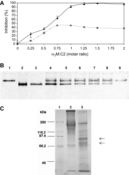
In order to study the interaction between congopain and the protease inhibitor, α2M, the extent of inhibition of C2 activity by α2M was assayed against both high (hide powder azure) and low (Bz-Pro-Phe-Arg-pNA) mol. wt. substrates. When proteases are trapped within the α2M tetramer, high mol. wt. substrates are excluded from interaction with the protease, resulting in complete inhibition, whereas low mol. wt. substrates are able to access the protease’s active site, resulting in partial inhibition [6]. Consistent with this observation bovine α2M inhibited 99% of the proteolytic activity of C2 towards hide powder azure and 45% of the peptidolytic activity against Bz-Pro-Phe-Arg-pNA (Fig. 1A). In contrast to cathepsin L and papain (result not shown), the peptide substrate experienced restricted access to the active site of congopain (15% inhibition at equimolar concentrations with α2M), and to a greater extent to that of C2, when in the α2M complex. A similar observation was made for cruzipain when assayed for inhibition with human α2M using the same peptide substrate [31]. From the inhibition assays it appeared that the enzyme and inhibitor interact in an equimolar ratio, since no further inhibition of C2 activity was observed above this ratio.
Figure 1.
The interaction of the recombinant catalytic domain of congopain (C2) with bovine α2M. (A) The effect of increasing amounts of α2M on the activity of C2 against hide powder azure (▲) and Bz-Pro-Phe-Arg-pNA (□). Different concentrations of α2M were incubated with C2 (100 pmol for hide powder azure assay; 20 pmol for Bz-Pro-Phe-Arg-pNA assay) at molar ratios of 0.25:1, 0.5:1, 0.75:1, 1:1, 1.25:1, 1.5:1 and 2:1 for 20 min at 37 °C. Proteolytic activity was determined by the extent of hydrolysis of substrate compared to that of a C2 control with no α2M (100% activity), and expressed as a percentage inhibition. Error bars represent the ± SEM (n = 3). (B) Non-denaturing PAGE (5% gel) analysis of the interaction between bovine α2M and activated C2. Lanes 1 and 9 500 ng α2M; lanes 2–8, α2M (500 ng) was incubated with increasing amounts of C2 (11–91 ng), corresponding to molar ratios of α2M:C2 of 0.25:1, 0.5:1, 0.75:1, 1:1, 1.25:1, 1.5:1 and 2:1 in the respective lanes. Proteins were silver stained. (C) Reducing SDS-PAGE (7.5% gel) after reaction of bovine α2M with activated C2. Lane 1, Bio-Rad molecular weight markers consisting of myosin (200 kDa), β-galactosidase (116.2 kDa), phosphorylase b (97.4 kDa), bovine serum albumin (66.2 kDa), and ovalbumin (45 kDa); lane 2, α2M (10 μg); lane 3, C2-α2M complex (10.372 μg). Proteins were stained with Coomassie blue R-250. Arrows represent bands at 95 and 85 kDa.
The transformation of α2M from the native “slow” to the “fast” form after interaction with C2 (i.e. the change of α2M to a more compact conformation after cleavage of the bait region) was illustrated by non-denaturing PAGE (Fig. 1B). There was a clearly observed increase in the mobility of α2M in the gel upon reaction with C2 at α2M:C2 molar ratios of 0.25:1 and 0.5:1 (lanes 2 and 3). For α2M:C2 molar ratios between 0.75:1 and 2:1 (lanes 4–8), decreasing proportions of α2M interacted with C2 as shown by the decreasing amounts of the “slow” compared to “fast” forms of α2M. Reducing SDS-PAGE of the α2M-C2 complex revealed that the α2M bait region had been cleaved by the enzyme C2 (Fig. 1C) as evidenced by the presence of protein bands at approximately 95 and 85 kDa, representing the sizes of the fragments produced under reducing conditions when the bait region has been cleaved [35].
3.2. Evaluation of rabbit anti-C2 antibody production by ELISA
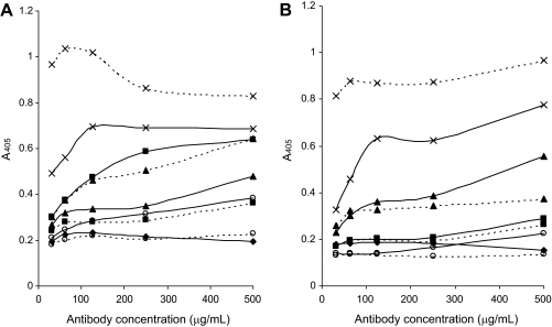
In order to compare the levels of antibodies produced over time in rabbits immunised with C2 in the absence and presence of adjuvants, ELISA plates were coated with C2 or native congopain and indirect ELISA conducted. Rabbits A1 and A2, immunised with C2 without the use of adjuvant, showed no discernible levels of anti-C2 IgG (Fig. 2), except at week 6 for rabbit A1 (Fig. 2A), when a low level of antibodies was produced. Rabbits B1 and B2, immunised with bovine α2M-C2 complexes, showed moderate levels of anti-C2 IgG throughout the test period (Fig. 2). Rabbits F1 and F2, immunised with C2 in Freund’s adjuvant, consistently showed the highest levels of anti-C2 IgG, with the exception of F1 at week 3 (result not shown). Rabbits R1 and R2, immunised with rabbit α2M-C2 complexes, showed differing anti-C2 IgG responses. At week 3, rabbit R1 showed high levels of anti-C2 IgG whereas R2 showed no anti-C2 IgG (result not shown). At week 6, both rabbits showed moderate levels of anti-C2 IgG, with levels in R1 higher than those in R2 (Fig. 2A), a trend that was repeated at week 9 (result not shown). At week 11, the anti-C2 IgG levels in R1 had decreased to become level with those in R2 (Fig. 2B).
Figure 2.
ELISA of anti-C2 antibodies produced at week 6 (A) and 11 (B) by rabbits immunised with C2 alone, or in combination with different adjuvants. C2 was coated onto ELISA plates (1 μg/mL in 50 mM carbonate buffer, pH 6, 16 h, 4 °C) and probed for recognition by IgG collected at weeks 6 and 11 from rabbit A1 (○; solid line), A2 (○; broken line), B1 (▲; solid line), B2 (▲; broken line), F1 (×; solid line), F2 (×; broken line), R1 (■; solid line), and R2 (■; broken line), and non-immune IgG (♦). Absorbance readings at 405 nm represent the average of two experiments. Goat anti-rabbit IgG-HRPO labelled antibodies were used for detection and ABTS/H2O2 as substrate as described in Materials and methods.
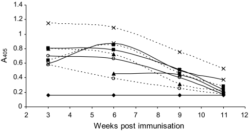
Recognition of native congopain by anti-C2 antibodies collected over the immunisation period was assayed in an ELISA using a single antibody concentration of 100 μg/mL (Fig. 3). The anti-C2 antibodies showed good recognition of native congopain. The highest antibody levels were observed for weeks 3 and 6, after which antibody levels declined. Generally, these results agree with those shown in Figure 2 for the recognition of C2 in an ELISA, except that there appears to be slightly better recognition of native congopain by antibodies from rabbits immunised with C2 without any adjuvant.
Figure 3.
Recognition of native congopain in an ELISA by anti-C2 antibodies produced at week 3, 6, 9 and 11 by rabbits immunised with C2 alone, or in combination with different adjuvants. Native congopain was coated onto ELISA plates (1 μg/mL in 50 mM carbonate buffer, pH 6, 16 h, 4 °C) and probed for recognition by IgG (100 μg/mL) collected at weeks 3, 6, 9 and 11 from rabbit A1 (○; solid line), A2 (○; broken line), B1 (▲; solid line), B2 (▲; broken line), F1 (×; solid line), F2 (×; broken line), R1 (■; solid line), and R2 (■; broken line), and non-immune rabbit IgG (♦). Absorbance readings at 405 nm represent the average of two experiments. Goat anti-rabbit IgG-HRPO labelled antibodies were used for detection and ABTS/H2O2 as substrate as described in Materials and methods. Rabbit B1 was not bled at week 3.
3.3. Inhibition of C2 and congopain activity by rabbit anti-C2 antibodies
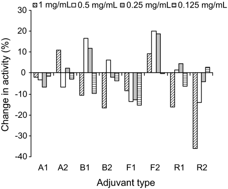
In order to determine the effect of the antibodies on C2 activity, the unactivated enzyme was incubated with the respective purified rabbit IgG samples before assaying for residual activity with the fluorescent substrate Z-Phe-Arg-AMC. In control incubations, purified non-immune rabbit IgG was incubated with C2. Negative percentage change in activity values (Fig. 4) suggest that the addition of anti-C2 IgG resulted in reduced hydrolysis of the substrate compared to samples containing non-immune IgG, i.e. decreased enzyme activity. Assays of the effect of antibody concentration on enzyme inhibition generally showed a decrease in % inhibition with a decrease in antibody concentration (Fig. 4).
Figure 4.
Effect on C2 activity of varying concentrations of anti-C2 antibodies from weeks 6 (A) and 11 (B) in the immunisation protocol. C2 (80 ng) was incubated with anti-C2 antibodies (0.125 to 1 mg/mL) from rabbits A1, A2, B1, B2, F1, F2, R1 and R2 collected at weeks 6 and 11 at pH 7.2 (15 min at 37 °C). Residual activity against Z-Phe-Arg-AMC was determined as described in Materials and methods. Each plot represents the mean of two experiments. Negative % change in activity indicates inhibition of enzyme activity.
Week 3 IgG generally had little effect on the activity of C2 with antibodies from rabbits immunised with bovine α2M-C2 complexes (B2) showing the only significant inhibition of C2 activity (result not shown). In the assay of week 6 IgG (Fig. 4A), IgG from all rabbits resulted in inhibition of the activity of C2 against the substrate, although the inhibitory effect of IgG from rabbits immunised with C2 and Freund’s adjuvant (F2) was minimal. The best inhibition was seen for IgG from rabbits immunised with rabbit α2M-C2 complexes (R1 and R2), followed by IgG from rabbits immunised with bovine α2M-C2 complexes (B1 and B2). IgG from rabbits immunised with C2 triturated with Freund’s adjuvant (F1 and F2) and immunised without adjuvant (A1 and A2) comparably showed the least inhibition. In the assay of week 9 IgG (result not shown), inhibition of C2 activity against the substrate was only observed for IgG from rabbits immunised with rabbit α2M-C2 complexes (R1 and R2) and bovine α2M-C2 complexes (B1), and to a small degree, IgG from rabbits immunised with C2 and Freund’s adjuvant (F1) and without any adjuvant (A1). The IgG from the other rabbits appeared to enhance the activity of C2 against the substrate. In the assay of week 11 IgG (Fig. 4B), the inhibition profile was very similar to that seen for week 6 IgG. The only exception was that week 11 IgG from rabbits immunised with C2 without adjuvant (A1 and A2) enhanced the activity of C2 to a small degree.
It was deemed important to determine whether IgG that inhibited Z-Phe-Arg-AMC hydrolysis by C2 would also be inhibitory towards the proteolytic activity of congopain, the native form of the enzyme purified from trypanosome lysates. The amount of congopain available was limited, therefore only IgG from week 6 was assayed. As was seen for the assay of the effect of week 6 IgG on C2 (Fig. 4A), IgG from rabbits immunised with rabbit α2M-C2 complexes (R1 and R2) was the most inhibitory, followed by IgG from rabbits immunised with bovine α2M-C2 complexes (B1 and B2) (Fig. 5).
Figure 5.
Effect on native congopain activity of varying concentrations of anti-C2 antibodies from weeks 6 in the immunisation protocol. Native congopain (100 ng) was incubated with anti-C2 antibodies (0.125 to 1 mg/mL) from rabbits A1, A2, B1, B2, F1, F2, R1 and R2 collected at weeks 3, 6, 9 and 11 at pH 7.2 (15 min at 37 °C). Residual activity against Z-Phe-Arg-AMC was determined as described in Materials and methods. Each plot represents the mean of two experiments. Negative % change in activity indicates inhibition of enzyme activity.
The highest percentage of inhibition of C2 hydrolysis of Z-Phe-Arg-AMC obtained in the present study were 61% and 65%, which were observed for week 11 IgG isolated from rabbits immunised with C2 complexed to rabbit and bovine α2M respectively (Fig. 4B). Antibodies produced by rabbits immunised with C2 in Freund’s adjuvant were generally found to weakly inhibit congopain and C2 with a maximum of 40%. Curiously the same antibodies from the one rabbit enhanced the activity of congopain against Z-Phe-Arg-AMC (Fig. 5, F2). From the data obtained, it appeared that in general IgG fractions isolated from rabbits immunised with rabbit α2M-C2 complexes were the most inhibitory towards the proteolytic activity of C2 and congopain.
4. DISCUSSION
There is currently no vaccine available for trypanosomosis in cattle, and although there are control strategies in place such as trypanocidal drugs and tsetse control, the disease is still a constraint to livestock production in many parts of Africa [25]. Due to the ability of the parasite to evade the host’s immune system by antigenic variation and immunosuppression [37], the development of an anti-disease vaccine, that would limit the pathogenic effects of infection rather than suppress parasite growth, constitutes an attractive alternative to conventional vaccination [4, 20]. Congopain has been identified as a pathogenic factor of trypanosomosis [3], and the purpose of the present study was to identify an adjuvant system that would elicit an antibody response to congopain to inhibit its enzyme activity and thus its pathogenic effects.
Our investigation into the interaction of C2, the truncated catalytic domain of congopain, with bovine α2M showed that active C2 cleaves the bait region of bovine α2M. This cleavage resulted in inhibition of C2 by trapping of the protease within the α2M tetramer. C2 and bovine α2M appear to interact in an equimolar ratio. This information was used for the preparation of complexes of C2 with bovine α2M and rabbit α2M for the production of anti-C2 antibodies in rabbits.
All eight rabbits were immunised with the same amount of C2, i.e. 10 μg per immunisation. However, the titres of anti-C2 antibodies, as determined by ELISA, varied according to the type of adjuvant used in the immunisation procedure. In the absence of adjuvant, the levels of anti-C2 antibodies were barely detectable. Freund’s adjuvant elicited a strong antibody response against the small amount of C2 used for immunisation, but the efficacy of these antibodies in inhibiting the activity of C2 in vitro was limited. The anti-C2 antibody titres observed where α2M (both bovine and rabbit) was used as an adjuvant were moderate, and yet, these antibodies showed the best inhibition of the C2 mediated hydrolysis of Z-Phe-Arg-AMC.
These anti-rabbit and bovine α2M complexed-C2 antibodies inhibited whole (native) congopain activity in addition to that of the (recombinant) catalytic domain of congopain, C2. This was imperative since it would be the native cysteine proteinase that these antibodies would encounter in vivo upon challenge with the parasite. Although the optimal pH values for C2 and native congopain activity are reported to be 6 and 6.4 respectively [9, 11], physiological pH was used in the present study to obtain results that would have application to a vaccine in vivo.
Rabbits immunised with rabbit α2M complexed with C2 produced antibodies with maximal enzyme inhibition, followed closely by antibodies from rabbits immunised with bovine α2M complexed with C2. Rabbits immunised with C2 mixed with Freund’s adjuvant produced antibodies that were only weakly inhibitory, and sometimes resulted in enhancement of enzyme activity. Binding of these “activating” antibodies to the enzyme may have caused conformational changes that favoured the reaction with the substrate. Activation of enzyme activity by Freund’s adjuvant generated antibodies has been reported in studies with β-lactamase and trypanopain-Tb [34, 41]. We were unable to explore why two rabbits immunised with the same immunogen (C2 and Freund’s adjuvant) produced antibodies that enhanced enzyme activity in the one instance and inhibited activity in the other. It would have been interesting to be able to measure the effects of antibodies collected at other time points in both rabbits. In summary, whilst immunisation using Freund’s adjuvant resulted in a higher level of antibody production, immunisation using rabbit or bovine α2M resulted in a qualitatively superior antibody production (i.e. antibodies that inhibited C2 activity). In the context of a vaccine to neutralise congopain activity, immunisation with C2 complexed to α2M seems a viable option since the antibodies produced were inhibitory towards the protease in vitro.
It is well established that α2M-antigen complexes are targeted to antigen presenting cells that express the α2M receptor, which in turn results in enhanced presentation of the antigen by MHC class II molecules to CD4+ T cells [13, 16, 26]. This antigen delivery has been shown to occur with monocytes and macrophages [1, 12, 26], recognised as amateur antigen presenting cells, and more recently professional antigen presenting cells such as CD11c+ lin− blood dendritic cells and Langerhans cells [16]. In the present study, this mechanism of antigen delivery appeared to result in antibodies that were better able to inhibit the activity of the enzyme than antibodies produced with the use of Freund’s adjuvant. This may be due to the preservation of the three-dimensional conformation of the enzyme when it is trapped inside the α2M molecule. With Freund’s adjuvant, the free enzyme is exposed to denaturation by the very low pH of the adjuvant and receives no such protection as in the α2M complexes. In separate (unpublished) experiments in mice, active C2 was significantly more immunogenic than the corresponding inactive enzyme (active site Cys mutated to Ala). This is consistent with current results since inactive C2 does not bind to natural host inhibitors such as α2M (unpublished observation). Where the inhibition of congopain by anti-peptide antibodies has been investigated, antibodies were generally found to enhance the activity of the enzyme (unpublished observations). Results for evaluating the effect of anti-peptide antibodies on the activity of trypanopain from T. b. brucei have been shown to range from enhanced activity (176%) to inhibition of up to 85% [41].
In a previous study where cattle were immunised with C2 and RWL™, a proprietary adjuvant from Smith Kline Beecham, the anti-C2 antibodies produced resulted in 50–60% inhibition of C2 activity in vitro [4]. Whilst immunisation had no effect on the establishment of infection or the development of acute anaemia, immunised cattle managed to maintain or even gain weight during infection. Studies are currently underway in a mouse model system to establish whether the humoral immune response induced by α2M-congopain complexes confers protection following experimental infection with T. congolense.
Another apparent benefit to using α2M as an alternative to conventional adjuvants, is its ability to induce antibody responses to lower doses of antigen, as observed for HIV envelope subunit peptides [22]. This is a critical aspect for a vaccine since the amount of antigen required will greatly affect the practicality of the anti-disease vaccine approach.
There are other molecules besides congopain that contribute towards the pathology of trypanosomosis and which may be worth associating in a multicomponent “vaccine”. Other potential pathogenic factors identified thus far include oligopeptidase B from T. brucei, T. congolense and T. evansi [14, 27, 28], and a surface located acid phosphatase [39]. Although these enzymes do not cleave the bait region of α2M, they could be incorporated into α2M using congopain or via a non-proteolytic method [15].
The findings of the present study are a step towards improving the efficacy of immunisation with cysteine proteases in a vaccine strategy aimed at mitigating the pathological consequences of trypanosome infections.
Acknowledgments
This work was supported by grants from the Commission of the European Communities’ Sixth Framework Programme TRYPADVAC2 (INCO Contract number 003716) and the South African National Research Foundation.
References
- 1.Anderson R.B., Cianciolo G.J., Kennedy M.N., Pizzo S.V.. Alpha 2-macroglobulin binds CpG oligodeoxynucleotides and enhances their immunostimulatory properties by a receptor-dependent mechanism. J. Leukoc. Biol. 2008;83:381–392. doi: 10.1189/jlb.0407236. [DOI] [PubMed] [Google Scholar]
- 2.Authié E., Muteti D.K., Mbawa Z.R., Lonsdale-Eccles J.D., Webster P., Wells C.W.. Identification of a 33-kilodalton immunodominant antigen of Trypanosoma congolense as a cysteine protease. Mol. Biochem. Parasitol. 1992;56:103–116. doi: 10.1016/0166-6851(92)90158-g. [DOI] [PubMed] [Google Scholar]
- 3.Authié E.. Trypanosomiasis and trypanotolerance in cattle: a role for congopain? Parasitol. Today. 1994;10:360–364. doi: 10.1016/0169-4758(94)90252-6. [DOI] [PubMed] [Google Scholar]
- 4.Authié E., Boulangé A., Muteti D., Lalmanach G., Gauthier F., Musoke A.J.. Immunisation of cattle with cysteine proteinases of Trypanosoma congolense: targeting the disease rather than the parasite. Int. J. Parasitol. 2001;31:1429–1433. doi: 10.1016/s0020-7519(01)00266-1. [DOI] [PubMed] [Google Scholar]
- 5.Balaban N., Waithaka H.K., Njogu A.R., Goldman R.. Intracellular antigens (microtubule-associated protein copurified with glycosomal enzymes) possible vaccines against trypanosomiasis. J. Infect. Dis. 1995;172:845–850. doi: 10.1093/infdis/172.3.845. [DOI] [PubMed] [Google Scholar]
- 6.Barrett A.J., Starkey P.M.. The interaction of α2-macroglobulin with proteases. Characteristics and specificity of the reaction, and a hypothesis concerning its molecular mechanism. Biochem. J. 1973;133:709–724. doi: 10.1042/bj1330709. [DOI] [PMC free article] [PubMed] [Google Scholar]
- 7.Barrett A.J., Brown M.A., Sayers C.A.. The electrophoretically “slow” and “fast” forms of the α2-macroglobulin molecule. Biochem. J. 1979;181:401–418. doi: 10.1042/bj1810401. [DOI] [PMC free article] [PubMed] [Google Scholar]
- 8.Barrett A.J., Kembhavi M.M., Brown M.A., Kirschke H., Knight C.G., Tamai M., Hanada K.. L-trans-epoxysuccinylleucylamido(4-gaunidino)butane (E-64) and its analogues as inhibitors of cysteine proteinases including cathepsins B, H and L. Biochem. J. 1982;201:189–198. doi: 10.1042/bj2010189. [DOI] [PMC free article] [PubMed] [Google Scholar]
- 9.Boulangé A., Serveau C., Brillard M., Minet C., Gauthier F., Diallo A.. et al. Functional expression of the catalytic domains of two cysteine proteinases from Trypanosoma congolense. Int. J. Parasitol. 2001;13:1435–1440. doi: 10.1016/s0020-7519(01)00267-3. [DOI] [PubMed] [Google Scholar]
- 10.Cazzulo J.J., Frasch A.C.. SAPA/trans-sialidase and cruzipain: two antigens from Trypanosoma cruzi contain immunodominant but enzymatically inactive domains. FASEB J. 1992;14:3259–3264. [PubMed] [Google Scholar]
- 11.Chagas J.R., Authié E., Serveau C., Lalmanach G., Juliano L., Gauthier F.. A comparison of the enzymatic properties of the major cysteine proteases from Trypanosoma congolense and T. cruzi. Mol. Biochem. Parasitol. 1997;88:85–94. doi: 10.1016/s0166-6851(97)00085-6. [DOI] [PubMed] [Google Scholar]
- 12.Chu C.T., Pizzo S.V.. Receptor-mediated antigen delivery into macrophages. Complexing antigen to α2-macroglobulin enhances presentation to T cells. J. Immunol. 1993;150:48–58. [PubMed] [Google Scholar]
- 13.Chu C.T., Oury T.D., Enghild J.J., Pizzo S.V.. Adjuvant-free in vivo targeting. Antigen delivery by α2-macroglobulin enhances antibody formation. J. Immunol. 1994;152:1538–1545. [PubMed] [Google Scholar]
- 14.Coetzer T.H., Goldring J.P., Huson L.E.. Oligopeptidase B: a processing peptidase involved in pathogenesis. Biochimie. 2008;90:336–344. doi: 10.1016/j.biochi.2007.10.011. [DOI] [PubMed] [Google Scholar]
- 15.Grøn H., Pizzo S.V.. Nonproteolytic incorporation of protein ligands into human α2-macroglobulin: implications for the binding mechanism of α2-macroglobulin. Biochemistry. 1998;37:6009–6014. doi: 10.1021/bi973027c. [DOI] [PubMed] [Google Scholar]
- 16.Hart J.P., Gunn M.D., Pizzo S.V.. A CD91-positive subset of CD11c+ blood dendritic cells: characterization of the APC that functions to enhance adaptive immune responses against CD91-targeted antigens. J. Immunol. 2004;172:70–78. doi: 10.4049/jimmunol.172.1.70. [DOI] [PubMed] [Google Scholar]
- 17.Hudson L., Hay F.C. Hudson L., Hay F.C. Practical immunology. London: Blackwell Scientific publications; 1980. Molecular weights and special properties of immunoglobulins and antigens of immunological interest; p. 347. [Google Scholar]
- 18.Imber M.J., Pizzo S.V.. Clearance and binding of two electrophoretic “fast” forms of human α2-macroglobulin. J. Biol. Chem. 1981;256:8134–8139. [PubMed] [Google Scholar]
- 19.Laemmli U.K.. Cleavage of structural proteins during the assembly of the head of bacteriophage T4. Nature. 1970;277:680–685. doi: 10.1038/227680a0. [DOI] [PubMed] [Google Scholar]
- 20.Lalmanach G., Boulangé A., Serveau C., Lecaille F., Scharfstein J., Gauthier F., Authié E.. Congopain from Trypanosoma congolense: drug target and vaccine candidate. Biol. Chem. 2002;383:739–749. doi: 10.1515/BC.2002.077. [DOI] [PubMed] [Google Scholar]
- 21.Li S.Q., Fung M.C., Reid S.A., Inoue N., Lun Z.R.. Immunization with recombinant beta-tubulin from Trypanosoma evansi induced protection against T. evansi, T. equiperdum and T. b. brucei infection in mice. Parasite Immunol. 2007;29:191–199. doi: 10.1111/j.1365-3024.2006.00933.x. [DOI] [PubMed] [Google Scholar]
- 22.Liao H., Cianciolo G.J., Staats H.F., Scearce R.M., Lapple D.M., Stauffer S.H.. et al. Increased immunogenicity of HIV envelope subunit complexed with α2-macroglobulin when combined with monophosphoryl lipid A and GM-CSF. Vaccine. 2002;20:2396–2403. doi: 10.1016/s0264-410x(02)00090-7. [DOI] [PubMed] [Google Scholar]
- 23.Lubega G.W., Byarugaba D.K., Prichard R.K.. Immunization with a tubulin-rich preparation from Trypanosoma brucei confers broad protection against African trypanosomosis. Exp. Parasitol. 2002;102:9–22. doi: 10.1016/s0014-4894(02)00140-6. [DOI] [PubMed] [Google Scholar]
- 24.Lubega G.W., Ochola D.O., Prichard R.K.. Trypanosoma brucei: anti-tubulin antibodies specifically inhibit trypanosome growth in culture. Exp. Parasitol. 2002;102:134–142. doi: 10.1016/s0014-4894(03)00035-3. [DOI] [PubMed] [Google Scholar]
- 25.McDermott J.J., Coleman P.G.. Comparing apples and oranges – model based assessment of different tsetse-transmitted trypanosomosis control strategies. Int. J. Parasitol. 2001;31:603–609. doi: 10.1016/s0020-7519(01)00148-5. [DOI] [PubMed] [Google Scholar]
- 26.Morrot A., Strickland D.K., de Lourdes Higuchi M., Reis M., Pedrosa R., Scharfstein J.. Human T cell responses against the major cysteine proteinase (cruzipain) of Trypanosoma cruzi: role of the multifunctional α2-macroglobulin receptor in antigen presentation by monocytes. Int. Immunol. 1997;9:825–834. doi: 10.1093/intimm/9.6.825. [DOI] [PubMed] [Google Scholar]
- 27.Morty R.E., Authié E., Troeberg L., Lonsdale-Eccles J.D., Coetzer T.H.T.. Purification and characterisation of a trypsin-like serine oligopeptidase from Trypanosoma congolense. Mol. Biochem. Parasitol. 1999;102:145–155. doi: 10.1016/s0166-6851(99)00097-3. [DOI] [PubMed] [Google Scholar]
- 28.Morty R.E., Lonsdale-Eccles J.D., Morehead J., Caler E.V., Mentele R., Auerswald E.A.. et al. Oligopeptidase B from Trypanosoma brucei, a new member of an emerging subgroup of serine oligopeptidases. J. Biol. Chem. 1999;274:26149–26156. doi: 10.1074/jbc.274.37.26149. [DOI] [PubMed] [Google Scholar]
- 29.Murray M., Morrison W.I., Whitelaw D.D.. Host susceptibility to African trypanosomiasis: trypanotolerance. Adv. Parasitol. 1982;21:1–68. doi: 10.1016/s0065-308x(08)60274-2. [DOI] [PubMed] [Google Scholar]
- 30a.Nagasawa S., Sugihara H., Han B.H., Suzuki T.. Studies on α2-macroglobulin in bovine plasma. I. Purification, physical properties, and chemical compositions. J. Biochem. 1970;67:809–819. doi: 10.1093/oxfordjournals.jbchem.a129313. [DOI] [PubMed] [Google Scholar]
- 30b.Polson A., Potgieter G.M., Largier J.F., Mears E.G.F., Joubert F.J.. The fractionation of protein mixtures by linear polymers of high molecular weight. Biochim. Biophys. Acta. 1964;82:463–475. doi: 10.1016/0304-4165(64)90438-6. [DOI] [PubMed] [Google Scholar]
- 31.Ramos A.M., Duschak V.G., Gerez de Burgos N.M., Barboza M., Remedi M.S., Vides M.A., Chiabrando G.A.. Trypanosoma cruzi: cruzipain and membrane-bound cysteine proteinase isoform(s) interacts with human alpha(2)-macroglobulin and pregnancy zone protein. Exp. Parasitol. 2002;100:121–130. doi: 10.1016/S0014-4894(02)00007-3. [DOI] [PubMed] [Google Scholar]
- 32.Rasooly R., Balaban N.. Trypanosome microtubule-associated protein p15 as a vaccine for the prevention of African sleeping sickness. Vaccine. 2004;22:1007–1015. doi: 10.1016/j.vaccine.2003.08.041. [DOI] [PubMed] [Google Scholar]
- 33.Read S.M., Northcote D.H.. Minimization of variation in the response to different proteins of the Coomassie blue G dye-binding assay for protein. Anal. Biochem. 1981;116:53–64. doi: 10.1016/0003-2697(81)90321-3. [DOI] [PubMed] [Google Scholar]
- 34.Richmond M.H. Salton M.R.J. Immunochemistry of enzymes and their antibodies. New York: Wiley; 1977. β-Lactamase/anti-β-lactamase interactions; pp. 39–55. [Google Scholar]
- 35.Salvesen G., Enghild J.J.. α-Macroglobulins: detection and characterization. Methods Enzymol. 1993;223:121–141. doi: 10.1016/0076-6879(93)23041-k. [DOI] [PubMed] [Google Scholar]
- 36.Serveau C., Boulangé A., Lecaille F., Gauthier F., Authié E., Lalmanach G.. Procongopain from Trypanosoma congolense is processed at basic pH: an unusual feature among cathepsin L-like cysteine proteinases. Biol. Chem. 2003;384:921–927. doi: 10.1515/BC.2003.103. [DOI] [PubMed] [Google Scholar]
- 37.Taylor K.A.. Immune responses of cattle to African trypanosomes: protective or pathogenic? Int. J. Parasitol. 1998;28:219–240. doi: 10.1016/s0020-7519(97)00154-9. [DOI] [PubMed] [Google Scholar]
- 38.Taylor K.A., Authié E. Maudlin I., Holmes P.H., Miles M.A. The trypanosomiases. Wallingford: CABI Publishing; 2004. Pathogenesis of animal trypanosomiasis; pp. 331–353. [Google Scholar]
- 39.Tosomba O.M., Coetzer T.H.T., Lonsdale-Eccles J.D.. Localisation of acid phosphatase activity on the surface of bloodstream forms of Trypanosoma congolense. Exp. Parasitol. 1996;84:429–438. doi: 10.1006/expr.1996.0131. [DOI] [PubMed] [Google Scholar]
- 40.Troeberg L., Pike R.N., Morty R.E., Berry R.K., Coetzer T.H.T., Lonsdale-Eccles J.D.. Proteases from Trypanosoma brucei brucei. Purification, characterisation and interactions with host regulatory molecules. Eur. J. Biochem. 1996;238:728–736. doi: 10.1111/j.1432-1033.1996.0728w.x. [DOI] [PubMed] [Google Scholar]
- 41.Troeberg L., Pike R.N., Lonsdale-Eccles J.D., Coetzer T.H.T.. Production of anti-peptide antibodies against trypanopain-Tb from Trypanosoma brucei brucei: effects of antibodies on enzyme activity against Z-Phe-Arg-AMC. Immunopharmacology. 1997;36:295–303. doi: 10.1016/s0162-3109(97)00034-9. [DOI] [PubMed] [Google Scholar]


