Abstract
Objective
To evaluate the utility and advantages of the coaxial snare technique in the retrieval of tubular foreign bodies.
Materials and Methods
Using the coaxial snare technique, we attempted to retrieve tubular foreign bodies present in seven patients. The bodies were either stents which were malpositioned or had migrated from their correct position in the vascular system (n=2), a fragmented venous introducer sheath (n=1), fragmented drainage catheters in the biliary tree (n=2), or fractured external drainage catheters in the urinary tract (n=2). After passing a guidewire and/or a dilator through the lumina of these foreign bodies, we introduced a loop snare over the guidewire or dilator, thus capturing and retrieving them.
Results
In all cases, it was possible to retrieve or reposition the various items, using a minimum-sized introducer sheath or a tract. No folding was involved. In no case were surgical procedures required, and no complications were encountered.
Conclusion
The coaxial snare technique, an application of the loop snare technique, is a useful and safe method for the retrieval of tubular foreign bodies, and one which involves minimal injury to the patient.
Keywords: Catheters and catheterization complications, Foreign bodies, Interventional procedures
With the increasing use of indwelling catheters and interventional devices, interventional radiologists are frequently confronted with the problem of percutaneous removal of foreign bodies or iatrogenically placed objects. Items retrieved from the vascular system include wires, catheter fragments, malpositioned or migrated stents, coils, and caval filters (1-9). Likewise, there is often the need to extract items from the urinary system, gastrointestinal tract, and biliary or bronchial tree (4, 7, 10-13). A large variety of techniques for the nonsurgical manipulation of intravascular and extravascular foreign bodies has been developed and described. Devices used for the retrieval of these objects include the loop snare, basket, grasping forceps, tip-deflecting wire, pincher device, oversize catheter or sheath, and balloon catheter (1, 3, 4, 8, 14-17). Of these, the loop snare technique has in most cases been the method of choice. In the case of large foreign bodies, however, the simple snare technique is used for folding, prior to retrieval; for extraction from the body, the much larger bore sheath is required. The process of folding foreign objects and inserting a large introducer sheath is traumatic to the patient, and during retrieval, rigid foreign bodies are particularly prone to cause traumatic injury to the vessel wall or access route. In this report, we assess the utility and advantages of the coaxial snare technique in the retrieval of tubular foreign bodies.
MATERIALS AND METHODS
Between February 1997 and May 2000, seven patients in whom tubular foreign bodies were present underwent removal procedures involving the coaxial snare technique (Table 1). The foreign bodies were either stents which were malpositioned or had migrated from their correct position in the vascular system (n=2), a fragmented venous introducer sheath (n=1), or fragmented external drainage catheters in the biliary tree (n=2) or urinary tract (n=2).
Table 1.
Summary of Retrieved or Relocated Items
Note.-TIPS: transjugular intrahepatic portosystemic shunt, PTA: percutaneous transluminal angioplasty, PTBD: percutaneous transhepatic biliary drainage
All procedures were performed with the loop snare coaxially oriented in relation to the guidewire, dilator, or catheter, and to describe them, we use the term 'coaxial snare technique'. The devices used for the procedure were a nitinol gooseneck snare (Amplatz Gooseneck; Microvena, Vadnais Heights, Minn., U.S.A.), an angiographic catheter, and a 0.035-inch superelastic guidewire (Glidewire; Terumo, Tokyo, Japan). To facilitate smooth engagement and withdrawal of the foreign bodies, a vascular sheath dilator was used in five patients.
In cases involving malpositioned or misplaced vascular stents, the guidewire remained within the lumen of the stent, and the loop snare was carefully advanced coaxially over the guidewire until it was positioned around the stent's proximal end. The loop was then tightened and the stent was withdrawn.
In the case involving a rigid broken sheath in the right atrium, a loop snare was used to capture the broken fragment, which was then pulled back into the inferior vena cava. The femoral vein was punctured a second time, and using an angiographic catheter and guidewire, the lumen of the broken fragment was located. A vascular sheath dilator was then advanced over the guidewire, and after tight engagement of the dilator with the sheath fragment, the whole system was withdrawn simultaneously.
In the other cases, involving the biliary or urinary systems, direct snaring either failed or was not attempted. Instead, a guidewire was passed through the central lumen of each fragmented external drainage catheter; a loop snare was then introduced coaxially over the extracorporeal end of the guidewire or catheter, which had already been passed through the fragmented catheter. After the barrel of the object was snared, the loop was closed around it and a vascular sheath dilator was introduced over the guidewire, facilitating tight engagement with the end of the catheter fragment. The whole system was then withdrawn simultaneously.
In addition, we conducted an ex-vivo experiment to demonstrate how the coaxial snare technique works. Three items retrieved in our clinical cases, namely a vascular stent (10 mm in diameter, 8 cm in length), a small-bore catheter fragment (5-F), and a large-bore sheath fragment (8-F in internal diameter and 10-F in outer diameter), were used, together with a nitinol gooseneck snare (20 mm in loop diameter), two angiographic catheters (4-F and 5-F), a 0.035-inch guidewire, a 12-F dilator, and two vascular introducer sheaths (10 and 12-F).
RESULTS
The retrieval or repositioning of various items present in the vascular, biliary, or urinary system was successful in all cases. In the two cases involving a misplaced vascular stent retrieval or repositioning was possible without changing the vascular sheath or additional vascular access. The fractured rigid venous sheath was retrieved using a minimum-sized vascular sheath (12-F), without folding. In the remaining four cases involving fractured catheters in the biliary or urinary system, the coaxial snare technique succeeded with the help of a coaxially engaged dilator. In no case were surgical procedures required, and no procedure-related complications were encountered.
Ex-vivo manipulations
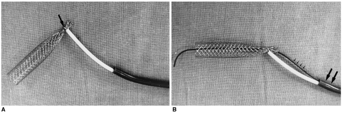
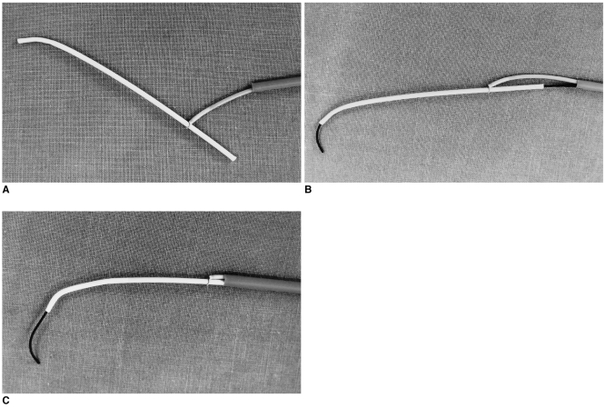
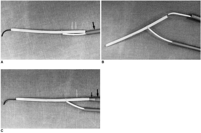
Simple snaring of a stent and catheter fragment resulted in the formation of a right angle between them and the snare axis (Figs. 1A, 2A). Owing to this angulation problem, it was necessary to fold the foreign objects prior to their retrieval by means of a simple snare technique; consequently, a much larger sheath was required for their extraction. In contrast, the coaxial snare technique greatly reduced the angle between the foreign objects and the snare axis, and allowed safe and smooth engagement between them and the sheath (Figs. 1B, 2B, 2C). Where a larger-bore venous sheath fragment was used, however, the extent to which the angle was reduced was insufficient to permit retrieval through the sheath (Fig. 3A). Although a larger-bore sheath can solve the problem, it carries the risk of vascular injury. As a variant of the coaxial snare technique, we used two access routes, the shorter one for a loop snare and the longer one for the retrieval of the sheath fragment. After capturing the fragment with a loop snare, an angiographic catheter and guidewire were used to locate its central lumen (Fig. 3B). In order to eliminate the shoulder between the guidewire and the fragment , and to facilitate smooth extraction, a dilator was then introduced through the sheath over the guidewire, and was tightly engaged with the end of the fragment (Fig. 3C).
Fig. 1.
Ex-vivo study of coaxial snaring of a vascular stent.
A. Simple snaring (arrow) of a Niti-S stent results in the formation of a right angle between the stent and the snare axis. Retrieval of the stent in this configuration would therefore be traumatic.
B. Coaxial snaring of the stent while a guidewire (small arrows) runs through it reduces the angle between the stent and the introducer sheath (large arrows), thus allowing repositioning of the stent through the TIPS tract.
Fig. 2.
Ex-vivo study of coaxial snaring of a small-bore catheter fragment (5-F).
A. Simple snaring of the catheter fragment also causes an angulation problem.
B. Coaxial snaring reduces the angle between the catheter fragment and the snaring catheter. Note that the guidewire was positioned through the lumen of the catheter fragment.
C. There is smooth engagement between the catheter fragment and the 10-F sheath.
Fig. 3.
Ex-vivo study of the retrieval of a large-bore sheath fragment (8-F internal diameter and 10-F outer diameter) using a modified coaxial snare technique.
A. In the large-bore fragmented vascular sheath, the simple coaxial snare technique does not allow engagement between the fragment and the 10-F or 12-F introducer sheath. The overall profile of the coaxial retrieval system is more than 14-F, with a shoulder between the 10-F introducer sheath (black arrow) and the sheath fragment (white arrows).
B. The loop snare introduced through the smaller sheath (10-F) captures the fractured vascular sheath. Separate access is obtained via the larger sheath (12-F), and the central lumen of the fractured sheath is located with an angiographic catheter and guidewire.
C. After engaging a dilator (small black arrow) at its end, the fragmented sheath (white arrow) is seen to be smoothly aligned with the introducer sheath (large black arrow).
Illustrative cases
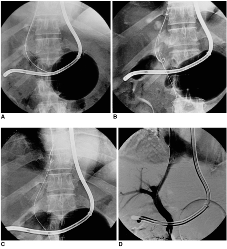
Case 1. A 48-year-old man with liver cirrhosis which led to massive variceal bleeding was referred to us for a transjugular intrahepatic portosystemic shunt (TIPS) procedure. After accessing the portal venous system via the right internal jugular route using a Colapinto transjugular cholangiography/liver biopsy set (Cook, Bloomington, Ind., U.S.A.), we dilated the parenchymal tract between the middle hepatic and left portal vein using a 10 mm-diameter Ultrathin Diamond balloon catheter (Medi-Tech/Boston Scientific, Watertown, Mass., U.S.A.). We tried to place a 10 mm-diameter, 8 cm-long Niti-S stent (Taewoong, Seoul, Korea) in the tract, but during deployment it migrated caudad to the main portal vein (Fig. 4A). A guidewire was inserted into the stent's central lumen, and an Amplatz gooseneck snare, its loop 25 mm in diameter, was introduced over it and positioned around the stent (Fig. 4B). After capturing and pulling the far proximal end of the stent by tightening the loop (Fig. 4C), we were able to relocate the stent to its appropriate position (Figs. 4C, 4D).
Fig. 4.
Relocation of a stent employed in a TIPS procedure using the coaxial snare technique.
A. The deployed nitinol stent has migrated caudad to the main portal vein. The inflated gastric balloon signifies that the patient is actively bleeding.
B. The loop of an Amplatz gooseneck snare is introduced over the preexisting guidewire and positioned around the stent.
C. After a loop snare was used to squeeze the proximal end of the stent, this was pulled back and relocated to its appropriate position.
D. A digital subtraction angiogram obtained after relocation shows rapid flow from the superior mesenteric vein to the right atrium via the created TIPS tract.
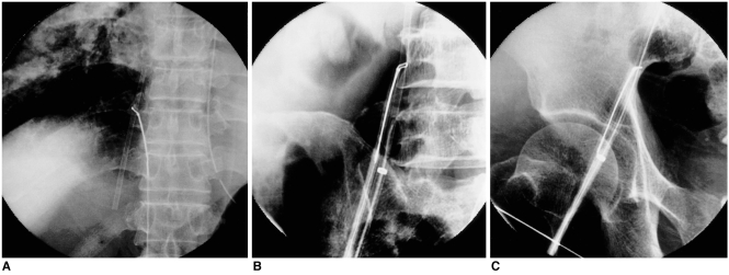
Case 3. A 42-year-old man with chronic renal failure underwent internal jugular venous catheterization at the bedside. During the procedure, the tip of the 8-F internal diameter venous introducer sheath fractured and migrated to the right atrium. Because the fragment moved vigorously in accordance with the patient's heartbeat, it was necessary to prevent its migration to the right ventricle or pulmonary circulation. Under fluoroscopic guidance, we inserted a 10-mm gooseneck snare through the right common femoral vein, successfully grasping the fragment and carefully pulling it back into the inferior vena cava (Fig. 5A). Because the fragment was very stiff and its outer diameter was 10-F, it had to be removed without folding. To this end, we used the modified coaxial technique illustrated in the ex-vivo study (Figs. 3B, 3C), inserting another 12-F vascular sheath (13 cm in length). The puncture site was more cranial to the previous one, used for insertion of the loop snare; the central lumen of the fragment was carefully located using an angiographic catheter and guidewire, and the dilator for the vascular sheath was then introduced over the guidewire (Fig. 5B). The entire retrieval system mirrored the one used in the ex-vivo experiment. After the dilator and the end of the sheath fragment were tightly engaged, the whole system was pulled back until the end of the fragment appeared at the femoral access site. It was grasped with forceps and successfully extracted after release of the loop snare and careful compression of the puncture site (Fig. 5C).
Fig. 5.
Retrieval of a large-bore sheath fragment using a modified coaxial snare technique.
A. During bedside placement of an internal jugular venous catheter, the tip of this sheared off and lodged in the right atrium. The fragment was captured by a gooseneck snare and pulled back into the inferior vena cava.
B. The femoral vein was punctured a second time, just above the puncture site for the loop snare, and a 12-F vascular sheath was introduced. With the assistance of a loop snare, the fragment has successfully engaged with the dilator of the vascular sheath.
C. The whole system was slowly withdrawn until the sheath fragment appeared at the puncture site. The linear mark represents the puncture site for the loop snare.
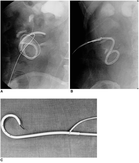
Case 6. A 51-year-old man with renal tuberculosis underwent periodic tube change under fluoroscopic guidance. The external part of the nephrostomy catheter was resected close to the point at which it entered the body, and during the procedure, the remaining catheter fragment was inadvertently dislodged, entering the nephrostomy tract. Several attempts to capture it caused its further migration into the pelvocalyceal system. We used a homemade loop snare and Amplatz gooseneck snare, also employing the 'in-situ loop snare' technique, as described by Savader et al. (18), but because the fragment was tightly wedged against the wall, were unable to withdraw any part of it (Fig. 6A). During the various attempts, we managed to pass the guidewire through the central lumen of the catheter fragment, inserting the Amplatz loop snare over the guidewire and easily capturing the proximal tip of the catheter fragment coaxially (Fig. 6B). A dilator was introduced over the guidewire to eliminate the shoulder between it and the cut catheter end and to allow smooth extraction with minimal injury to the access route. The entire system was successfully extracted by simultaneously withdrawing the snare and dilator through the tract (Fig. 6C). The patient was discharged uneventfully after placement of another nephrostomy catheter.
Fig. 6.
Retrieval of a catheter fragment (10.2-F) from the renal pelvis using the coaxial snare technique.
A. Because the fragmented catheter was tightly attached to the wall of the pelvocalyceal system, other method failed to capture it.
B. After passing a guidewire through the catheter lumen, the end of the catheter was easily captured by a loop snare introduced coaxially over the guidewire. A second small fragmented catheter tip is also seen.
C. Photograph of the entire extracted retrieval system. The catheter fragment is firmly grasped by the retracting loop snare, and for smooth extraction, a 10-F dilator has been introduced into the catheter fragment over the guidewire.
DISCUSSION
With the increasing use of indwelling catheters and interventional devices, the percutaneous retrieval of iatrogenic foreign bodies has become more common. Various techniques for retrieving unwanted objects from various systems have been reported: the devices used include the wire loop snare, retrieval basket, grasping forceps, tip-deflecting wire, pincher device, oversize sheath or catheter, and balloon catheter (1, 3, 4, 8, 14-17). Since each technique has its own advantages and limitations, the decision as to which one to use in a particular case is reached only after extremely careful consideration.
Retrieval baskets have proved useful in foreign body removal (1, 8), though there must be a free end or doubled-over segment, and they cannot be guided. Many types of grasping forceps have been tried (4, 19), a major advantage of these over a snare or basket being their ability to seize a foreign body at its mid portion as well as its free ends. Any part of the fragment is caught by alligator jaws and pulled out through a catheter or sheath. However, their large size and rigidity, as well as the potential for damage to the vessel wall, have limited their popular use. Oversize sheaths or catheters can be used for the retrieval of foreign bodies such as iatrogenically placed guidewires or their fragments, but their applications are more limited than those of other devices (16, 20).
The large number of reports on a variety of retrieval instruments and devices indicate that no particular technique has proven superior in all cases. However, loop snares are the devices most commonly used in retrieving foreign bodies, in part because they are inexpensive and simple, and can be assembled in any angiographic laboratory (1, 2). The snaring technique combines a high degree of success with very little risk of damage, but suffers certain limitations. To ensure a quick retrieval, experience and skill are required, and the outcome is successful only when the foreign body has a free-floating end or doubled-over segment that can be surrounded by the wire loop. Because the nature, location and shape of a foreign body varies, so too does the snaring technique applied in a particular case (1, 16, 18, 21-23). When snaring of the free tip of a foreign body has failed, preliminary manipulation may be able to reorient or move the object by rotating and pulling it to a more easily accessible position (3, 24). Successful preliminary manipulation may involve the use of a catheter, a steerable wire, or grasping forceps.
The coaxial snare technique, as used in our cases, is a variant of the loop snare technique, and a number of reports have described its use (8, 9, 25, 26). In most of those cases, however, the guidewires used for deployment were still in place within the lumina of the misplaced stents or catheters. In five of our seven cases, we had no safety guidewire within the lumina of the objects involved, so actively located the central lumen of each object using an angiographic catheter and guidewire. According to our clinical experience and the results of the ex-vivo experimental study, the placement of a guidewire through the center of the objects not only allowed their rapid capture with a loop snare but also greatly reduced the angle between them and the snare axis. Not only the use of the coaxial snare technique, but also the engagement of a dilator with the end of a fragmented catheter, eliminated the shoulder between the guidewire and the catheter and allowed its safe and smooth extraction without injury to the access route. However, it is sometimes very difficult to locate the lumen of a free-floating fragment within a greater patulous space; where patency is impaired, as with a chronically indwelling catheter fragment, it may be impossible.
For retrieval of an intravascular foreign body using the coaxial snare technique, the minimum inner diameter of the vascular sheath should be greater than the sum of the outer diameter of the foreign body and the catheter used with the loop snare. In order to minimize the profile of the retrieval system for large-bore intravascular foreign objects, the use of a coaxial system using dual vascular access, as in our Case 3, should be considered. Using this modified technique, it was possible -although an extra vascular access site was required for the loop snare- to retrieve a large-bore rigid intravascular foreign body without folding it. In this system, since the engaged dilator permits smooth extraction of the fragment through the access site, complete engagement between the fragment and the sheath is not mandatory. The profile of an introducer sheath can thus be minimized: one which is only slightly larger than the outer diameter of the foreign body is suitable.
Metallic stents are important as remedial components of the vascular, biliary, bronchial, urinary, and gastrointestinal systems. As with any procedure, however, there are potential complications, one of which is stent misplacement or migration. The literature includes cases of stent migration involving the biliary tree (12, 13) and the arterial system (9, 28), and of those which occur during TIPS procedures (25-27), and we have described the use of a loop snare to relocate or retrieve stents misplaced during a TIPS or PTA procedure. The ability to retrieve or relocate misplaced stents improves the safety and efficacy of stenting procedures, and it is important, in case misplacement occurs, to keep the guidewire in place within the stent. Technically, snaring and removing a stent by means of the coaxial snare technique is quite simple. To prevent the stent escaping completely, and in order to facilitate passage of the snare loop around it, it is essential that the guidewire remains within the stent throughout the procedure. After snaring its barrel, the stent can be either relocated to the desired position or removed. In some instances, the snare cannot be positioned around the leading margin of the stent, a problem which is usually solved by inflating a balloon at its mouth, thus allowing the snare to pass more easily over the margin.
Broken diagnostic or indwelling catheter is a not uncommon problem, and one which may well arise if a catheter is reused, or stored for a long time. Where catheters are broken, or foreign bodies with lumina are present, the coaxial snare technique can be safely used for retrieval. If direct snaring is possible and there is no chance of traumatic injury, the coaxial technique is not necessary. If, however, the lumina of the objects to be retrieved by the guidewire can be located, placement of the loop snare over the objects will be easier, and removal will be safer and smoother, as shown in our ex-vivo study and clinical cases.
In conclusion, the coaxial snare technique, an application of the loop snare technique, allowed the safe and effective percutaneous retrieval of tubular foreign bodies or stents, without folding, and traumatic injury to the patient during the procedure was minimized.
References
- 1.Dotter CT, Rosch J, Bilbao MC. Transluminal extraction of catheter and guidewire fragments from the heart and great vessels: 29 collected cases. AJR. 1971;111:467–472. doi: 10.2214/ajr.111.3.467. [DOI] [PubMed] [Google Scholar]
- 2.Bloomfield DA. The nonsurgical retrieval of intracardiac foreign bodies: an international survey. Cathet Cardiovasc Diagn. 1978;4:1–14. doi: 10.1002/ccd.1810040102. [DOI] [PubMed] [Google Scholar]
- 3.Uflaker R, Lima S, Melichar AC. Intravascular foreign bodies: percutaneous retrieval. Radiology. 1986;160:731–735. doi: 10.1148/radiology.160.3.3737911. [DOI] [PubMed] [Google Scholar]
- 4.Selby JB, Tegtmeyer CJ, Bittner GM. Experience with new retrieval forceps for foreign body removal in the vascular, urinary, and biliary systems. Radiology. 1990;176:535–538. doi: 10.1148/radiology.176.2.2367671. [DOI] [PubMed] [Google Scholar]
- 5.Dondeliger RF, Lepontre B, Kurdziel JC. Percutaneous vascular foreign body retrieval: experience of an 11-year period. Eur J Radiol. 1991;12:4–10. doi: 10.1016/0720-048x(91)90124-e. [DOI] [PubMed] [Google Scholar]
- 6.Siegel EL, Robertson EF. Percutaneous transfemoral retrieval of a free-floating titanium Greenfield filter with an Amplatz gooseneck snare. J Vasc Interv Radiol. 1993;4:565–568. doi: 10.1016/s1051-0443(93)71923-5. [DOI] [PubMed] [Google Scholar]
- 7.Cekirge S, Weiss JP, Foster RG, Neiman HL, McLean GK. Percutaneous retrieval of foreign bodies: experience with the nitinol gooseneck snare. J Vasc Interv Radiol. 1993;4:805–810. doi: 10.1016/s1051-0443(93)71978-8. [DOI] [PubMed] [Google Scholar]
- 8.Egglin TKP, Dickey KW, Rosenblatt M, Pollak JS. Retrieval of intravascular foreign bodies: experience in 32 cases. AJR. 1995;164:1259–1264. doi: 10.2214/ajr.164.5.7717243. [DOI] [PubMed] [Google Scholar]
- 9.Hartnell GG, Jordan SJ. Percutaneous removal of a misplaced Palmaz stent with a coaxial snare technique. J Vasc Interv Radiol. 1995;6:799–801. doi: 10.1016/s1051-0443(95)71188-5. [DOI] [PubMed] [Google Scholar]
- 10.Coleman CC, Kimura Y, Castaneda-Zuniga WR. Interventional techniques in the ureter. Semin Intervent Radiol. 1984;1:24–37. [Google Scholar]
- 11.LeRoy AJ, Williams HJ, Jr, Segura JW, Patterson DE, Benson RC., Jr Indwelling ureteral stents: percutaneous management of complications. Radiology. 1986;158:219–222. doi: 10.1148/radiology.158.1.3510020. [DOI] [PubMed] [Google Scholar]
- 12.Abramson AF, Javit DJ, Mitty HA, Train JS, Dan SJ. Wallstent migration following deployment in right and left bile ducts. J Vasc Interv Radiol. 1992;3:463–465. doi: 10.1016/s1051-0443(92)71990-3. [DOI] [PubMed] [Google Scholar]
- 13.Johanson JF, Schmalz MJ, Geenen JE. Incidence and risk factors for biliary and pancreatic stent migration. Gastrointest Endosc. 1992;38:341–346. doi: 10.1016/s0016-5107(92)70429-5. [DOI] [PubMed] [Google Scholar]
- 14.Nemcek AA, Jr, Vogelzang RL. Modified use of the tip-deflecting wire in manipulation of foreign bodies. AJR. 1987;149:777–779. doi: 10.2214/ajr.149.4.777. [DOI] [PubMed] [Google Scholar]
- 15.Boren SR, Dotter CT, McKinney M, Rosch J. Percutaneous removal of ureteral stents. Radiology. 1984;152:230–231. doi: 10.1148/radiology.152.1.6729125. [DOI] [PubMed] [Google Scholar]
- 16.Park JH, Yoon DY, Han JK, Kim SH, Han MC. Retrieval of intravascular foreign bodies with the snare and catheter capture technique. J Vasc Interv Radiol. 1992;3:581–582. doi: 10.1016/s1051-0443(92)72020-x. [DOI] [PubMed] [Google Scholar]
- 17.Katske FA, Celis P. Technique for removal of migrated double-J ureteral stent. Urology. 1991;37:579. doi: 10.1016/0090-4295(91)80330-a. [DOI] [PubMed] [Google Scholar]
- 18.Savader SJ, Brodkin J, Osterman FA. In-situ formation of a loop snare for retrieval of a foreign body without a free end. Cardiovasc Intervent Radiol. 1996;19:298–301. doi: 10.1007/BF02577656. [DOI] [PubMed] [Google Scholar]
- 19.Millan VG. Retrieval of intravascular foreign bodies using a modified bronchoscopic forceps. Radiology. 1978;129:587–589. doi: 10.1148/129.3.587. [DOI] [PubMed] [Google Scholar]
- 20.Kim SH, Song IS, Kim JH, Park JH, Han MC. Retrieval of a guidewire introducer by catheter-capture from the proximal inferior vena cava: technical note. Cardiovasc Intervent Radiol. 1991;14:252–253. doi: 10.1007/BF02578473. [DOI] [PubMed] [Google Scholar]
- 21.Curry JL. Recovery of detached intravascular catheter or guide wire fragments: a proposed method. AJR. 1969;105:894–896. doi: 10.2214/ajr.105.4.894. [DOI] [PubMed] [Google Scholar]
- 22.Fisher RG, Ferreyro R. Evaluation of current techniques for nonsurgical removal of intravascular iatrogenic foreign bodies. AJR. 1978;130:541–548. doi: 10.2214/ajr.130.3.541. [DOI] [PubMed] [Google Scholar]
- 23.Fjalling M, List AR. Transvascular retrieval of an accidentally ejected tip occluder and wire. Cardiovasc Intervent Radiol. 1982;5:34–36. doi: 10.1007/BF02552101. [DOI] [PubMed] [Google Scholar]
- 24.Rossi P. Hook catheter technique for transfemoral removal of foreign body from right side of the heart. AJR. 1970;108:101–106. doi: 10.2214/ajr.109.1.101. [DOI] [PubMed] [Google Scholar]
- 25.Sanchez RB, Roberts AC, Valji K, Lengle S, Bookstein JJ. Wallstent misplaced during transjugular placement of an intrahepatic portosystemic shunt: retrieval with a loop snare. AJR. 1992;159:129–130. doi: 10.2214/ajr.159.1.1609686. [DOI] [PubMed] [Google Scholar]
- 26.Cekirge S, Foster RG, Weiss JP, McLean GK. Percutaneous removal of an embolized Wallstent during a transjugular intrahepatic portosystemic shunt procedure. J Vasc Interv Radiol. 1993;4:559–560. doi: 10.1016/s1051-0443(93)71921-1. [DOI] [PubMed] [Google Scholar]
- 27.Cohen GS, Ball DS. Delayed wallstent migration after a transjugular intrahepatic portosystemic shunt procedure: relocation with a loop snare. J Vasc Interv Radiol. 1993;4:561–563. doi: 10.1016/s1051-0443(93)71922-3. [DOI] [PubMed] [Google Scholar]
- 28.Foster-Smith KW, Garratt KN, Higano ST, Holmes DR. Retrieval techniques for managing flexible intracoronary stent misplacement. Cathet Cardiovasc Diagn. 1985;11:623–628. doi: 10.1002/ccd.1810300116. [DOI] [PubMed] [Google Scholar]


