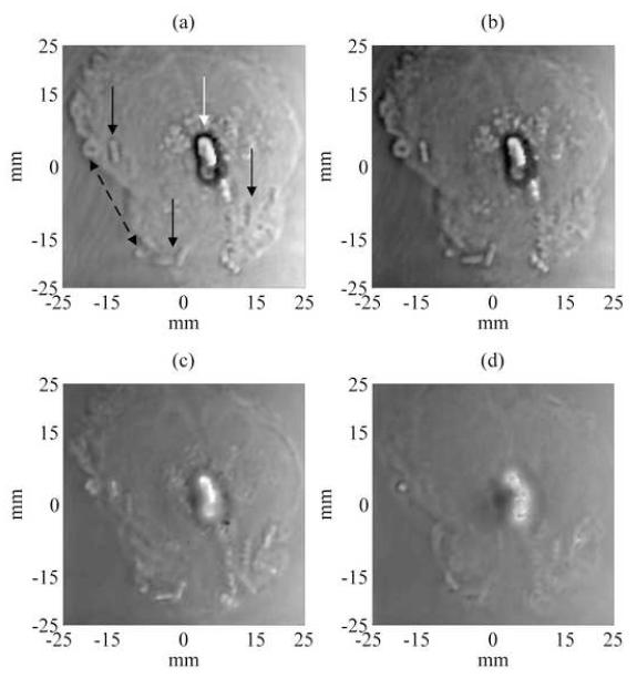
Figure 4.
Experimental unwrapped phase VA images of the excised prostate showing seed location for 4 different depths at 1, 5, 10 and 15 mm (Figs. (a)-(d) respectively) deep from the surface of the prostate gland. Displaying the unwrapped phase of the acoustic emission provides another source of image contrast for examining the prostate tissue and embedded objects. The unwrapped phase of the acoustic emission from prostate tissue is relatively constant. The unwrapped phase contrast of the calcification differs with the depth of the focal plane. The border of the calcification is well-defined and the brachytherapy seeds are well visualized at focal depths of 1-10 mm (i.e. Figs. 4-(a)-(c)).

