Abstract
Knowledge of the morphology and kinematics of the male and female knees is important for understanding gender related dimorphism in knee pathology and improvement of related surgical treatments. Twelve male and twelve female subjects with healthy knees were recruited and each subject performed a single leg lunge while images of the knee were recorded by two fluoroscopes. Tibiofemoral joint motion was then reproduced using bony models matched to the fluoroscopic images. Femoral trochlear groove orientation was also measured in each knee.
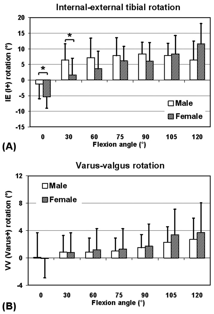
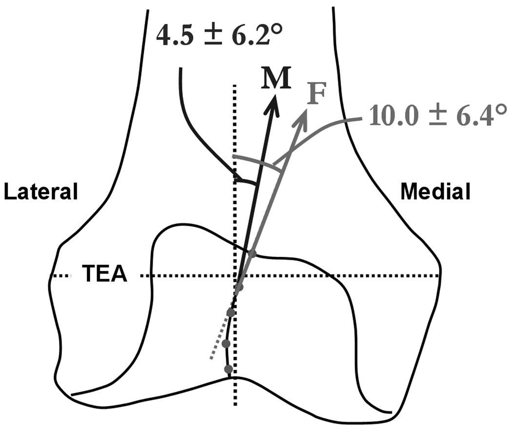
While many of the measured parameters were found to be similar between the genders, few interesting differences were also noted. Females showed greater external tibial rotation at 0° (−5.4° vs. −1.3°, p = 0.03), smaller internal rotation at 30° flexion (1.7° vs. 6.4°, p =0.04) and greater range of tibial rotation (18.2° vs. 12.4°, p = 0.01) compared to males. Female knees also had a more medially oriented proximal trochlear groove (10.0° vs. 4.5°, p = 0.04). These gender differences in rotational kinematics and trochlear groove orientation may warrant further studies to determine implications for surgical treatments such as total knee arthroplasty, and gender related dimorphism in certain knee injuries and pathologies, like ACL injury and patellofemoral problems.
Keywords: Gender difference, Trochlear orientation, Knee kinematics, Fluoroscopy, Magnetic resonance imaging
INTRODUCTION
Recently, gender related dimorphism in the knee has received increased attention in sports medicine (1–3) and total knee arthroplasty (TKA) (4–6). Certain soft tissue injuries, particularly ACL injuries, have a higher prevalence in young female athletes compared to their male counterparts (3,7, 8). Patellofemoral pain, recurrent patellar dislocations and patellofemoral arthritis have been reported to be more common in females (9,10). Women above 50 years of age have a higher incidence of osteoarthritis and associated disability, and account for about two-thirds of TKA patients (5, 11, 12).
These differences in the pathophysiology of the knee have been related to differences in muscle recruitment, passive and dynamic knee stiffness, neuromuscular control, training, femoral notch width, ACL thickness and lower limb alignment (1, 7, 10, 13). Gender specific TKA implant designs have been proposed based on reported morphological differences between the male and female knees (6). Morphological measurements have shown that female knees are narrower than male knees for the same anteroposterior dimension, and have a less prominent anterior condyle (6, 14, 15). Nonetheless, the need and efficacy of gender specific TKA continues to be a matter of debate (4, 5).
Few studies have delineated gender differences in the six degrees-of-freedom (six-DOF) tibiofemoral kinematics during functional weight-bearing activities. This knowledge is essential to understanding of mechanisms underlying gender related differences in knee injuries and pathology. Additionally, this will help determine if there exists a justification for gender specific TKA beyond achieving better anatomical match. Previous studies have looked at the effect of gender on various morphological parameters of the knee (6, 14, 15). However, gender differences in the orientation of the trochlear groove, have not been clearly delineated. In the native knee, trochlear groove orientation plays a major role in patellar tracking (16). Therefore, the study of trochlear groove orientation in male and female knees is crucial to the understanding of the patellofemoral pathophysiology. In the TKA knee, trochlear groove orientation of the femoral component has major effect on patellar tracking (17, 18). Non-physiologic forces resulting from changes in patellofemoral motion can contribute to patellofemoral pain, patellar component loosening and wear (17, 18). Consequently TKA designs have sought to achieve physiological tracking by manipulating the orientation of the femoral trochlear groove (17, 19). Therefore, knowledge of trochlear groove orientation in the native male and female knees is also important for the design of TKA implants.
We hypothesized that there exists a gender difference in the in vivo six-DOF tibiofemoral motion and trochlear groove orientation. The objective of this study was to investigate gender differences in the coronal plane orientation of the femoral trochlear groove and the six-DOF tibiofemoral kinematics during weight-bearing knee flexion, using 3D MR image based models of the knee and a dual fluoroscopic imaging system.
MATERIALS AND METHODS
Subject Recruitment and Statistics
A sample of convenience was utilized in this cross-sectional study. Twelve male and twelve female subjects were recruited. Each subject signed an informed consent for the study, which was approved by our institutional review board. The average (and standard deviation) age of the male subjects was 33.0 ± 7.3 years and the average age of the female subjects was 29.0 ± 10.5 years. There was no significant difference in age between the genders (p = 0.29). However, male subjects were taller (71 ± 2 in vs. 66 ± 3 in, p = 0.01) and weighed significantly more than female subjects (186 ± 18 lbs vs. 138 ± 23 lbs, p < 0.001). All knees included in the study were healthy, without symptoms of osteoarthritis or soft tissue injury, as verified via clinical examination and MR imaging. Criteria for exclusion included body mass index greater than 30.
Creation of 3D Knee Model
Magnetic resonance (MR) image scans of each knee were obtained using a 3.0 Tesla magnet (Siemens, Germany) and fat suppressed 3D spoiled gradient-recalled sequence (20). During the scan, the subject was supine with his or her knee in a relaxed and extended position. Parallel sagittal plane images were obtained (resolution 512 × 512 pixels) with a field of view of 160×160 mm and spaced 1 mm apart. These images were then segmented using a 3D modeling software (Rhinoceros®, Robert McNeel and Associates, Seattle, WA) to construct 3D models of the knee, including the tibia and femur (20).
Measurement of In Vivo Tibiofemoral Kinematics
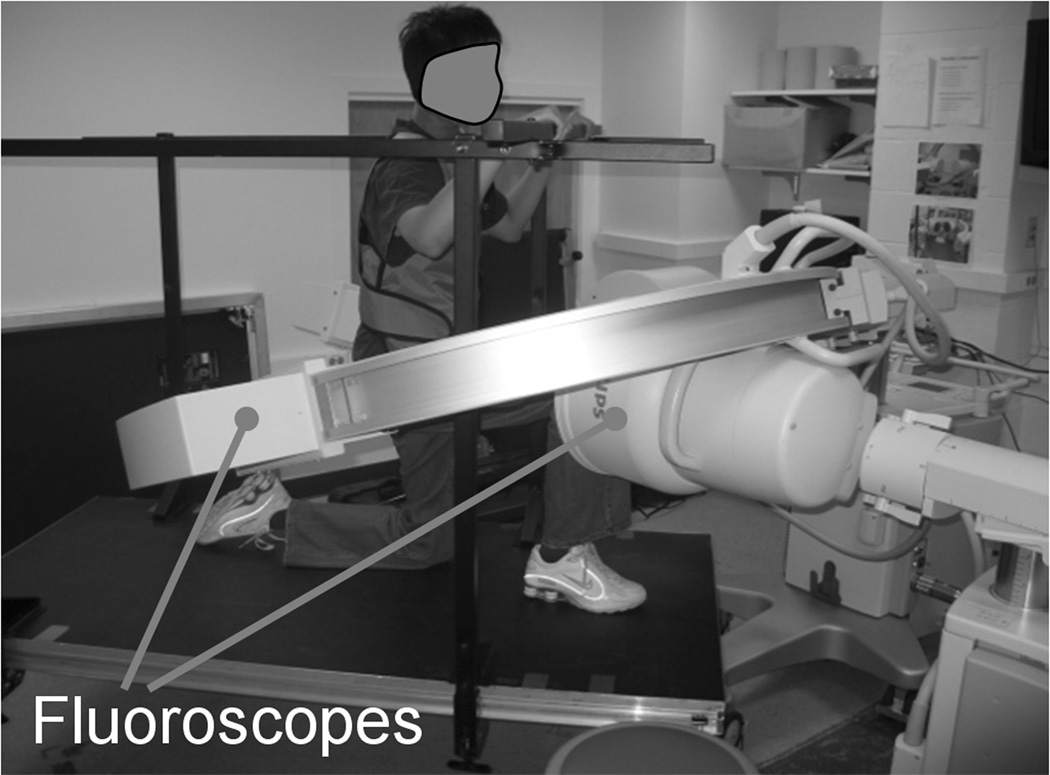
Following the MR scan, the subjects performed a single leg lunge with their knee in the field of view of two orthogonally placed fluoroscopes (BV Pulsera, Philips, Bothell, WA) (Fig. 1). The subject was instructed to flex his or her knee through different flexion angles from full extension to maximum flexion, while being monitored using a goniometer. The subjects held their position at specified flexion angles for a brief moment while the fluoroscopic images were captured. These fluoroscopic images were then imported into a virtual fluoroscopic setup in the solid modeling software (Rhinoceros®) and were placed at their respective positions (20). Next, the 3D MR image-based models of the tibia and femur were imported into the software. The models were then individually manipulated in six-DOF, until they matched their projections on the orthogonal fluoroscopic images captured during the actual weight-bearing activity. Thus, the in vivo knee motion was represented by a series of 3D knee models at different flexion angles (20).
Figure 1.
Subject performing weight-bearing single leg lunge while being imaged by two orthogonally placed fluoroscopes.
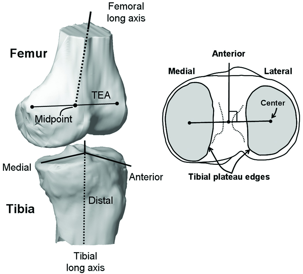
Six-DOF kinematics of the tibiofemoral joint was directly obtained from the series of 3D models, using coordinate systems embedded in the distal femur and proximal tibia (Fig. 2) (21, 22). The tibial long axis passed through the middle of the tibial spines and was oriented parallel to the posterior wall of the tibial shaft in the sagittal plane. In the coronal plane it was angled equally with respect to the medial and lateral edges of the tibial shaft. An orthogonal coordinate system was placed on the tibia with the mediolateral axis obtained by projecting a line passing through the centers of the medial and lateral tibial plateaus onto a plane perpendicular to the tibial long axis. The center of the medial/lateral tibial plateau was defined as the centroid of the closed curve formed by tracing the edges of the plateau. The midpoint of the tibial mediolateral axis was defined as the origin of the tibial coordinate system. The proximal-distal axis of the tibia was parallel to tibial long axis. The tibial anteroposterior axis was defined to be perpendicular to the tibial mediolateral and proximal-distal axes (21, 22). For the femur, two axes were defined, the transepicondylar axis and a line passing through midpoint of the transepicondylar axis, and parallel to the femoral long axis (21, 22). The femoral long axis passed through the most distal point on the femoral intercondylar notch. In the sagittal and coronal planes the femoral long axis was angled equally with respect to the anterior/posterior and medial/lateral edges of the femoral shaft, respectively.
Figure 2.
Coordinate systems embedded in the proximal tibia and distal femur, used to measure tibiofemoral kinematics.
Knee flexion was defined as the angle between the femoral and tibial long axis projected onto the tibial sagittal plane (21, 22). Internal-external rotation of tibia was defined as the angle between the transepicondylar axis and the tibial mediolateral axis, projected onto the transverse plane of the tibia. Varus-valgus rotation was defined as the angle between the transepicondylar axis and the tibial mediolateral axis, projected onto the coronal plane of the tibia. Flexion, internal tibial rotation and varus rotation were defined to be positive. The midpoint of the transepicondylar axis was used to measure femoral translations in the anteroposterior, mediolateral and proximal-distal directions with respect to the tibial coordinate system. Anterior, proximal and medial femoral translations were defined to be positive.
The accuracy of this combined MR and dual fluoroscopic imaging technique for measuring translations has been validated under in vitro conditions to be better than 0.1 mm (21, 23). Repeatability of the technique for measuring translations and rotations has been validated to be better than 0.1 mm and 0.3° (21, 23). To account for the effect of knee size, tibiofemoral translations were also normalized by the anteroposterior and mediolateral dimensions of the tibia. The methodology for measuring tibial and femoral dimensions is described in Appendix A and the measurements are listed in Table A1 of the appendix.
Measurement of Trochlear Groove Orientation
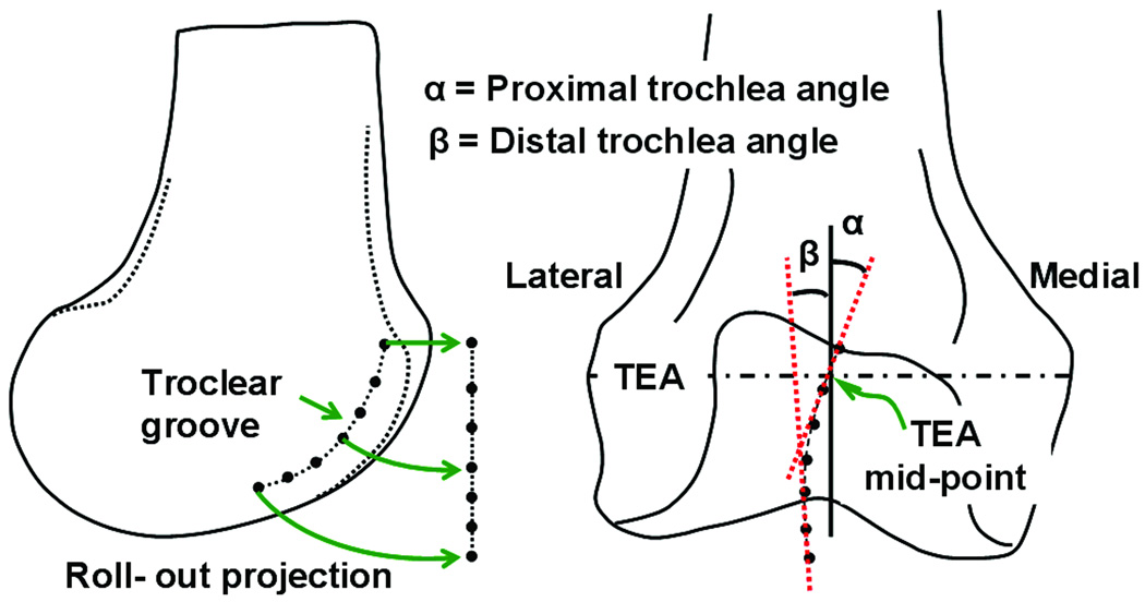
The coronal plane orientations of the proximal and distal portions of the trochlear groove were measured by importing the 3D model of the femur into the solid modeling software. The deepest points on the trochlear groove were traced and projected onto the coronal plane of the femur using a rollout projection (Fig. 3). The rollout projection was used to preserve the length of the trochlear groove similar to method used by Barink et al. (24). Linear regression was used to fit a second order polynomial curve to the projected points to form the trochlear line. The slopes of the proximal and distal halves of the trochlear line (measured at middle of each half), were measured relative to a line perpendicular to the transepicondylar axis to determine the proximal and distal trochlear groove angles. A positive angular measurement indicates medial orientation.
Figure 3.
Measurement of coronal plane orientation of trochlear groove. α = distal trochlear groove orientation and β = proximal trochlear groove orientation.
Two-tailed t-test for independent samples was used to compare the six-DOF tibiofemoral kinematics (at each flexion angle) and trochlea groove orientation between the male and female knees. The level of statistical significance was set at p < 0.05.
RESULTS
Tibiofemoral Kinematics
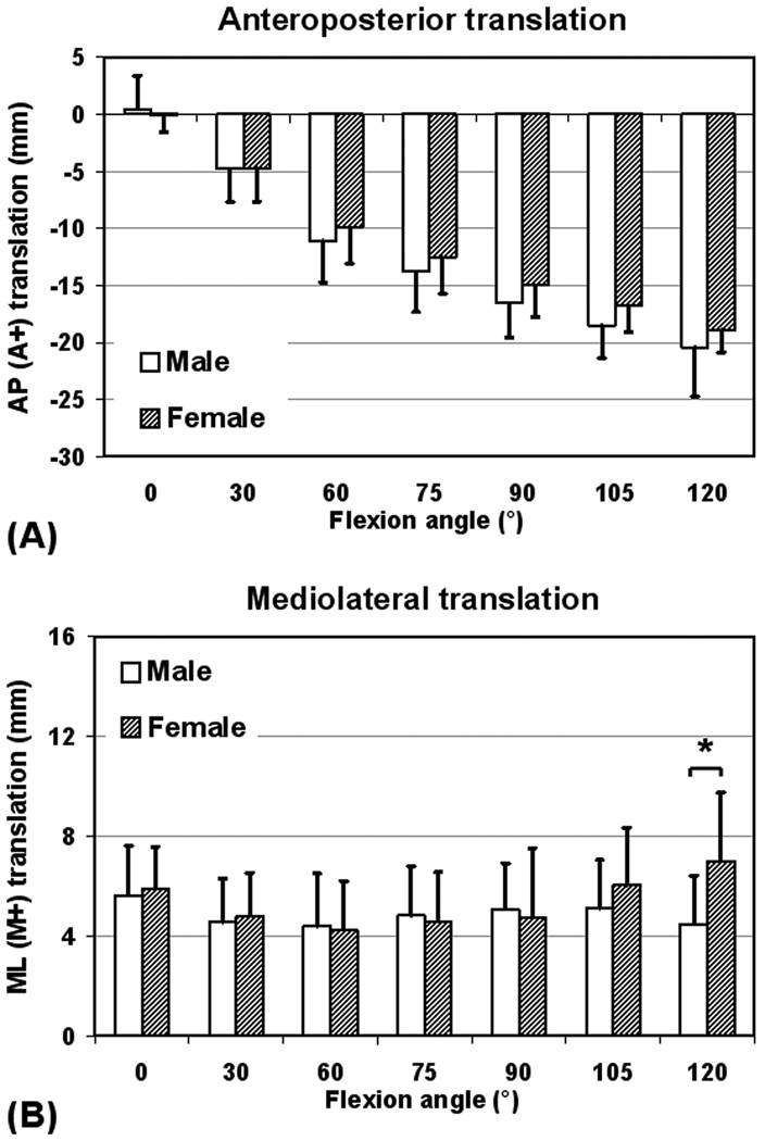
Both male and female knees showed consistent femoral rollback with flexion and there was no significant difference between the genders (p ≥ 0.15, power ≤ 0.28, Fig. 4A). When normalized by anteroposterior size of the tibia, again no significant gender differences were noted (p ≥ 0.67, power ≤ 0.07, Fig. A3 - Appendix A). In the mediolateral direction, no significant gender differences were noted except at 120° flexion (p ≥ 0.32, power ≤ 0.18 for 0–105° flexion, Fig. 4B). At 120° flexion, female knees had a significantly greater medial position compared to male knees (7.0 ± 2.7 mm vs. 4.5 ± 1.9 mm, p = 0.03). Upon normalization by mediolateral size of tibia, female knees tended to show a more medial position (Fig. A4 - Appendix A). However, difference between the genders was not significant except at 120° flexion (p ≥ 0.1, power ≤ 0.38 for 0–105° flexion).
Figure 4.
(A) Anteroposterior and (B) mediolateral femoral translation as a function of flexion for male and female knees (* =p<0.05). Positive values indicate anterior and medial translations.
Female knees showed significantly greater external tibial rotation at 0° (−5.4 ± 3.6° vs. −1.3 ± 4.7°, p = 0.03) and smaller internal rotation at 30° flexion (1.7 ± 5.4° vs. 6.4 ± 5.2°, p = 0.04, Fig. 5A). The difference between genders at higher flexion angles was not statistically significant (p ≥ 0.09). Female knees also showed slightly greater varus rotation (Fig. 5B), at 105° (3.4° vs. 2.3°) and 120° (3.7° vs. 2.7°) flexion. However, difference between genders was not statistically significant at any flexion angle (p ≥ 0.41, power ≤ 0.13).
Figure 5.
(A) Internal-external tibial rotation and (B) varus-valgus knee rotation as a function of flexion angle, for male and female knees (* = p<0.05). Positive values correspond to internal tibial and varus knee rotations.
The range of knee motion between 0 and 120° of flexion was also compared between the male and female knees (Table 1). There was no significant difference in the range of anteroposterior translation, mediolateral translation, and varus-valgus rotation between the male and female knees. However, female knees had significantly greater range of tibial rotation compared to male knees (18.2 ± 5.8° vs. 12.4 ± 4.1°, p = 0.01).
Table 1.
Range of tibiofemoral motion between 0° and 120° of knee flexion for twelve male and eleven female knees
| AP (mm) |
ML (mm) |
AP *100/ Tibial AP Size (%) |
ML *100/ Tibial ML Size (%) |
IE (°) | VV (°) | ||
|---|---|---|---|---|---|---|---|
| Male | Mean | 20.7 | 3.1 | 46.4 | 3.8 | 12.4 | 5.5 |
| SD | 3.8 | 1.3 | 10.4 | 1.6 | 4.1 | 3.8 | |
| Female | Mean | 18.3 | 3.0 | 46.1 | 4.4 | 18.2 | 5.2 |
| SD | 1.9 | 1.5 | 5.2 | 2.3 | 5.5 | 2.4 | |
| t-test | pvalue | 0.11 | 0.9 | 0.93 | 0.46 | 0.01* | 0.95 |
(p<0.05). Range of anteroposterior and mediolateral femoral translations as a percentage of the corresponding tibial dimensions are also listed.
AP = anteroposterior, ML = mediolateral, IE = internal-external, VV = varus-valgus and SD = standard deviation.
Trochlear Groove Orientation
The average distal trochlear angle for male knees was 1.7 ± 6.7° and for female knees was −0.6 ± 5.8°. No significant difference was seen in the distal trochlear orientation between the male and female knees (p = 0.39). However, the average proximal trochlear angle for female knees was 10.0 ± 6.4° and for male knees was 4.5 ± 6.2° (Fig. 6). The proximal portion of the trochlear groove was oriented significantly more medially in females compared to males (p = 0.04).
Figure 6.
Coronal plane orientation of proximal portion of femoral trochlear groove in male (M) and female (F) knees.
DISCUSSION
In this study we investigated the six-DOF tibiofemoral kinematics and coronal plane orientation of the femoral trochlear groove, in male and female knees. The results of this study showed that while many of the measured parameters were similar between genders, there exist gender differences in the coronal plane orientation of the trochlear groove and internal-external rotation of the tibia relative to the femur.
In our study we did not find significant gender difference in the anteroposterior knee translation, particularly upon normalization by anteroposterior tibial dimension. Mediolateral translations also did not show any significant gender difference (except at 120° flexion), both with and without normalization by mediolateral tibial dimension. However, in female knees the normalized mediolateral translation data showed a trend towards more medial position compared to males. This warrants further investigation with a larger sample size.
Compared to males, females showed greater external tibial rotation at 0° flexion, smaller internal rotation at 30° flexion and greater range of tibial rotation, during the weight bearing activity. These data are consistent with the reported lower passive torsional stiffness and higher rotary knee joint laxity in females (2, 25). From a biomechanical point of view, increased external rotation correlates to an increase in the effective Q angle during weight-bearing activity (26, 27). This could lead to shift of contact pressure towards the lateral facet of the patellofemoral joint (7–9, 28). This hypothesis and its relation to the reported higher incidence of patellofemoral problems in females still need to be investigated (7–10). In literature, increased passive joint laxity has been linked with heightened risk of ACL injury in females (3, 8). How the increased active rotation of the female knee seen in this study affects the ACL function, warrants further investigation. The increased external tibial rotation at low flexion angles could predispose the female knee to increased ACL impingement with the lateral femoral condyle during pivoting motions associated with ACL injuries. If this hypothesis is true then the increased external tibial rotation could represent a disadvantage in protecting the ACL from injury (13, 29–31).
In our study males and females showed similar varus-valgus rotation with no significant gender difference at any flexion angle, although females showed slightly greater varus rotation at 105° and 120°. Studies on dynamic activities such as landing from a jump have shown females to have significantly greater valgus rotation (32). The differences between results of our study and other reports may indicate that differences in knee stiffness and neuromuscular control during specific dynamics activities are the dominant cause for gender variation in varus-valgus motion, rather than any inherent varus or valgus bias in the male or female knee.
The overall medial orientation of the trochlear groove is consistent with previous reports in literature (24, 33, 34). This also explains the lateral patellar translation with knee flexion, once the patella engages the femoral trochlea (24, 34). While there was no significant gender difference in the distal trochlear orientation, the proximal trochlea in females was found to be oriented significantly more medially in this study. This difference in proximal trochlear groove orientation might lead to differences in patellar tracking. Gender difference in patellar tracking may not necessarily imply a disadvantage with regards to patellofemoral problems. However, if it exists, it could have important implications for design of TKA implants. The data showing more medial orientation of the trochlear groove in females also contradicts the perception of a more lateral trochlea in females, which is based on greater Q angle of female knees (6, 35). Recently, gender specific TKA designs have been proposed to accommodate the geometric features of female knees (5, 6). Our data on trochlear orientation in male and female knees could enable improvement of the gender specific total knee and patellofemoral arthroplasty designs. Although it is too early to speculate on implications of the differences in rotational kinematics for total knee or patellofemoral arthroplasty procedures, future studies are warranted to investigate this issue further. In determining these implications it is also important to consider gender related differences together with the inherent subject-to-subject variations.
One factor that could lead to reduction in accuracy of the dual fluoroscopic technique compared to values obtained from in vitro validation is image blurring due to unwanted motion of the subject’s knee. This was not a major issue for this study since the relatively young subjects possessed sufficient muscle strength to balance during the lunge activity, and the test setup included hand rails for support. Normal motions of small magnitudes, occurring with limited velocities, do not significantly affect the accuracy of the dual fluoroscopic technique as has been confirmed by our previous dynamic validations (36, 37). It should be noted that the system accuracy/repeatability values obtained during validations do not relate to inter/intra-observer variability in placement of coordinate axes. Specifying accuracy of axes placement is difficult, if not impossible, since there is no gold standard with which to compare a given choice of coordinate axes. Inter/intra-observer variability in axes placement is undesirable but unavoidable. This variability can lead to increase in the standard deviation of the measured kinematics (over the inherent subject-to-subject variation) making it harder to detect differences between the study groups. However, by using a consistent protocol for axes placement, such variability can be minimized.
The aim of this study was to investigate any underlying gender differences in the weight-bearing motion of the knee from full extension to deep flexion, independent of factors such as landing strategy and training level (32). The lunge motion allows us to investigate the natural kinematics of the knee over a large range of motion (0–120°) in a controlled manner. Activities such as landing from a jump or pivoting motion may be more relevant for specific injuries such as ACL injuries, but have limited range of weight bearing flexion and inherently include factors such as differences in training level and landing strategy. In this study the subjects performed the lunge motion in a quasi-static manner, holding the knee position at specified flexion angles for a brief moment while the fluoroscopic images were captured. Differences in muscle activitation between quasi-static and dynamic knee motion may lead to changes in measured kinematics, which need to be kept in mind while interpreting results of this study. The advantage of a quasi-static study is minimal motion artifacts and reduced radiation exposure compared to dynamic fluoroscopy.
Another limitation of this study is that in determining trochlear groove orientation we utilized the bony morphology, neglecting the geometry of the articular cartilage. Shih et al. showed differences between the measurements of the bony and cartilaginous geometry of the trochlea (38). However, they found general trends to be similar in both measurements.
In conclusion, this study revealed that significant gender differences exist only in the tibiofemoral rotational kinematics and proximal trochlear groove orientation. In particular, female knees have greater external tibial rotation at low flexion angles and greater range of tibial rotation. The more externally rotated position of the tibia in females, particularly at low flexion angles, could increase the effective Q angle during weight-bearing activity. Additionally, the proximal trochlea in females is oriented more medially than in males. Further studies are warranted to determine implications of these data for surgical treatments, such as total knee or patellofemoral arthroplasty, and to investigate factors underlying increased risk of ACL injury and patellofemoral problems in females.
Supplementary Material
ACKNOWLEDGMENTS
This work was funded through grants received from the National Institutes of Health (R01-AR052408, R21-AR05107). The authors would also like to acknowledge Samuel Van de Velde, Ramprasad Papannagari, Louis E. DeFrate, Jefferey Bingham and Kyung Wook Nha for their technical assistance.
REFERENCES
- 1.Wojtys EM, Ashton-Miller JA, Huston LJ. A gender-related difference in the contribution of the knee musculature to sagittal-plane shear stiffness in subjects with similar knee laxity. J Bone Joint Surg Am. 2002;84-A:10–16. doi: 10.2106/00004623-200201000-00002. [DOI] [PubMed] [Google Scholar]
- 2.Hsu WH, Fisk JA, Yamamoto Y, et al. Differences in torsional joint stiffness of the knee between genders: a human cadaveric study. Am J Sports Med. 2006;34:765–770. doi: 10.1177/0363546505282623. [DOI] [PubMed] [Google Scholar]
- 3.Myer GD, Ford KR, Paterno MV, et al. The effects of generalized joint laxity on risk of anterior cruciate ligament Injury in young female athletes. Am J Sports Med. 2008:1–8. doi: 10.1177/0363546507313572. [DOI] [PMC free article] [PubMed] [Google Scholar]
- 4.Barrett WP. The need for gender-specific prostheses in TKA: does size make a difference? Orthopedics. 2006;29:S53–S55. [PubMed] [Google Scholar]
- 5.Greene KA. Gender-specific design in total knee arthroplasty. J Arthroplasty. 2007;22:27–31. doi: 10.1016/j.arth.2007.05.019. [DOI] [PubMed] [Google Scholar]
- 6.Conley S, Rosenberg A, Crowninshield R. The female knee: anatomic variations. J Am Acad Orthop Surg. 2007;15 Suppl 1:S31–S36. doi: 10.5435/00124635-200700001-00009. [DOI] [PubMed] [Google Scholar]
- 7.Hutchinson MR, Ireland ML. Knee injuries in female athletes. Sports Med. 1995;19:288–302. doi: 10.2165/00007256-199519040-00006. [DOI] [PubMed] [Google Scholar]
- 8.Loud KJ, Micheli LJ. Common athletic injuries in adolescent girls. Curr Opin Pediatr. 2001;13:317–322. doi: 10.1097/00008480-200108000-00005. [DOI] [PubMed] [Google Scholar]
- 9.Csintalan RP, Schulz MM, Woo J, et al. Gender differences in patellofemoral joint biomechanics. Clin Orthop Relat Res. 2002;402:260–269. doi: 10.1097/00003086-200209000-00026. [DOI] [PubMed] [Google Scholar]
- 10.Arendt EA. Dimorphism and patellofemoral disorders. Orthop Clin North Am. 2006;37:593–599. doi: 10.1016/j.ocl.2006.08.004. [DOI] [PubMed] [Google Scholar]
- 11.O'Connor MI. Osteoarthritis of the hip and knee: sex and gender differences. Orthop Clin North Am. 2006;37:559–568. doi: 10.1016/j.ocl.2006.09.004. [DOI] [PubMed] [Google Scholar]
- 12.Petterson SC, Raisis L, Bodenstab A, Snyder-Mackler L. Disease-specific gender differences among total knee arthroplasty candidates. J Bone Joint Surg Am. 2007;89:2327–2333. doi: 10.2106/JBJS.F.01144. [DOI] [PubMed] [Google Scholar]
- 13.Shelbourne KD, Davis TJ, Klootwyk TE. The relationship between intercondylar notch width of the femur and the incidence of anterior cruciate ligament tears. A prospective study. Am J Sports Med. 1998;26:402–408. doi: 10.1177/03635465980260031001. [DOI] [PubMed] [Google Scholar]
- 14.Mahfouz MR, Merkl BC, Fatah EE, et al. Automatic methods for characterization of sexual dimorphism of adult femora: distal femur. Comput Methods Biomech Biomed Engin. 2007;10:447–456. doi: 10.1080/10255840701552093. [DOI] [PubMed] [Google Scholar]
- 15.Hitt K, Shurman JR, 2nd, Greene K, et al. Anthropometric measurements of the human knee: correlation to the sizing of current knee arthroplasty systems. J Bone Joint Surg Am. 2003;85-A Suppl 4:115–122. [PubMed] [Google Scholar]
- 16.Ahmed AM, Duncan NA. Correlation of patellar tracking pattern with trochlear and retropatellar surface topographies. J Biomech Eng. 2000;122:652–660. doi: 10.1115/1.1322036. [DOI] [PubMed] [Google Scholar]
- 17.Barink M, Van de Groes S, Verdonschot N, De Waal Malefijt M. The difference in trochlear orientation between the natural knee and current prosthetic knee designs; towards a truly physiological prosthetic groove orientation. J Biomech. 2006;39:1708–1715. doi: 10.1016/j.jbiomech.2005.04.027. [DOI] [PubMed] [Google Scholar]
- 18.D'Lima DD, Chen PC, Kester MA, Colwell CW., Jr Impact of patellofemoral design on patellofemoral forces and polyethylene stresses. J Bone Joint Surg Am. 2003;85-A Suppl 4:85–93. doi: 10.2106/00004623-200300004-00010. [DOI] [PubMed] [Google Scholar]
- 19.Petersilge WJ, Oishi CS, Kaufman KR, et al. The effect of trochlear design on patellofemoral shear and compressive forces in total knee arthroplasty. Clin Orthop Relat Res. 1994;309:124–130. [PubMed] [Google Scholar]
- 20.DeFrate LE, Sun H, Gill TJ, et al. In vivo tibiofemoral contact analysis using 3D MRI-based knee models. J Biomech. 2004;37:1499–1504. doi: 10.1016/j.jbiomech.2004.01.012. [DOI] [PubMed] [Google Scholar]
- 21.DeFrate LE, Papannagari R, Gill TJ, et al. The 6 degrees of freedom kinematics of the knee after anterior cruciate ligament deficiency: an in vivo imaging analysis. Am J Sports Med. 2006;34:1240–1246. doi: 10.1177/0363546506287299. [DOI] [PubMed] [Google Scholar]
- 22.Li G, Papannagari R, Nha KW, et al. The coupled motion of the femur and patella during in vivo weightbearing knee flexion. J Biomech Eng. 2007;129:937–943. doi: 10.1115/1.2803267. [DOI] [PubMed] [Google Scholar]
- 23.Li G, Wuerz TH, DeFrate LE. Feasibility of using orthogonal fluoroscopic images to measure in vivo joint kinematics. J Biomech Eng. 2004;126:314–318. doi: 10.1115/1.1691448. [DOI] [PubMed] [Google Scholar]
- 24.Barink M, Van de Groes S, Verdonschot N, De Waal Malefijt M. The trochlea is bilinear and oriented medially. Clin Orthop Relat Res. 2003:288–295. doi: 10.1097/01.blo.0000069892.31220.26. [DOI] [PubMed] [Google Scholar]
- 25.Wojtys EM, Huston LJ, Schock HJ, et al. Gender differences in muscular protection of the knee in torsion in size-matched athletes. J Bone Joint Surg Am. 2003;85-A:782–789. doi: 10.2106/00004623-200305000-00002. [DOI] [PubMed] [Google Scholar]
- 26.Mizuno Y, Kumagai M, Mattessich SM, et al. Q-angle influences tibiofemoral and patellofemoral kinematics. J Orthop Res. 2001;19:834–840. doi: 10.1016/S0736-0266(01)00008-0. [DOI] [PubMed] [Google Scholar]
- 27.Malo M, Vince KG. The unstable patella after total knee arthroplasty: etiology, prevention, and management. J Am Acad Orthop Surg. 2003;11:364–371. doi: 10.5435/00124635-200309000-00009. [DOI] [PubMed] [Google Scholar]
- 28.Li G, DeFrate LE, Zayontz S, et al. The effect of tibiofemoral joint kinematics on patellofemoral contact pressures under simulated muscle loads. J Orthop Res. 2004;22:801–806. doi: 10.1016/j.orthres.2003.11.011. [DOI] [PubMed] [Google Scholar]
- 29.Harner CD, Paulos LE, Greenwald AE, et al. Detailed analysis of patients with bilateral anterior cruciate ligament injuries. Am J Sports Med. 1994;22:37–43. doi: 10.1177/036354659402200107. [DOI] [PubMed] [Google Scholar]
- 30.Lund-Hanssen H, Gannon J, Engebretsen L, et al. Intercondylar notch width and the risk for anterior cruciate ligament rupture. A case-control study in 46 female handball players. Acta Orthop Scand. 1994;65:529–532. doi: 10.3109/17453679409000907. [DOI] [PubMed] [Google Scholar]
- 31.Fung DT, Hendrix RW, Koh JL, Zhang LQ. ACL impingement prediction based on MRI scans of individual knees. Clin Orthop Relat Res. 2007;460:210–218. doi: 10.1097/BLO.0b013e31804d2339. [DOI] [PubMed] [Google Scholar]
- 32.Hewett TE, Myer GD, Ford KR. Decrease in neuromuscular control about the knee with maturation in female athletes. J Bone Joint Surg Am. 2004;86-A:1601–1608. doi: 10.2106/00004623-200408000-00001. [DOI] [PubMed] [Google Scholar]
- 33.Feinstein WK, Noble PC, Kamaric E, Tullos HS. Anatomic alignment of the patellar groove. Clin Orthop Relat Res. 1996;331:64–73. doi: 10.1097/00003086-199610000-00009. [DOI] [PubMed] [Google Scholar]
- 34.Nha KW, Papannagari R, Gill TJ, et al. In vivo patellar tracking: Clinical motions and patellofemoral indices. J Orthop Res. 2008;26:1067–1074. doi: 10.1002/jor.20554. [DOI] [PMC free article] [PubMed] [Google Scholar]
- 35.Livingston LA. The quadriceps angle: a review of the literature. J Orthop Sports Phys Ther. 1998;28:105–109. doi: 10.2519/jospt.1998.28.2.105. [DOI] [PubMed] [Google Scholar]
- 36.Li G, Van de Velde SK, Bingham JT. Validation of a non-invasive fluoroscopic imaging technique for the measurement of dynamic knee joint motion. J Biomech. 2008;41:1616–1622. doi: 10.1016/j.jbiomech.2008.01.034. [DOI] [PubMed] [Google Scholar]
- 37.Varadarajan KM, Moynihan AL, D'Lima D, et al. In vivo contact kinematics and contact forces of the knee after total knee arthroplasty during dynamic weight-bearing activities. J Biomech. 2008;41:2159–2168. doi: 10.1016/j.jbiomech.2008.04.021. [DOI] [PMC free article] [PubMed] [Google Scholar]
- 38.Shih YF, Bull AM, Amis AA. The cartilaginous and osseous geometry of the femoral trochlear groove. Knee Surg Sports Traumatol Arthrosc. 2004;12:300–306. doi: 10.1007/s00167-003-0414-3. [DOI] [PubMed] [Google Scholar]
Associated Data
This section collects any data citations, data availability statements, or supplementary materials included in this article.


