Abstract
A 37-year-old man presented with a three-week history of chest pain. Transthoracic echocardiography demonstrated a mitral valve prolapse and mild mitral insufficiency. Coronary angiography showed normal left main, circumflex, left anterior descending and right coronary arteries; however, the right ventricular branch of the right coronary artery had a separate ostium. Concomitant congenital heart abnormalities have been observed with coronary artery anomalies. Primary congenital coronary and valvular anomalies may have genetic heredity. In the present case, mitral valve prolapse was accompanied by a right ventricular coronary artery origin anomaly which, to the best of our knowledge, is the first report in the literature in which both anomalies presented together.
Keywords: Coronary anomaly, Mitral valve prolapse
Coronary artery anomalies are found in approximately 0.2% to 2% of adults undergoing coronary angiography procedures (1–3). Anomalous origin of the right ventricular branch of the right coronary artery is extremely rare (4). However, concomitant congenital heart abnormalities have been observed with coronary artery anomalies, for example, mitral valve prolapse syndrome occurrs in 2.4% of the population (5). To our knowledge, the present case is the first in the English literature presenting with a right ventricular branch of the right coronary artery arising from a separate ostium, together with mitral valve prolapse.
CASE PRESENTATION
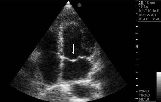
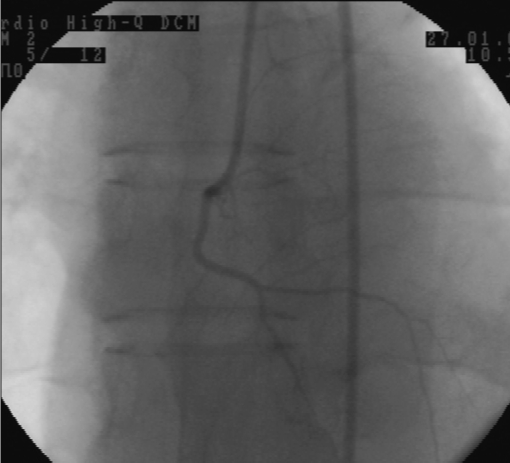
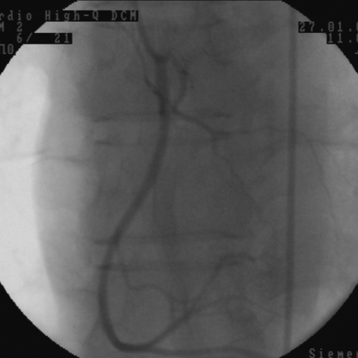
A 37-year-old man presented with a three-week history of chest pain. He complained of a stabbing pain in the chest that would last approximately 1 min to 2 min. He had a history of smoking of two packages of cigarettes daily for the past 15 years. The patient had no other risk factors for coronary artery disease. On admission, his heart rate was 73 beats/minute and blood pressure was 110/70 mmHg. There were no murmurs, friction rubs or gallops on cardiac auscultation. Physical examination of the other organ systems revealed no abnormal findings. His electrocardiogram showed normal sinus rhythm, incomplete right bundle branch block and poor R progression in leads V1 to V3 leads with no ST-T abnormalities in any leads. The patient’s chest x-ray was normal. Laboratory analysis revealed a total cholesterol of 4.22 mmol/L, triglyceride levels of 1.25 mmol/L and high density cholesterol of 0.98 mmol/L. Transthoracic echocardiography showed a left ventricular ejection fraction of 67% without any wall motion abnormality and a mitral valve prolapse (Figure 1) and mild mitral insufficiency. Coronary angiography was performed using the Judkins technique. The left coronary artery projections showed normal left main, circumflex and left anterior descending arteries. The right coronary projections showed that the right ventricular branch of the right coronary artery had a separate ostium in the right coronary sinus (Figure 2). The right coronary (Figure 3) and right ventricular arteries were normal.
Figure 1).
Echocardiogram showing mitral valve prolapse (arrow)
Figure 2).
Coronary angiogram showing the right ventricular branch of right coronary artery had a separate ostium in the right coronary sinus
Figure 3).
Coronary angiogram showing the right coronary and right ventricular arteries
DISCUSSION
Most coronary anomalies are found incidentally during coronary angiography procedures because they often do not cause any signs or symptoms. Although rare, they can cause chest pain and other serious symptoms. Knowledge of possible variations in the coronary circulations and their anatomical differences are important for therapeutic and diagnostic reasons.
The most common irregularity of the coronary arteries is the anomalous origin of the right coronary artery (1,3,6). In the present case, the right ventricular artery had a separate, aberrant ostium arising from the right coronary sinus, and concomitant mitral valve prolapse was also detected. Atypical chest pain may be seen in patients with mitral valve prolapse. Mitral valve prolapse is frequently seen in patients with connective tissue disorders, such as Marfan syndrome and Ehlers-Danlos syndrome, and congenital malformations, such as atrial septal defect of the ostium secundum variety, pulmonary, aortic valve prolapse and Ebstein anomaly. A review of anomalous coronary arteries by Topaz et al (6) demonstrated that these patients frequently had concomitant congenital heart abnormalities, the most common being bicuspid aortic valve and mitral valve prolapse. Primary congenital coronary and valvular anomalies may have genetic heredity. In the present case, mitral valve prolapse was accompanied by a right ventricular coronary artery origin anomaly which, to the best of our knowledge, is the first report in the literature in which both anomalies presented together.
REFERENCES
- 1.Click RL, Holmes DR, Jr, Vlietstra RE, Kosinski AS, Kronmal RA. Anomalous coronary arteries: Location, degree of atherosclerosis and effect on survival – A report from the Coronary Artery Surgery Study. J Am Coll Cardiol. 1989;13:531–7. doi: 10.1016/0735-1097(89)90588-3. [DOI] [PubMed] [Google Scholar]
- 2.Yamanaka O, Hobbs RE. Coronary artery anomalies in 126,595 patients undergoing coronary arteriography. Cathet Cardiovasc Diagn. 1990;21:28–40. doi: 10.1002/ccd.1810210110. [DOI] [PubMed] [Google Scholar]
- 3.Garg N, Tewari S, Kapoor A, Gupta DK, Sinha N. Primary congenital anomalies of the coronary arteries: A coronary arteriographic study. Int J Cardiol. 2000;74:39–46. doi: 10.1016/s0167-5273(00)00243-6. [DOI] [PubMed] [Google Scholar]
- 4.Uyan C, Akdemir R, Uyan AP, Erbilen E. Aberrant origin of the right ventricular coronary artery: A case report. Exp Clin Cardiol. 2003;8:108–9. [PMC free article] [PubMed] [Google Scholar]
- 5.Freed LA, Levy D, Levine RA, et al. Prevalence and clinical outcome of mitral-valve prolapse. N Engl J Med. 1999;341:1–7. doi: 10.1056/NEJM199907013410101. [DOI] [PubMed] [Google Scholar]
- 6.Topaz O, DeMarchena EJ, Perin E, Sommer LS, Mallon SM, Chahine RA. Anomalous coronary arteries: Angiographic findings in 80 patients. Int J Cardiol. 1992;34:129–38. doi: 10.1016/0167-5273(92)90148-v. [DOI] [PubMed] [Google Scholar]


