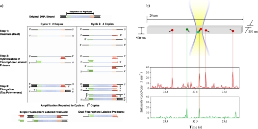
Figure 3.
(a) Schematic of PCR amplification process. Target DNA molecules were denatured by heating, and primers labeled with Alexa Fluor 488 and Alexa Fluor 594 were hybridized to the target strands. Taq polymerase was used for elongation. After several cycles, double labeled two color products were the majority product. (b) Schematic of the submicrometer fluidic channel, laser excitation profile and PCR primer and amplicon detection. The channel was illuminated in an approximately uniform manner through its width and depth, resulting in a focal volume of 180 aL. As single labeled primers and double labeled amplicons were driven electrophoretically through the focal volume, bursts of fluorescence were detected in the two color channels. Primers were detected as single color red or green bursts. Amplicons were detected as simultaneous bursts of fluorescence in both color channels.

