Abstract
The plasmepsins are the aspartic proteases of malaria parasites. Treatment of aspartic protease inhibitor inhibits hemoglobin hydrolysis and blocks the parasite development in vitro suggesting that these proteases might be exploited their potentials as antimalarial drug targets. In this study, we determined the genetic variations of the aspartic proteases of Plasmodium vivax (PvPMs) of wild isolates. Two plasmepsins (PvPM4 and PvPM5) were cloned and sequenced from 20 P. vivax Korean isolates and two imported isolates. The sequences of the enzymes were highly conserved except a small number of amino acid substitutions did not modify key residues for the function or the structure of the enzymes. The high sequence conservations between the plasmepsins from the isolates support the notion that the enzymes could be reliable targets for new antimalarial chemotherapeutics.
Keywords: Plasmodium vivax, aspartic proteases, antimalarial drug
INTRODUCTION
The plasmepsins are a group of aspartic proteases found in the malaria parasites. In the case of Plasmodium falciparum, more than 10 genes possibly encoding plasmepsins or related enzymes have been identified (Coombs et al., 2001; Dame et al., 2003). Plasmepsins 1, 2, and 4 (PfPM1, PfPM2, and PfPM4) and the closely related histoaspartic protease (PfHAP) are localized in the food vacuole and are believed to be involved in hemoglobin hydrolysis (Goldberg et al., 1990; Francis et al., 1994; Banerjee et al., 2002). PfPM1 facilitates an initial cleavage of the α-globin chain of hemoglobin, which presumably leads to the unraveling of the molecule, and further proteolysis by PfPM2, PfPM4, PfHAP, falcipains and facilysin (Francis et al., 1994; Eggleson et al., 1999). The small peptides formed by the action of these enzymes are converted to free amino acids by aminopeptidases and other enzymes (Kolakovich et al., 1997; Gavigan et al., 2001). PfPM2 and PfPM4 also appear to participate in the remodeling of the erythrocyte cytoskeleton by cleaving spectrin at neutral pH (Le Bonniec et al., 1999; Wyatt and Berry, 2002). The exact roles of the other six plasmepsins (PfPM5-10) remain unclear, but they are believed to be associated with the maintenance of the asexual erythrocytic stage or in the development of different stages (Banerjee et al., 2002). Treatment with aspartic protease inhibitor inhibits hemoglobin hydrolysis and blocks the parasite development in vitro (Francis et al., 1994; Rosenthal, 1995; Haque et al., 1999; Nezami et al., 2002) and it has been suggested that at least one of the plasmepsins are essential for parasite survival. Thus, they present attractive targets for the development of new antimalarial chemotherapeutics.
P. vivax is the most prevalent human malaria parasite which causes significant morbidity worldwide together with P. falciparum (Mendis et al., 2001). The parasite is responsible for over 60% episodes of human malaria, which totals several hundred million cases annually. Recent reports from several countries of Asian and South American countries have strongly suggested the presence of chloroquine-resistant P. vivax malaria (Fryauff et al., 1998; Ruebush et al., 2003). Thus, it is urgently required to develop new drugs against vivax malaria. One of the plausible candidates is an inhibitor that specifically blocks the functions of plasmodial aspartic proteases. Therefore, it is also crucial to determine the genetic diversities of the enzymes in wild isolates obtained from various geographical localities because of if the enzymes have great genetic diversities, the task of developing specific inhibitors becomes appreciably more difficult. However, comprehensive genetic and biochemical studies on P. vivax aspartic proteases have been largely limited, because in vitro culture of P. vivax has not been established.
To expand our knowledge on P. vivax aspartic proteases and evaluate the feasibility of using these enzymes as antimalarial drug targets, we cloned two plasmepsins (PvPM4 and PvPM5) and analyzed the genetic diversities of the enzymes in 20 wild type P. vivax Korean isolates collected during 2001-2003 and in two imported isolates.
MATERIALS AND METHODS
Extraction of parasite DNA
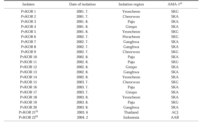
Blood samples were collected from patients residing in Gyonggi-do, a malaria endemic region in Korea, during 2001-2003 (Table 1). In addition, two imported vivax malaria patients infected in Thailand and Indonesia were also included. All patients were diagnosed by microscopic examination at the National Institute of Health, Korea. P. vivax genomic DNAs were extracted from the patients' blood samples by using a QIAamp DNA Blood Kit (Qiagen, Valencia, USA).
Table 1.
Plasmodium vivax isolates used in this study
Cloning and sequence analyses of plasmepsins
Nucleotide sequences encoding the plasmepsins, PvPM4 and PvPM5, of P. vivax were retrieved from BLAST searches of available malaria genome sequence database (The Institute for Genomic Research through the website at http://www.tigr.org) using the nucleotide sequences of P. falciparum plasmepsins as query sequences. PCR was performed with 50 ng of P. vivax genomic DNA by using specific oligonucleotide primers for each gene: PvPM4 (5'-ATGGATATAGCAGTGAAAGAACAAGACTACTCAAA-3' and 5'-TTAATTCTTTGCGATGGCAAAACCGACACTCTC-3') and PvPM-5 (5'-ATGGTCGGAGCGAGCTTGGGGCCCCCCGGT-3' and 5'-CTACGCATCCGCGGGCGCCTTGCCCTCGGAGG-3'). Amplified products were gel-purified, ligated into the pGEM T-Easy vector (Promega, Madison, USA) and transformed into competent E. coli DH5α cells. Sequencing reactions were performed using the BigDye Terminator Cycle Sequencing Ready Reaction Kit in an ABI 377 automatic DNA sequencer (Applied Biosystems, Foster City, USA). Nucleotide and deduced amino acid sequences were analyzed using the SeqEd.V1.0.3 program and Clustal of the Megalign program, a multiple-alignment program of the DNASTAR package (DNASTAR, Madison, USA). To verify the sequences, sequence analysis was performed by analyzing at least 3 plasmid clones which containing each gene insert. The nucleotide sequences encoding each gene have been submitted to GenBank under the accession numbers AY584069-AY584111.
Polymorphic analysis
Polymorphic regions of the genes encoding apical membrane antigen-1 (AMA-1) of P. vivax isolates were analyzed as polymorphic molecular markers to determine population diversity. The polymorphic region of the AMA-1 gene, corresponding to nucleotides 324-735 (aa 108-245), was amplified as described previously (Chung et al., 2003). Each product amplified by PCR was cloned and sequenced, respectively, as described above.
RESULTS
Amplification of plasmepsins
The 20 genomic DNAs of the P. vivax Korean isolates and the two genomic DNAs of the imported isolates from Thailand and Indonesia were amplified by PCR using specific primers for PvPM4 and PvPM5 to isolate each plasmepsin gene. The PCR products appeared at the expected sizes of 1,353 and 1,661 bp, which corresponded well with PvPM4 and PvPM5 (data not shown). Each PCR products were cloned and sequenced, respectively.
Genetic diversities of the plasmepsins
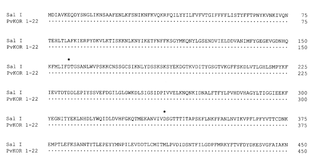
The 22 PCR products of PvPM4 were cloned, sequenced and the deduced amino acid sequences were aligned with that of P. vivax strain Salvador I (Sal I). All the 22 clones did not contain intron within the flanking region. The 22 sequences had well conserved essential residues required for active site formation of aspartic proteases and the amino acids characteristically found in aspartic proteases. Although the Korean isolates were classified into two genotypes (SAG and SAK) based on AMA-1 sequences, the 20 clones showed exactly the same sequences of the PvPM4 gene as P. vivax strain Sal I at the nucleotide and amino acid levels (Fig. 1). In the cases of two imported isolates, one was classified into ACJ and the other as AAR based on AMA-1 sequence analysis. However, they also had the same PvPM4 sequences as Sal I strain and showed no amino acid substitution.
Fig. 1.
Alignment of the deduced amino acid sequences of the PvPM4s of wild isolates and Sal I strain. Korean isolates were classified into two genotypes (SAG and SAK) based on their AMA-1 sequences, and the 20 Korean clones revealed exactly the same sequences with PvPM4 gene of the P. vivax strain Sal I. The two imported isolates also showed the same PvPM4 sequences. The two active-site aspartic acid residues are marked with asterisks.
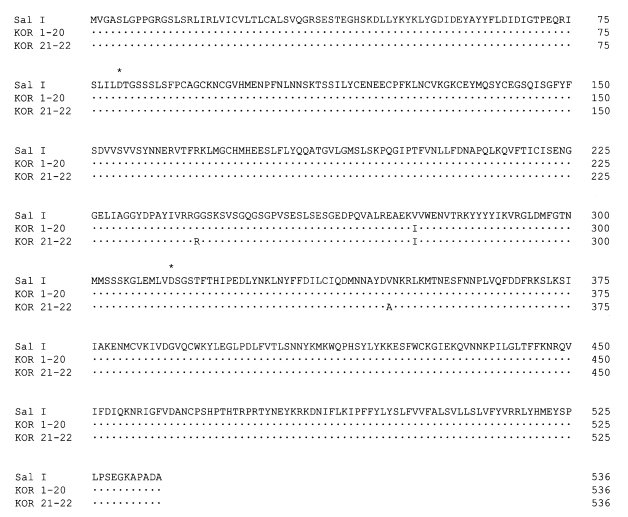
Sequence analysis of the PvPM5 gene also demonstrated that all 20 Korean isolates shared a high degree of identity. A single amino acid substitution at 275 (Val to Ile), which was conserved in all 20 Korean isolates, was identified when compared to the PvPM5 sequence of P. vivax strain Sal I (Fig. 2). Both imported isolates showed two additional substitutions at amino acid 243 (Gly to Ala) and 347 (Val to Ala) along with the substitution at amino acid 276 (Val to Ile). However, the amino acid residues required for active site formation and the amino acids characteristic for aspartic proteases were well conserved in all isolates analyzed.
Fig. 2.
Alignment of the deduced amino acid sequences of the PvPM5s of wild isolates and Sal I strain. A single amino acid substitution at 276 (Val to Ile) was found in the 20 Korean isolates. Both imported isolates showed two additional substitutions at amino acid 243 (Gly to Ala) and at 347 (Val to Ala) along with a substitution at amino acid 276 (Val to Ile). The two active-site aspartic acid residues are marked with asterisks.
DISCUSSION
In this study, we investigated the genetic diversities of the PvPMs of P. vivax wild isolates. Although the Korean isolates examined in this study were classified into two genotypes based on genetic variations of the AMA-1 genes as previously reported (Chung et al., 2003), the PvPM4 sequences were identical in both genotypes of Korean isolates. The two imported isolates also possessed an identical sequence with PvPM4 of Sal I strain. Sequence identity of PvPM4 between New World and Old World isolates indicated that the gene is highly conserved in P. vivax and it can be a promising antimalarial drug target. A recent investigation identified and compared orthologs of plasmepsins in human infecting malaria parasites as well as rodent and primate malaria parasites (Dame et al., 2003). Interestingly, non-falciparum species lacked genes encoding either PM1, PM2 or HAP, but all encoded an ortholog of PfPM4. This result suggested strongly that PfPM4 is a common plasmepsin to all Plasmodium species. However, it is not certain whether PfPM4 orthologs played the same functional roles in all human infecting species. Nevertheless, it seems likely to play an important role in human infecting Plasmodium species due to the ubiquitous presence of this ortholog in all other species.
PfPM5 orthologs are also found in all Plasmodium species (Dame et al., 2003). These molecules are localized to other regions of the asexual erythrocytic stage parasite and are thought to be expressed in a different stage of the parasite's life cycle (Banerjee et al., 2002). The 20 Korean isolates and 2 imported isolates examined revealed the substitution of a small number of amino acids compared to Sal I. However, the two aspartic acid residues critically required for active site formation were tightly conserved. These minor variations in PvPM5 may not effect its enzymic properties and thus would not be expected to complicate drug development efforts targeting aspartic proteases.
In conclusion, this study demonstrated that a very low genetic variation was found among plasmepsins of P. vivax wild isolates analyzed. The sequences of PvPM4, one of food vacuole proteases and found in all Plasmodium species, were completely identical in all isolates examined. Although PvPM5 showed a small number of amino acid substitutions between isolates, these substitutions do not affect the essential amino acids required for active site formation of the enzyme. Thus, the present study indicates that PvPMs are highly conserved in wild isolates and they are promising targets for structure-based drug development. Studies are underway to characterize these enzymes biochemically and to determine their biological roles in P. vivax.
Footnotes
This work was supported by a grant from the Korea Health 21 R&D Project, Ministry of Health and Welfare, Republic of Korea (03-PJ1-PG1-CH01-0001).
References
- 1.Banerjee R, Liu J, Beatty W, Pelosof L, Klemba M, Goldberg DE. Four plasmepsins are active in the Plasmodium falciparum food vacuole, including a protease with an active-site histidine. Proc Natl Acad Sci USA. 2002;99:990–995. doi: 10.1073/pnas.022630099. [DOI] [PMC free article] [PubMed] [Google Scholar]
- 2.Chung JY, Chun EH, Chun JH, Kho WG. Analysis of the Plasmodium vivax apical membrane antigen-1 gene from re-emerging Korean isolates. Parasitol Res. 2003;90:325–329. doi: 10.1007/s00436-002-0777-2. [DOI] [PubMed] [Google Scholar]
- 3.Coombs GH, Goldberg DE, Klemba M, Berry C, Kay J, Mottram JC. Aspartic proteases of Plasmodium falciparum and other parasitic protozoa as drug targets. Trends Parasitol. 2001;17:532–537. doi: 10.1016/s1471-4922(01)02037-2. [DOI] [PubMed] [Google Scholar]
- 4.Dame JB, Yowell CA, Omara-Opyene L, Carlton JM, Cooper RA, Li T. Plasmepsin 4, the food vacuole aspartic proteinase found in all Plasmodium spp. infecting man. Mol Biochem Parasitol. 2003;130:1–12. doi: 10.1016/s0166-6851(03)00137-3. [DOI] [PubMed] [Google Scholar]
- 5.Eggleson KK, Duffin KL, Goldberg DE. Identification and characterization of falcilysin, a metallopeptidase involved in hemoglobin catabolism within the malaria parasite Plasmodium falciparum. J Biol Chem. 2000;274:32411–32417. doi: 10.1074/jbc.274.45.32411. [DOI] [PubMed] [Google Scholar]
- 6.Figtree M, Pasay CJ, Slade R, et al. Plasmodium vivax synonymous substitution frequencies, evolution and population structure deduced from diversity in AMA1 and MSP1 genes. Mol Biochem Parasitol. 2000;108:53–66. doi: 10.1016/s0166-6851(00)00204-8. [DOI] [PubMed] [Google Scholar]
- 7.Francis SE, Gluzman IY, Oksman A, et al. Molecular characterization and inhibition of a Plasmodium falciparum aspartic hemoglobinase. EMBO J. 1994;13:306–317. doi: 10.1002/j.1460-2075.1994.tb06263.x. [DOI] [PMC free article] [PubMed] [Google Scholar]
- 8.Fryauff DJ, Tuti S, Mardi A, et al. Chloroquine-resistant Plasmodium vivax in transmigration settlements of West Kalimantan, Indonesia. Am J Trop Med Hyg. 1998;59:513–518. doi: 10.4269/ajtmh.1998.59.513. [DOI] [PubMed] [Google Scholar]
- 9.Gavigan CS, Dalton JP, Bell A. The role of aminopeptidasesin haemoglobin degradation in Plasmodium falciparum-infected erythrocytes. Mol Biochem Parasitol. 2001;117:37–48. doi: 10.1016/s0166-6851(01)00327-9. [DOI] [PubMed] [Google Scholar]
- 10.Goldberg DE, Slater AF, Cerami A, Henderson GB. Hemoglobin degradation in the malaria parasite: an ordered process in a unique organelle. Proc Natl Acad Sci USA. 1990;87:2931–2935. doi: 10.1073/pnas.87.8.2931. [DOI] [PMC free article] [PubMed] [Google Scholar]
- 11.Haque TS, Skillman AG, Lee CE, et al. Potent, low-molecular-weight non-peptide inhibitors of malarial aspartyl protease plasmepsin II. J Med Chem. 1999;42:1428–1440. doi: 10.1021/jm980641t. [DOI] [PubMed] [Google Scholar]
- 12.Kolakovich KA, Gluzman IY, Duffin KL, Goldberg DE. Generation of hemoglobin peptides in the acidic digestive vacuole of Plasmodium falciparum implicates peptide transport in amino acid production. Mol Biochem Parasitol. 1997;87:123–135. doi: 10.1016/s0166-6851(97)00062-5. [DOI] [PubMed] [Google Scholar]
- 13.Le Bonniec S, Deregnaucourt C, Redeker V, et al. Plasmepsin II, an acidic hemoglobinase from the Plasmodium falciparum food vacuole, is active at neutral pH on the host erythrocyte membrane skeleton. J Biol Chem. 1999;274:14218–14223. doi: 10.1074/jbc.274.20.14218. [DOI] [PubMed] [Google Scholar]
- 14.Mendis K, Sina BJ, Marchesini P, Carter R. The neglected burden of Plasmodium vivax malaria. Am J Trop Med Hyg. 2001;64:97–106. doi: 10.4269/ajtmh.2001.64.97. [DOI] [PubMed] [Google Scholar]
- 15.Nezami A, Luque I, Kimura T, Kiso Y, Freire E. Identification and characterization of allophenylnorstatine-based inhibitors of plasmepsin II an antimalarial target. Biochemistry. 2002;41:2273–2280. doi: 10.1021/bi0117549. [DOI] [PubMed] [Google Scholar]
- 16.Rosenthal PJ. Plasmodium falciparum: effects of proteinase inhibitors on globin hydrolysis by cultured malaria parasites. Exp Parasitol. 1995;80:272–281. doi: 10.1006/expr.1995.1033. [DOI] [PubMed] [Google Scholar]
- 17.Ruebush TK, Jr, Zegarra J, Cairo J, et al. Chloroquine-resistant Plasmodium vivax malaria in Peru. Am J Trop Med Hyg. 2003;69:548–552. [PubMed] [Google Scholar]
- 18.Wyatt DM, Berry C. Activity and inhibition of plasmepsin IV, a new aspartic proteinase from the malaria parasite, Plasmodium falciparum. FEBS Lett. 2002;513:159–162. doi: 10.1016/s0014-5793(02)02241-x. [DOI] [PubMed] [Google Scholar]


