Abstract
Activity level in childhood has long been of interest to researchers in psychology, personality, and pediatrics. With the current epidemic in child obesity, even greater attention is being paid to physical activity and its role in preventing excess weight gain. Activity can be rated by observers as well as recorded more objectively by mechanical devices, however, some research suggests that estimates from these two sources do not always correlate. Via a demonstration with a human infant and a doll, the present report suggests that for younger infants at least, much of their measured activity may be confounded by their caregivers’ movements.
Motor activity in infancy and early childhood has long been considered a core dimension of behavioral individuality, or temperament, by pediatricians, psychologists, and personality theorists (Buss & Plomin, 1984; Rothbart, 1981; Thomas et al., 1968). With the increased interest in factors that may be contributing to the current child obesity problem, activity level has received additional attention as a proxy for energy expenditure (Wells et al., 1996). Activity may be quantified as early as the newborn period, and is thought to be the most consistent of the temperament traits in demonstrating stability with age (Hubert et al., 1982). Activity has the added advantage of being measurable over the life span through a variety of objective means, such as doubly-labeled water, whole body calorimetry, heart rate monitoring, and movement sensors such as actometers or accelerometers (Parsons et al., 1999). These approaches can be relatively expensive, so questionnaires and rating scales, admittedly more subjective, are frequently used to quantify activity, especially with infants and children.
In two early reports that totaled fewer than 100 infants, Rose and colleagues showed that ratings of movement and quantitative assessments of activity (as measured by modified calendar watches) were better predictors of body size at 5-months than was caloric intake (Rueda-Williamson & Rose, 1962; Rose & Mayer, 1968). More recently, Li et al. (1995) showed that percentage of body fat was inversely related to activity level (as rated by observers) in 31 infants who had been formula-fed, and the correlations became stronger by 12-months. Rapid weight gain in the first months of life has been shown to predict child overweight (Stettler et al., 2002), but in general, studies that have examined either activity (Berkowitz et al., 1985; Ku et al., 1981; O’Callaghan et al., 1997) or energy expenditure (Davies et al., 1991; Wells et al., 1996) in pre-walking infants have failed to show early activity to be predictive of fatness in childhood. Early motor activity may not prove to be any more than marginally predictive of later fatness, but some of the uncertainty may in fact be due to the difficulty of quantifying movements in younger infants.
For example, Rothbart (1986) found no association between mother- and observer-rated activity at 3-months, but did so at 6-and 9-months. At 12-months, Carnicero et al. (2000) found agreement between maternal and observer ratings of activity, but did not during the preceding 3-, 6- or 9-month assessments. Interestingly, the convergence between maternal and mechanical ratings of activity shows a parallel trend. In separate reports, Eaton and colleagues found no associations between maternal ratings and actometer measures of activity in two studies of infants ranging from 3–4½ months (Eaton & Dureski, 1986), but did find a significant correlation with 6½-month-olds (Saudino & Eaton, 1991). Similarly, Worobey and Islas-Lopez (2008) found that maternal ratings and actometer scores did not correlate in their sample of infants at 3-months, but did so when the babies were measured again at 6-months.
A long-acknowledged limitation to the actometer approach, particularly when pitted against maternal ratings, lies in the window of observation. An actometer measures movements over a limited, specified time interval. Although rating scales may ask the caregiver to think about behaviors observed over the previous week (Rothbart, 1981) or even day (Eaton & Dureski, 1986) it is likely the case that a mother’s ratings, in particular, may be colored by the general impression that she has gleaned from a few months of daily contact with her infant. Although she may not equate “goodness” with level of activity at 3-months, her objectivity may nonetheless be questioned.
In contrast, an actometer may be totally objective over the limited time period that it records activity, but it may not be very selective. In other words, it will measure movement bereft of emotion or attachment, but will not discriminate based on who is doing the moving. Tulve et al. (2007) recently showed that caregivers could categorize levels of activity correctly relative to accelerometers about 70% of the time, however, their judgments were restricted to defined periods of sleeping, eating, quiet play and active play. The present report attempts to illustrate the possible confounding of infant movement with maternal movement, at least in studies where the infant may be monitored all day while being carried about or moved by a caregiver.
Method
Subjects
Two women, a 31-year-old mother and a 23-year-old graduate research assistant (NRV)); a baby girl, “Emily,” age 3 months, and a girl baby doll, “Maria,” age unknown.
Measures and Procedures
To measure motor activity over a 24-hour period, both the infant and doll wore a Micro Mini Motionlogger (Ambulatory Monitoring, Inc., Ardsley, NY). These instruments were chosen because of their small size (2.5 cm in diameter) and weight (14 g), being waterproof, and their ability to distinguish instances of activity during sleep states from movements exhibited while in awake states. These motionloggers use a piezoelectric bimorph-ceramic beam that generates a voltage every time the device is moved. The voltage next passes to the analog circuitry where the signal is amplified, filtered and accumulated, and then stored in 1-minute epoch lengths.
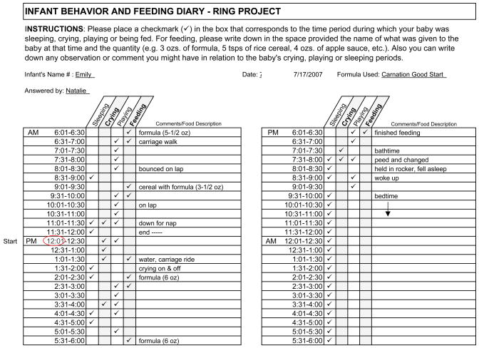
The devices were attached to the infant’s and doll’s left ankle at approximately 11:45 AM on a Tuesday morning, and programmed to begin recording movements at 12 noon. While the mother was told to handle, feed and care for her baby just as she normally would, the research assistant (RA) was instructed to mimic with the doll whatever maneuvers she observed the mother to do whenever in contact with her infant. For example, when the mother fed the infant, the RA would hold the doll and a baby bottle in a similar manner at the same time, and move the doll to a burping position when the mother did so. When the infant cried, the doll was soothed in the same manner. Likewise, as the infant was held or put down for a diaper change, so was the doll. Although the doll did not actually eat or wet, the RA would remove and replace the doll’s diaper in direct imitation of what the mother did with her infant. When the infant was laid to sleep, the doll was placed down on a cushion in the nursery, and left undisturbed until the infant demanded attention upon awakening. At approximately 12:15 the following day, the motionloggers were removed, ensuring a 24-hour sample of behavior. The diary kept by the RA that delineates how the infant’s time was spent appears in Figure 1 (Note that recording began on the PM 12:01–12:30 line in the left column, and finished up the next morning on the 11:31–12:00 line just above the previous start time).
Figure 1.
Results
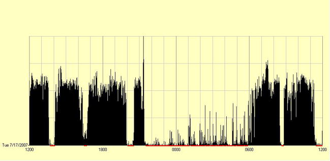
Figure 2 displays the pattern of Emily’s activity in a histogram format as minute-by-minute movements over the 24-hour period (noon until noon the next day), with higher frequencies reflected by higher vertical lines. As seen in Figure 2, her movements from the afternoon through early evening indicate a relatively high level of wakeful activity, with reduced movement in the 1:30–2 PM, 4:30–5 PM, and 8–8:30 PM intervals, where sleep is indicated by the motionlogger (bars at base of graph) and corroborated by the diary. Although sporadic movements are evident from 9:30 PM until 6 AM, the motionlogger registered the infant as being asleep, again validated by the diary. Awake activity is then displayed from approximately 6:30–11 AM, with brief periods of sleep around 8:30 AM and 11AM. Considering the context of activity as provided by the diary, it is evident that more pronounced movements occurred in conjunction with her crying, being fed, being bathed (or changed– though not generally noted), and when engaged in play with her mother. In contrast, markedly reduced levels of movement were recorded when Emily was asleep.
Figure 2.
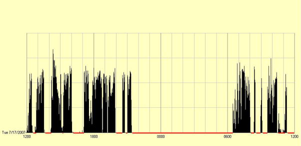
As the Maria doll was incapable of initiating movement on its own, one would predict that the motionlogger would register a continuous sleep state, with no measurable activity. However, as shown in Figure 3, appreciable levels of activity were recorded at various times throughout the 24-hour period. Although the pattern resembles a somewhat skeletal version of Figure 2, the vertical lines do appear within the boundaries of the awake segments as defined by the infant. In contrast, while the infant slept through the night from 9:30 PM until 6:30 AM, the doll was left undisturbed, and the motionlogger duly reflects the complete absence of movement.
Figure 3.
Table 1 displays the proportion of time that the infant and doll were estimated to be in wake versus sleep states, as well as their average levels of activity over the 24-hour period. As would be expected, the frequencies of the doll’s movements were substantially lower than those of the infant’s, with the doll characterized as spending more time in sleep than did the infant, and more time asleep than awake.
Table 1.
Activity Comparison for Infant vs. Doll
| Infant | Doll | |
|---|---|---|
| Activity Mean/Minute (SD) | 144.21 (123.89) | 79.06 (14.29) |
| Wake minutes | 854 | 632 |
| Percent of time “awake” | 59% | 44% |
| Sleep minutes | 586 | 808 |
| Percent of time “asleep” | 41% | 56% |
Discussion
The purpose of this exercise was not to pit an infant against a doll in a contest of motor activity—barring the use of a wind-up or battery-operated toy, a human infant would assuredly be favored to beat an inanimate doll in terms of spontaneous activity. And Maria was nothing more than an old-fashioned cloth and vinyl doll that could not move of her own accord. That she displayed any movement at all, as registered by the motionlogger, must be attributed to the shifting, lifting and concomitant handling that she received as the RA feigned caregiving and play with her. Rather, the purpose was to demonstrate the level of confounding that apparently occurs when a motionlogger or actometer is used to estimate infant activity, at least in a young infant.
The term “measurement error” may not be appropriate, because we have no cause to question the ability of the device to accurately detect movements. The problem instead is that moving an infant, and unavoidably her limbs, results in the motionlogger detecting motion. The more the caregiver is instrumental in moving the infant’s limbs, the greater the likelihood that it is her activity, and not the infant’s, that is being registered. This is not to say that the actometer is not detecting true infant activity as well. After all, approximately 45% more activity was detected for the infant relative to the doll. But when one steps back to reflect on this disparity, it is still noteworthy that any movement at all could be credited to the doll. Taken another way, if one were presented with only Figure 3, and not told that it was a doll that wore the motionlogger, an observer might very well have concluded that the histogram represented the movements of an actual infant, albeit one who slept quite soundly.
From this one exercise we cannot estimate the degree of “maternal enhancement” of movement that is attributable to a caregiver’s handling and playing with an infant. Whether it would be much less for an infant who is not handled very much, or as high as 50% or more for an infant who is hauled about in a sling or frontal carrier is an empirical question. Likewise, the present illustration was not meant to dismiss the use of motionloggers, actometers or accelerometers with infants or children. Numerous reports attest to their validity in assessing sleep versus wake activity in infants under a year of age (Gnidovic et al., 2002; Sadeh et al., 1995), and many studies have successfully employed such devices with preschoolers and older children (e.g., Acebo et al., 2005; Moore et al., 2003; Reilly et al., 2003; Stevens et al., 1978). However, when assessing convergence between actometer devices and maternal ratings, the handful of studies that have included infants under a year indicate that agreement increases with the age of the baby (Saudino & Eaton, 1991; Worobey & Islas-Lopez, 2008). By 6-months of age, many infants are beginning to crawl on their own, and with walking by some at 1-year, more and more of the infants’ movements are controlled by themselves. For infants who do not yet sit up on their own, however, a significant amount of the disagreement between caregiver ratings and objective actometers may be due to the confounding of maternal movement with infant activity. If infant activity is to be explored as a possible correlate of early weight gain, researchers who seek to quantify activity in subjects less than 6-months would be advised to also monitor the caregiver’s handling of the infant, and perhaps measure their activity level as well.
Acknowledgments
The authors wish to thank Carrie Ciaburri and Carolina Espinosa. This work was supported by NICHD Grant HD47338 to the first author.
References
- Acebo C, Sadeh A, Seifer R, Tzichinsky O, hafer A, Carskadon MA. Sleep/wake patterns derived from activity monitoring and maternal report for healthy 1- to 5-year-old children. Sleep. 2005;28(12):1568–1577. doi: 10.1093/sleep/28.12.1568. [DOI] [PubMed] [Google Scholar]
- Berkowitz RI, Agras WS, Korner AF, Kraemer HC, Zeanah CH. Physical activity and adiposity: A longitudinal study from birth to childhood. Journal of Pediatrics. 1985;106(5):734–738. doi: 10.1016/s0022-3476(85)80345-0. [DOI] [PubMed] [Google Scholar]
- Buss AH, Plomin R. Temperament: Early developing personality traits. Hillsdale, NJ: Erlbaum; 1984. [Google Scholar]
- Carnicero JAC, Perez-Lopez J, Salinas MDCG, Martinez-Fuentes MT. A longitudinal study of temperament in infancy. European Journal of Personality. 2000;14:21–37. [Google Scholar]
- Davies PSW, Day J, Lucas A. Energy expenditure in early infancy and later body fatness. International Journal of Obesity. 1991;15:727–731. [PubMed] [Google Scholar]
- Eaton WO, Dureski CM. Parent and actometer measures of motor activity level in the young infant. Infant Behavior & Development. 1986;9:383–393. [Google Scholar]
- Gnidovic B, Neubauer D, Zidar J. Actographic assessment of sleep-wake rhythm during th first 6 months of life. Clinical Neurophysiology. 2002;113:1815–181. doi: 10.1016/s1388-2457(02)00287-0. [DOI] [PubMed] [Google Scholar]
- Ku LC, Shapiro LR, Crawford PB, Huenemann RL. Body composition and physical activity in 8-year-old children. American Journal of Clinical Nutrition. 1981;34:2770–2775. doi: 10.1093/ajcn/34.12.2770. [DOI] [PubMed] [Google Scholar]
- Li R, O’Connor L, Buckley D, Specker B. Relation of activity levels to body fat in infants 6-to 12-months of age. Journal of Pediatrics. 1995;126(3):353–357. doi: 10.1016/s0022-3476(95)70447-7. [DOI] [PubMed] [Google Scholar]
- Moore LL, Gao D, Bradlee ML, Cupples LA, Sundarajan-Ramamurti A, Proctor MH, Hood MY, Singer MR, Ellison RC. Does early physical activity predict body fat change throughout childhood? Preventive Medicine. 2003;37:10–17. doi: 10.1016/s0091-7435(03)00048-3. [DOI] [PubMed] [Google Scholar]
- O’Callaghan MJ, Williams GM, Andersen MJ, Bor W, Najman JM. Prediction of obesity in children: A cohort study. Journal of Paediatric Child Health. 1997;3:311–316. doi: 10.1111/j.1440-1754.1997.tb01607.x. [DOI] [PubMed] [Google Scholar]
- Parsons TJ, Power C, Logan S, Summerbell CD. Childhood predictors of adult obesity: A systematic review. International Journal of Obesity. 1999;23(Suppl B):S1–S107. [PubMed] [Google Scholar]
- Reilly JJ, Coyle J, Kelly L, Burke G, Grant S, Paton JY. An objective method for measurement of sedentary behavior in 3-to 4-year olds. Obesity Research. 2003;11(10):1155–1158. doi: 10.1038/oby.2003.158. [DOI] [PubMed] [Google Scholar]
- Rose HE, Mayer J. Activity, calorie intake, fat storage, and the energy balance of infants. Pediatrics. 1968;41(1):18–29. [PubMed] [Google Scholar]
- Rothbart MK. Measurement of temperament in infancy. Child Development. 1981;52:569–578. [Google Scholar]
- Rothbart MK. Longitudinal observation of infant temperament. Developmental Psychology. 1986;22:356–365. [Google Scholar]
- Rueda-Williamson R, Rose H. Growth and nutrition of infants. Pediatrics. 1962;30:639–653. [PubMed] [Google Scholar]
- Sadeh A, Acebo C, Seifer R, Aytur S, Carskadon MA. Activity-based assessment of sleep-wake patterns during the 1st year of life. Infant Behavior & Development. 1995;18:329–337. [Google Scholar]
- Saudino K, Eaton WO. Infant temperament and genetics: An objective twin study of motor activity level. Child Development. 1991;62:1167–1174. [PubMed] [Google Scholar]
- Stettler N, Zemel BS, Kumanyika S, Stallings VA. Infant weight gain and childhood overweight status in a multicenter, cohort study. Pediatrics. 2002;109(2):194–199. doi: 10.1542/peds.109.2.194. [DOI] [PubMed] [Google Scholar]
- Stevens TM, Kupst M, Suran BG, Schulman JL. Activity level: A comparison between actometer scores and observer ratings. Journal of Abnormal Psychology. 1978;6(2):163–173. doi: 10.1007/BF00919122. [DOI] [PubMed] [Google Scholar]
- Tulve NS, Jones PA, McCurdy T, Croghan CW. A pilot study using an accelerometer to evaluate a caregiver’s interpretation of an infant or toddler’s activity level as recorded in a time activity diary. Research Quarterly for Exercise and Sport. 2007;78(4):375–383. doi: 10.1080/02701367.2007.10599435. [DOI] [PubMed] [Google Scholar]
- Thomas A, Chess S, Birch HG. Temperament and behavior disorders in children. New York: New York University Press; 1968. [Google Scholar]
- Wells JCK, Stanley M, Laidlaw AS, Day JM, Davies PSW. The relationship between components of infant energy expenditure and childhood body fatness. International Journal of Obesity. 1996;20:848–853. [PubMed] [Google Scholar]
- Worobey J, Islas-Lopez M. Temperament measures of African-American infants: Change and convergence with age. Early Child Development and Care. 2009:107–112. doi: 10.1080/03004430802279926. [DOI] [PMC free article] [PubMed] [Google Scholar]


