Figure 3.
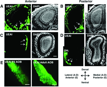
Fucα(1−2)Gal sugars are enriched in the developing OB and are present in the ONL and glomerular layers on all faces of the developing OB. (A−D) Coronal sections of the MOB from wild-type P3 mice (A, B) and adult mice (C, D) were labeled with UEAI conjugated to fluorescein and imaged by confocal fluorescence microscopy. Nuclei were stained with DAPI. Extensive staining of the ONL and glomerular layer was observed in the developing MOB, whereas labeling was confined to small clusters of glomeruli in the adult MOB. The arrows indicate the presence of glomeruli in the olfactory bulb. The scale bar is 200 μm. (E) Sagittal sections of the developing AOB were labeled with UEAI conjugated to fluorescein (green). Fucα(1−2)Gal sugars were enriched in the anterior AOB and were present in a gradient from anterior to posterior. The scale bar is 100 μm.

