Abstract
A high-frequency angled needle ultrasound transducer with an aperture size of 0.4 × 0.56 mm2 was fabricated using a lead zinc niobate-lead titanate (PZN-7%PT) single crystal as the active piezoelectric material. The single crystal was bonded to a conductive silver particle matching layer and a conductive epoxy backing material through direct contact curing. A parylene outer matching layer was formed by vapor deposition. Angled needle probe configuration was achieved by dicing at 45° to the single crystal poling direction to satisfy a clinical request for blood flow measurement in the posterior portion of the eye. The electrical impedance magnitude and phase of the transducer were 42 Ω and −63°, respectively. The measured center frequency and the fractional bandwidth at −6 dB were 43 MHz and 45%, respectively. The two-way insertion loss was approximately 17 dB. Wire phantom imaging using fabricated PZN-7%PT single crystal transducers was obtained and spatial resolutions were assessed.
I. INTRODUCTION
The narrowing of retinal arterioles has been identified as a precursor to hypertension, which may lead to occlusion of the microcirculation in the retina and eventually results in blindness due to retinal hemorrhages or edema. Retinal arteriole narrowing can be identified based on the decrease of velocity of blood flow, which can be detected by using a pulsed-wave Doppler system with high-frequency needle transducers [1]. A known angle between transducers and blood flow is a critical component for an accurate Doppler measurement. This leads to difficulties when the transducers have to be inserted into the eye and placed over the posterior region at certain locations. Therefore, angled needle transducers are the proposed solution for this particular application.
Medical imaging transducers usually utilize a piezoelectric material as the active element to transform the electric input into a mechanical wave propagating into the imaging target and then transform the echo back into an electric output signal. Due to its reasonable piezoelectric properties (d33 = 170–500 pC/N, and kt = 0.4–0.5), PZT ceramic is the predominant material used in medical imaging transducers. Recently, superior piezoelectric properties have been discovered for relaxor-based lead zinc niobate-lead titanate (PZN-PT) and lead magnesium niobate-lead titanate (PMN-PT) single crystals (d33 = 2000–3000 pC/N, k33 = 85–95%) [2], [3]. Low-frequency (3.5–4.6 MHz) transducers fabricated from PZN-PT single crystals using the mode have obtained two-way bandwidths of 74–141% [4]. However, for high-frequency single element transducers, thickness mode (kt mode) is usually employed rather than longitudinal mode (k33 or mode), which requires a high aspect ratio of length-to-in-plane dimensions, and poses difficulty in dicing for resonant frequencies higher than 30 MHz. For PZN-PT or PMN-PT single crystals operating in thickness mode, the electromechanical coupling factor kt is slightly higher than that of PZT. However, due to the much superior piezoelectric strain constant d33 and high k33, transducers made from these single crystals may have much higher sensitivity. In our previous work, a PMN-PT flat needle transducer for Doppler application was reported [5]. However, due to the stringent requirement of imaging of eyes, the imaging area is not always accessible from direct contact of a perpendicular probe. Therefore, an angled needle probe, with imaging area at a 45° angle to the shaft of the probe, is more desirable for high-frequency pulsed-wave Doppler and medical imaging applications.
The Curie temperature of PZN-PT single crystal (160°C) is higher than that of PMN-PT (130°C), and therefore we chose to use this material for the angled transducer because it is expected to better handle the thermal stresses caused by the Doppler application. The design, fabrication, and testing of a PZN-PT single crystal angled transducer is described in this paper. Results were compared with those obtained from the PMN-PT flat needle transducer as well as from PZFlex and PiezoCAD models.
II. DESIGN AND METHODS
PZN-7%PT single crystals were supplied by Microfine Materials Technologies P/L (Singapore). The crystals were grown using the high-temperature flux technique, the details of which can be found in [6]. The Laue back reflection technique was used to orient the crystal. The wafers were sliced in the (001) plane and polished to a thickness of 400 µm. The gold/palladium electrodes were sputter-coated on the (001) surfaces. The samples were poled with an electric field 0.8 kV/mm in silicone oil at room temperature. The properties of the samples were measured by using a high-frequency impedance analyzer (HP 4291B; Agilent Technologies, Englewood, CO) and are shown in Table I. The properties are consistent with the reported values [7].
TABLE I.
Material properties of a PZN-7%PT (001) poled single crystal.
| Property | Value |
|---|---|
| Electromechanical coupling coefficient (kt) | 0.53 |
| Piezoelectric constant d33 of wafer (pC/N) | 1720 |
| Relative clamped dielectric constant (εS/ε0) | 1000 |
| Dielectric loss | 0.003 |
| Density (g/cm3) | 8.35 |
| Longitudinal wave velocity (m/s) | 4060 |
| Acoustic impedance (MRayl) | 33.9 |
| Curie temperature (°C) | 160 |
To resonate at a thickness mode of f0 = 45 MHz, the piezo-layer should have a thickness of a half wavelength. Considering that the piezo-layer is attached with soft backing and matching materials while working as a transducer, a reduced thickness by lapping of 40 µm was chosen. A small probe size is required for easy access and precise placement of the transducer over the retina for measurement. However, the size of the piezo-material is limited by the requirement of having enough capacitance to match with the standard 50 Ω input of the pulser/receiver electronics. The capacitance of the piezo-layer at its resonance is approximately given by
| (1) |
where A is surface area of the piezo-layer, and is the clamped dielectric constant, which is about 1000 εo for single crystal PZN-7%PT, where εo is the permittivity of the vacuum. Therefore, A should be between 0.16 and 0.32 mm2 to provide a 50–100 Ω magnitude impedance at resonance, based on Z = 1/2πωC and (1). A square dimension of 0.40 × 0.56 mm2 was used in this study.
Due to the large difference of the acoustic impedance of the piezo-material (33.3 MRayl) and that of the load medium (1.5 MRayl), it was necessary to incorporate impedance matching layers to enhance the performance of the transducer. If two matching layers are to be used in this transducer, the design criteria for wideband transducer given by Desilets et al. [8] requires impedance Zm1 and Zm2 for the quarter wavelength matching layers as follows:
| (2) |
| (3) |
where Z1 is the acoustic impedance of PZN-7%PT in the thickness mode, and Z2 is that of the load medium. Zm1 = 8.8 MRayl and Zm2 = 2.3 MRayl are obtained from (2) and (3) for the two matching layers. The transducer fabrication process was referenced in previous work [9]. In this study, a 3-mm thickness is used for the backing, which provides sufficient attenuation and poses less difficulty in dicing.
To fabricate the 45° angle transducer, the stack of transducers above was diced in two steps. First, it was diced along the thickness direction into wafers 0.4 mm wide. Afterwards, the wafers were aligned with the width direction facing up, and then diced into 0.40 × 0.56mm2 posts, along a 45° angle to the thickness direction.
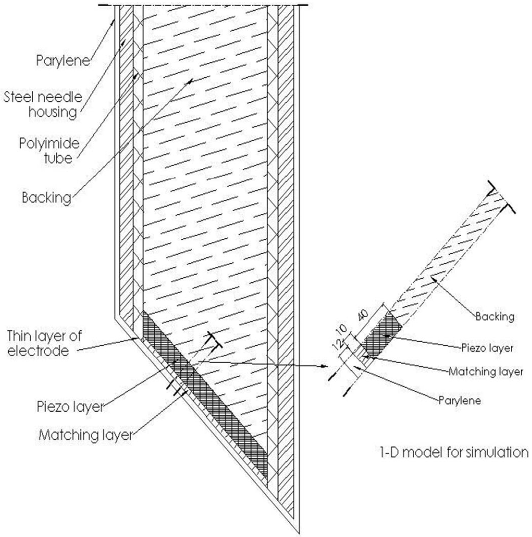
The PZFlex finite element modeling (FEM) package (Weidlinger Associates Inc., Mountain View, CA) was used to simulate the transducer performance before fabrication using the model shown in Fig. 1. Since the in-plane dimension is significantly larger than the thickness, the transducer working in the thickness mode can be simulated by using a 1-D model along the thickness direction. It was noticed that, for the thickness mode, the measured acoustic properties, υ and , for PZN-7%PT were close to those of PZN-4.5%PT [10]. It is reasonable to model the transducer performance based on the properties of PZN-4.5%PT due to the availability of the full matrix of the material properties [9], which is required by FEM software packages in simulation. For comparison with the FEM modeling result, the transducer was also designed with an analogous electrical circuit model (PiezoCAD; Sonic Concepts, Woodinville, WA), using the measured data of PZN-PT in Table I.
Fig. 1.
Design cross section of a needle transducer.
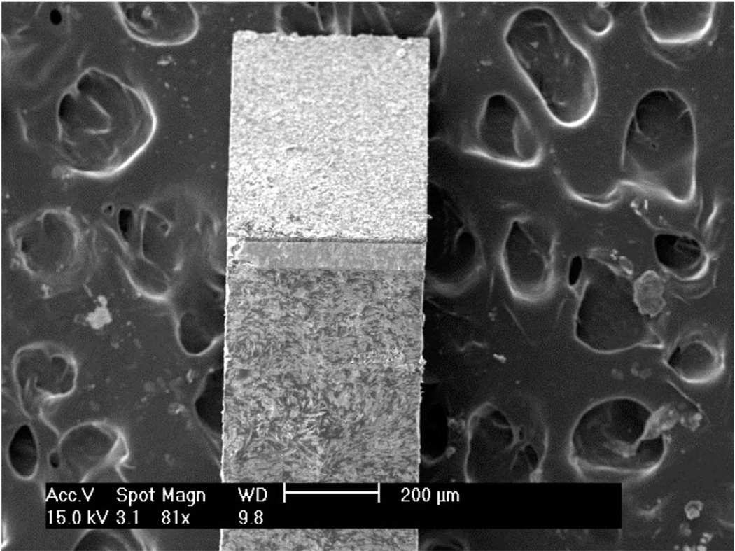
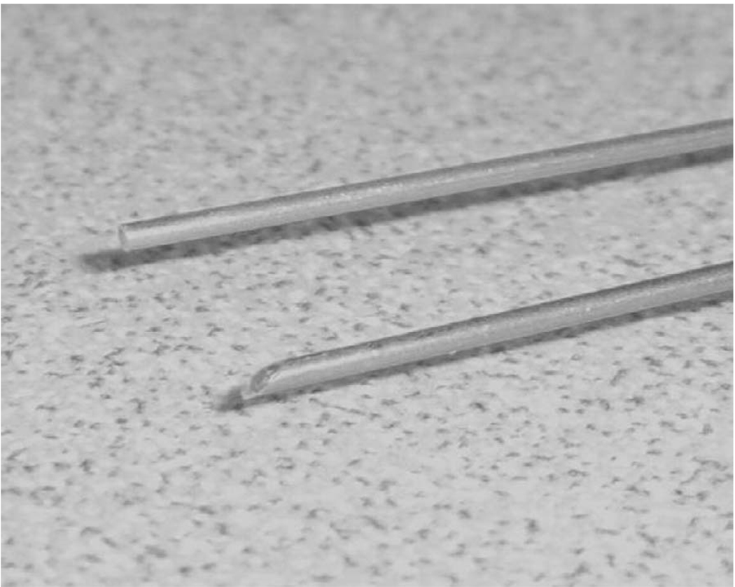
A cross section of the post observed by scanning electron microscopy (SEM) is shown in Fig. 2. The post was inserted into a polyimide tube with an inner diameter of 0.57 mm (MedSource Technologies, Trenton, GA), which serves as an insulating layer, and then the tube was inserted into the housing of a steel needle, which was also diced to have a 45° angle at the top side. An electrical connector was fixed to the conductive backing using a conductive epoxy. An electrode was sputtered across the silver matching layer and the needle housing to form the ground plane connection. The device was electrically shielded by a stainless steel needle housing connecting a sputtered chrome/gold (50/100 nm) layer to a silver particle matching layer. Vapor-deposited parylene (Specialty Coating Systems, Indianapolis, IN) with a thickness of 12 µm was used to coat the aperture and the needle housing. The finished angle and flat needle transducers are shown in Fig. 3. The diameter of the needle is 0.9 mm. The transducer was re-poled at room temperature in the final fabrication step because the single crystal was depoled after lapping.
Fig. 2.
SEM image of a cross section of the PZN-PT and matching layers.
Fig. 3.
Photograph of flat and angled PZN-PT needle transducers.
Electrical impedance measurements were taken using the HP4291B impedance analyzer. The piezoelectric properties of samples were measured by a YE 2730 A d33 meter (APC Products, Inc., Pleasant Gap, PA). The pulse-echo analysis consisted of measuring the received echo from the reflection of a flat quartz target in a deionized water bath. A Panametrics 5900PR pulser/receiver (Panametrics, Waltham, MA) was used to excite the transducer and receive the echo waveform. The received echo waveform was displayed on a LeCroy LC534 (LeCroy Corp., Chestnut Ridge, NY) oscilloscope set at 50-Ω coupling. The insertion loss was measured using the same approach as reported in [10]. Transducer excitation was achieved with a multi-cycle sine wave burst from a Sony/Tektronix model AFG2020 function generator (Tektronix, Inc., Beaverton, OR) with 50-Ω coupling. Approximately 20% of the receive pressure/voltage lost due to transmission into the quartz crystal was compensated for in the final insertion loss calculation. The signal loss due to attenuation in the water bath was also compensated for [11].
III. RESULTS AND DISCUSSION
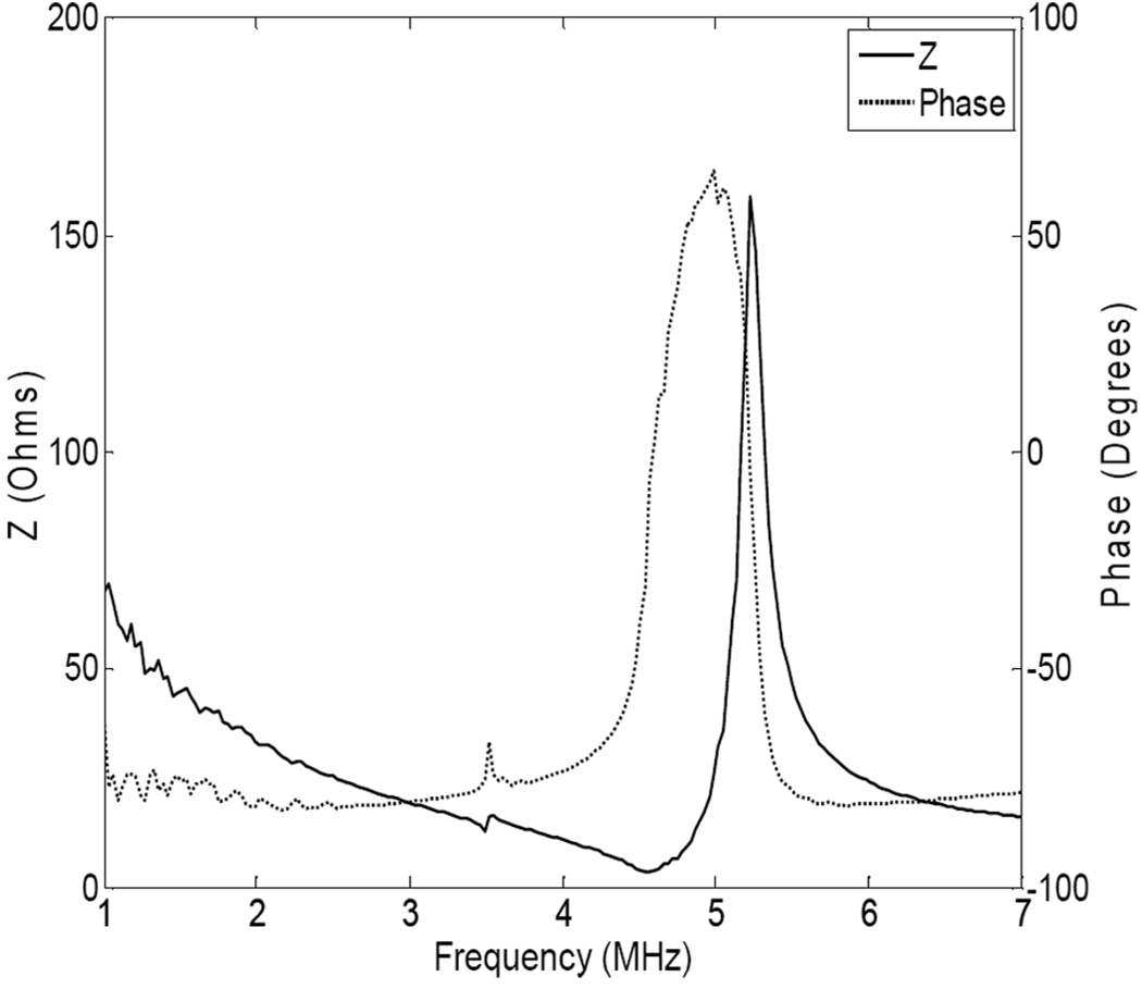
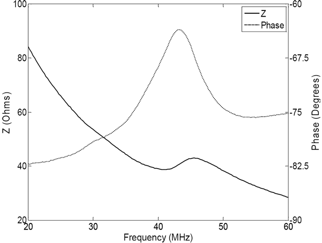
Fig. 4 shows the measured electrical impedance magnitude (solid line) and phase (dotted line) for a 400-µm-thick PZN-PT single crystal as a function of frequency in air. The first resonant peaks were observed near a frequency of approximately 5.0 MHz. The electromechanical coupling coefficient kt in the single crystal can be determined [12]. According to data from Fig. 4, kt is determined to be 0.53 ± 0.03. Fig. 5 shows the electrical impedance magnitude and phase of the 43 MHz needle transducer after housing and re-poling. At the resonant frequency, the electrical impedance was 42 Ω, which is very close to the 50 Ω required by electrical impedance matching of the system. The negative phase (−63°) shows the capacitive nature of the device. From the values of the resonant peaks, the effective electromechanical coupling coefficient keff for the transducer after re-poling was calculated to be about 0.52, which is close to previous data.
Fig. 4.
Electrical impedance magnitude and phase for a PZN-PT single crystal plate.
Fig. 5.
Electrical impedance magnitude and phase for a 43-MHz angled needle transducer.
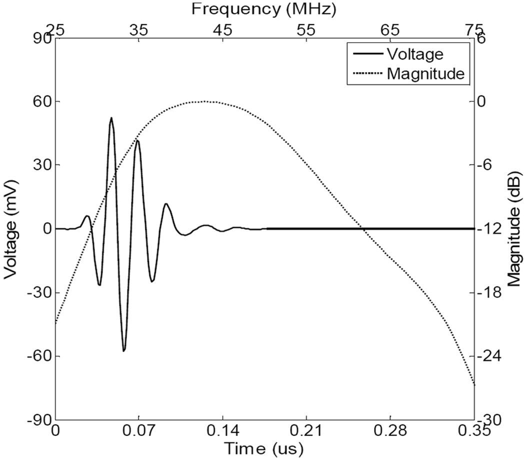
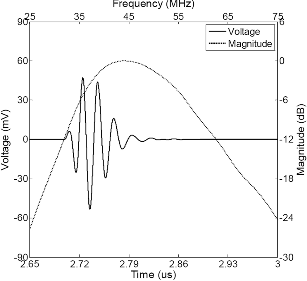
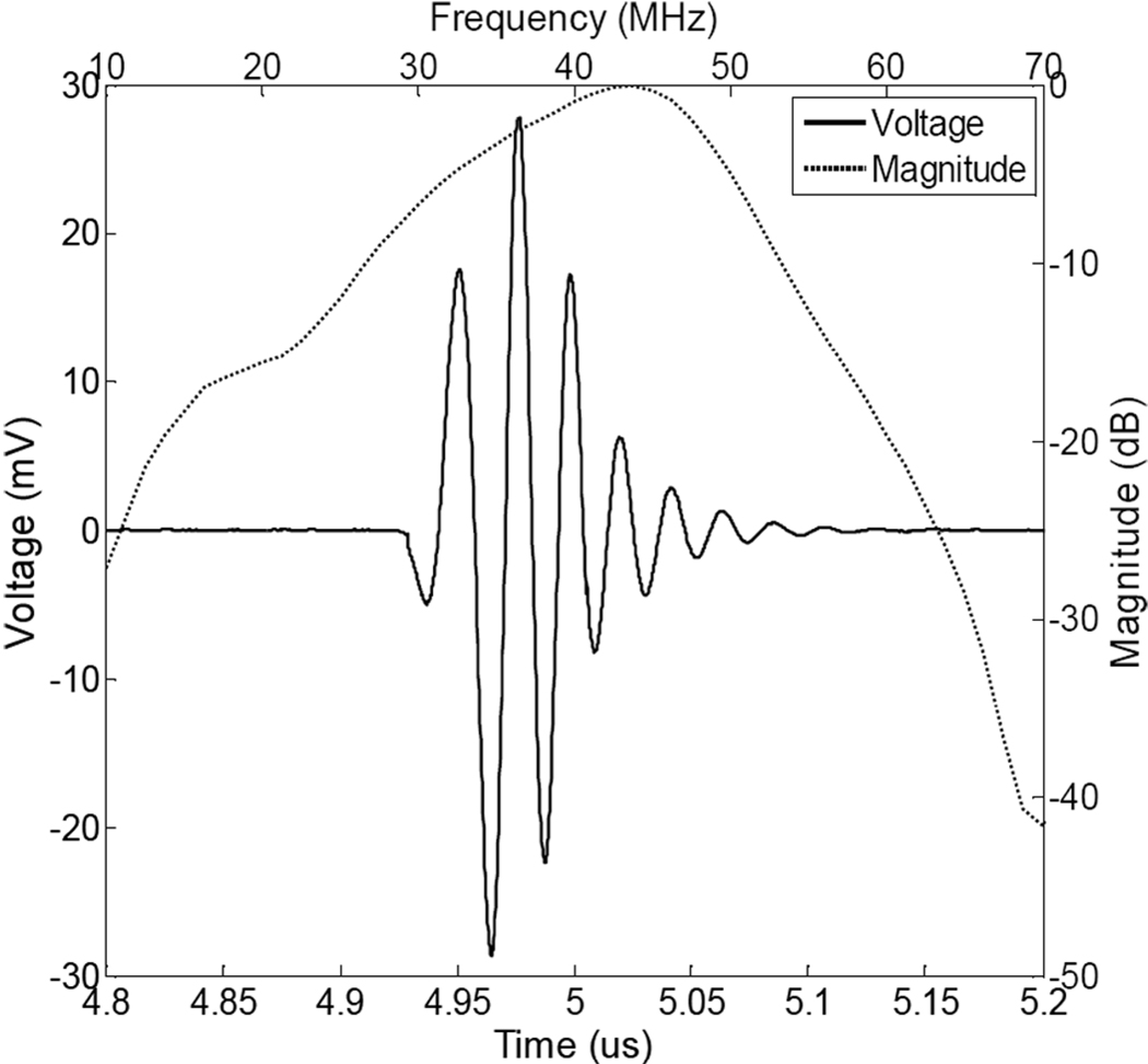
Fig. 6 and Fig 7 show a pulse-echo waveform and frequency spectrum of the PZN-PT needle transducer modeled using PZFlex and PiezoCAD, respectively. There is a good agreement between the two models. The center frequency of the transducer was around 43 MHz and the −6 dB fractional bandwidth was approximately 51%. Fig. 8 shows a measured pulse-echo waveform and frequency spectrum for the PZN-PT needle transducer. The center frequency of the transducer was around 43 MHz and the −6 dB fractional bandwidth was approximately 45%. The measured bandwidth is a little lower than that predicted by both models. This discrepancy was most likely caused by either thickness variability in the matching layers, due to the lapping process, or a degradation of the piezoelectric properties of the crystal during processing. The two-way insertion loss of the transducer was approximately 17 dB. These results show that the PZN-PT needle transducer was highly sensitive, due to excellent piezoelectric properties of this material. It is one of best alternative materials to a PMN-PT single crystal, especially when a higher Curie temperature is desired.
Fig. 6.
PiezoCAD-modeled pulse-echo waveform and frequency spectrum for a high-frequency PZN-PT.
Fig. 7.
PZFlex-modeled pulse-echo waveform and frequency spectrum for a high-frequency PZN-PT needle transducer.
Fig. 8.
Measured pulse-echo waveform and frequency spectrum for the 43-MHz PMN-PT angled needle transducer.
The high-frequency transducer made from single crystal PZN-7%PT utilizes the thickness mode and operates at 43 MHz for high-resolution ultrasonic Doppler flow analysis. The active material inside the transducer was placed at a 45° angle to the shaft of the probe. This feature provides the capability of the transducer during imaging of target areas that are very difficult to access. The bandwidth of the transducer is fairly wide, considering the thickness coupling coefficient of over 50% for state-of-the-art piezoelectric material, including PZT and single crystal PZN-PT and PMN-PT. It also agrees well with the result of FEM simulation and PiezoCAD modeling. The inferior performance of the transducer before re-poling is due to partial de-poling of the piezoelectric material during the lapping process. It was noticed that the transducer can be re-poled in situ after fabrication, resulting in a significant increase in the sensitivity of the poled transducer. However, the reason for the de-poling of the piezoelectric material may not be temperature-induced, but stress-induced. A similar result was observed during the fabrication of PMN-PT single crystal transducers [5]. Since the piezoelectric materials used are so thin, even a small amount of stress can cause a phase transformation which inherently degrades the dielectric and piezoelectric properties of the material. Future work will focus on developing 1–3 composite high-frequency PZN-PT needle transducers by MEMS technology and domain engineering to enhance piezoelectric properties.
IV. WIRE PHANTOM IMAGING
The wire phantom images were acquired by using both the flat and the angled needle transducers to assess and compare their spatial resolutions. In this study, the transducers were driven by a motor controller card (DMC-1802; Galil Motion Control Inc., Mountain View, CA), which also generated a signal to trigger data acquisition. The Panametrics 5900PR pulser/receiver was used to excite the transducer. The echoes were then digitized by an 8-bit digital oscilloscope (Tektronix, Inc.) at a sampling frequency of 250 MHz.
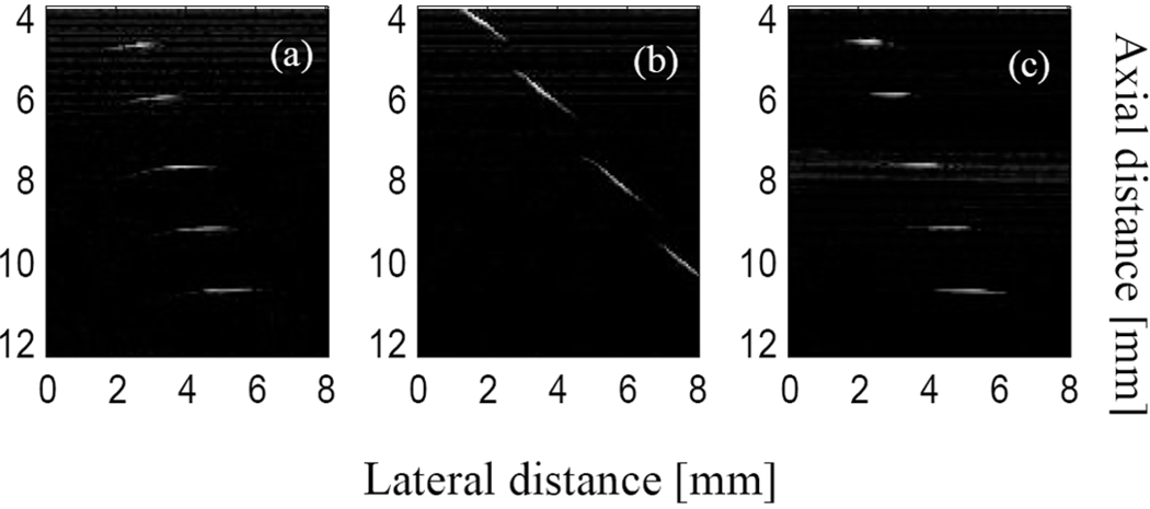
A 20-µm-diameter tungsten wire phantom was used for this measurement. The five phantom wires were arranged diagonally with equal distance in the axial (1.55 mm) and lateral (0.65 mm) directions. The pulse-echo image was acquired as the transducer scanned across the wire phantom placed in degassed water. Fig. 9 shows the images generated using the needle transducers. Fig. 9(a) is from a flat needle transducer. Fig. 9(b) is the image using an angled transducer when the angled transducer has the same mounting position as in Fig. 9(a). Fig. 9(c) is the image obtained by rotating the motor shaft by 45° so that the surface of the transducer is in parallel with the scanning direction. The images are displayed with the dynamic range of 50 dB. As shown in Fig. 9(b), when the angled transducer was scanned across the wires using a linear servo motor, which results in a 45° angle between the beaming pattern generated from the angled transducer and the wire surface, the steered cross-sectional image of the wires is observed. In this case, degraded lateral resolution is observed due to this angle. If the motor shaft is rotated roughly by 45°, the wire image [Fig. 9(c)] has the same orientation as shown in Fig. 9(a). Spatial resolutions can therefore be measured from the wire phantom images. At 4.6 mm, the −6 dB axial resolution and lateral resolution for the angled needle probe were 50 and 300 µm, respectively. Further development of focused high-frequency angled needle transducer will improve lateral resolution for medical Doppler application.
Fig. 9.
Wire phantom images using PZN-PT needle transducers: (a) flat transducer; (b) angled transducer with the same position as (a); (c) angled transducer with a 45° rotation of the motor shaft. All images are displayed with a 50-dB dynamic range.
V. CONCLUSIONS
A high-frequency (over 40 MHz) angled needle ultrasonic transducers with an aperture size of approximately 0.4 mm was successfully fabricated using a PZN-7%PT single crystal. The measured center frequency and bandwidth of this needle transducer were 43 MHz and 45%, respectively. The results were very close to PZFlex and Piezo-CAD simulations. The measured two-way insertion loss was around 17 dB. Phantom imaging of a high-frequency transducer with five wires in the axial direction was obtained. The −6 dB axial resolution and lateral resolution of the 43 MHz angled needle transducer were 50 and 300 µm, respectively. These results demonstrate that this type of PZN-PT single-crystal high-frequency transducer is suitable for Doppler flow measurements in the human eye.
Acknowledgments
This work was supported by NIH grant # P41-EB2182.
Contributor Information
Qifa Zhou, Email: qifazhou@usc.edu, NIH Resource on Medical Ultrasonic Transducer Technology, Department of Biomedical Engineering, University of Southern California, Los Angeles, CA 90089.
Dawei Wu, NIH Resource on Medical Ultrasonic Transducer Technology, Department of Biomedical Engineering, University of Southern California, Los Angeles, CA 90089.
Jing Jin, Microfine Materials Technologies P/L, Singapore.
Chang-hong Hu, NIH Resource on Medical Ultrasonic Transducer Technology, Department of Biomedical Engineering, University of Southern California, Los Angeles, CA 90089.
Xiaochen Xu, NIH Resource on Medical Ultrasonic Transducer Technology, Department of Biomedical Engineering, University of Southern California, Los Angeles, CA 90089.
Jay Williams, NIH Resource on Medical Ultrasonic Transducer Technology, Department of Biomedical Engineering, University of Southern California, Los Angeles, CA 90089.
Jonathan M. Cannata, NIH Resource on Medical Ultrasonic Transducer Technology, Department of Biomedical Engineering, University of Southern California, Los Angeles, CA 90089.
Leongchew Lim, Microfine Materials Technologies P/L, Singapore.
K. Kirk Shung, NIH Resource on Medical Ultrasonic Transducer Technology, Department of Biomedical Engineering, University of Southern California, Los Angeles, CA 90089.
REFERENCES
- 1.Christopher DA, Burns PN, Starkoski BG, Foster FS. A high-frequency pulsed-wave Doppler ultrasound system for the detection and imaging of blood flow in the microcirculation. Ultrasound Med. Biol. 1997;vol. 23(no 7):997–1015. doi: 10.1016/s0301-5629(97)00076-8. [DOI] [PubMed] [Google Scholar]
- 2.Park S-E, Shrout TR. Characteristics of relaxor-based piezoelectric single crystals for ultrasonic transducers. IEEE Trans. Ultrason., Ferroelect., Freq. Contr. 1997;vol. 44(no 5):1140–1147. [Google Scholar]
- 3.Rajan KK, Shanthi M, Chang WS, Jin J, Lim LC. Dielectric and piezoelectric properties of [0 0 1] and [0 1 1]-poled relaxor ferroelectric PZN-PT and PMN-PT single crystals. Sens. Actuators A. 2007;vol. 133(no 1):110–116. [Google Scholar]
- 4.Ritter TA, Geng X, Shung KK, Lopath PD, Park S-E, Shrout TR. Single crystal PZN-PT polymer composite for ultrasound transducer applications. IEEE Trans. Ultrason., Ferroelect., Freq. Contr. 2000;vol. 47:792–800. doi: 10.1109/58.852060. [DOI] [PubMed] [Google Scholar]
- 5.Zhou Q, Xu X, Gottlieb EJ, Sun L, Cannata JM, Ameri H, Humayun MS, Han P, Shung KK. PMN-PT single crystal, high-frequency ultrasonic needle transducers for pulsed-wave Doppler application. IEEE Trans. Ultrason., Ferroelect., Freq. Contr. 2007;vol. 54(no 3):668–675. doi: 10.1109/tuffc.2007.290. [DOI] [PubMed] [Google Scholar]
- 6.Lim LC, Rajan KK. High-homogeneity high-performance flux-grown Pb(Zn1/3Nb2/3)O3-(6–7)%PbTiO3 single crystals. J. Cryst. Growth. 2004;vol. 271(no 3–4):435–444. [Google Scholar]
- 7. www.microfine-piezo.com.
- 8.Selfridge AR. Ph.D. dissertation. Stanford, CA: Department of Electrical Engineering, Stanford University; 1983. Jul, The design and fabrication of ultrasonic transducers and transducer arrays. [Google Scholar]
- 9.Yin J, Jiang B, Cao W. Elastic, piezoelectric, and dielectric properties of 0.955Pb(Zn1/3Nb2/3)O3–0.45PbTiO3 single crystal with designed multidomains. IEEE Trans. Ultrason., Ferroelect., Freq. Contr. 2000;vol. 47(no 1):285–291. doi: 10.1109/58.818772. [DOI] [PubMed] [Google Scholar]
- 10.Cannata JM, Ritter TA, Chen WH, Silverman RH, Shung KK. Design of efficient, broadband single-element (20–80 MHz) ultrasound transducers for medical imaging applications. IEEE Trans. Ultrason., Ferroelect., Freq. Contr. 2003;vol. 50(no 11):1548–1557. doi: 10.1109/tuffc.2003.1251138. [DOI] [PubMed] [Google Scholar]
- 11.Lockwood GR, Turnbull DH, Foster FS. Fabrication of high frequency spherically shaped ceramic transducers. IEEE Trans. Ultrason., Ferroelect., Freq. Contr. 1994;vol. 41(no 2):231–235. [Google Scholar]
- 12.IEEE Standard on Piezoelectricity. ANSI/IEEE Std. 176–1987; New York: IEEE; 1987. [Google Scholar]


