Abstract
Objective
The purpose of this study was to compare the effectiveness of MR urography (MRU) with that of ultrasonography (US) in the evaluation of urinary tract when this failed to opacify during excretory urography (EXU).
Materials and Methods
Twelve urinary tracts in 11 patients were studied. In each case, during EXU, the urinary system failed to opacify within one hour of the injection of contrast media, and US revealed dilatation of the pelvocalyceal system. Patients underwent MRU, using a HASTE sequence with the breath-hold technique; multi-slice acquisition was then performed, and the images were reconstructed using maximal intensity projection. Each set of images was evaluated by three radiologists to determine the presence, level, and cause of urinary tract obstruction.
Results
Obstruction was present in all twelve cases, and in all of these, MRU accurately demonstrated its level. In this respect, however, US was successful in only ten. The cause of obstruction was determined by MRU in eight cases, but by US in only six. In all of these six, MRU also successfully demonstrated the cause.
Conclusion
MRU is an effective modality for evaluation of the urinary tract when this fails to opacify during EXU, and appears to be superior to US in demonstrating the level and cause of obstruction.
Keywords: Kidney, obstruction; Kidney, MR; Kidney, US; Urography; Magnetic resonance (MR), comparative studies
Excretory urography (EXU) is inexpensive, easy to perform, offers fair resolution, and reflects the functional status of the kidney, and has therefore been used as the primary imaging modality for the evaluation of urinary tract abnormalities. When the obstruction is severe, however, EXU sometimes fails to adequately visualize the urinary tract (1).
When opacification of the tract fails to occur during EXU, determination of the presence or absence, level, and cause of obstrution requires the use of a further imaging modality. Since it is non-invasive, easy to perform, and does not require contrast media injection and radiation, ultrasonography is usually chosen (2). It is, however, operator dependent and lacks objectivity, and because bowel gas interferes with sonic transmission, often fails to trace dilated ureters (1).
Magnetic resonance urography (MRU) offers good image quality, comparable to that achieved by EXU, and a diagnostic capability that is improving as imaging sequences become faster (3-8). Little has been reported, however, about the value of MRU and US when EXU fails to opacify the urinary tract due to obstruction (4, 6).
The aim of this study was to compare the effectiveness of MRU with that of US in the evaluation of urinary tract which failed to opacify during EXU.
MATERIALS AND METHODS
Subjects
Between May 1997 and May 1998, we prospectively studied twelve urinary tracts (right 5, left 5, bilateral 1) in 11 patients [five men and six women aged 31-77 (mean, 50) years]. In each patient, EXU failed to produce a nephrogram within one hour of contrast media injection. Though the level of obstruction was not identified by EXU, patients in whom this technique revealed an obvious cause of obstruction, such as a radiopaque ureteral stone were excluded from the study group.
Imaging techniques and study protocols
In each patient, EXU was performed after overnight fasting, with intravenous injection of 50 ml iohexol (Omnipaque, 300 mg/ml; Nycomed, New York, U.S.A.) and 15 minutes' abdominal compression after the injection of contrast media. In all the patients, the use of EXU failed to produce a nephrogram within one hour of contrast media injection, and US and MRU were therefore performed. All US studies were undertaken by one radiologist, unaware of the findings of EXU, and in all patients, US was performed prior to MRU.
MRU involved the use of a 1.0-T Magnetom Expert System (Siemens, Erlangen, Germany) with a body phased-array coil, and a HASTE (half-Fourier acquisition single-shot turbo spin-echo) sequence with an effective TE of 87 msec, one excitation, a TR of 10.9 msec, and a 240×256 matrix. The field of view was 25-35 cm. To suppress the signal from peritoneal fat, fat saturation was applied. Patients were instructed to hold their breath during the examination, and for this, a multi-slice acquisition technique with a slice thickness of 5 mm and seven to nine sequential sections was employed. The acquisition time was 14-20 seconds. For post-acquisition image processing after multislice scanning, maximum intensity projection (MIP) was used. No medication was administrated, and for MRU, external compression was not applied. Delayed EXU images were acquired until 24 hours after MRU, confirming that further opacification of the urinary tract had not occurred. The termination of delayed EXU was independently decided by a radiologist not involved in the study. The time needed to complete MRU was less than 10 minutes, and the total study time including EXU and US was 2 to 24 hours.
Image analysis
EXU and MRU were separately evaluated by three radiologists, working independently and unaware of the results of other imaging studies, in terms of their ability to reveal the presence, level, and cause of obstruction. US was evaluated by another radiologist. The findings of US were compared with those of MRU in terms of detection, level, and cause of obstruction; level of obstruction was classified as ureteropelvic junction, or proximal, middle or distal ureter. Our diagnosis of the cause of obstruction was based on findings relating to either the intrinsic or extrinsic mass, or specific morphologic findings of the pelvocalyceal system, such as congenital UPJ obstruction or tuberculosis. The diagnoses were confirmed by surgery in three patients, CT in four, cystoscopy in two, and retrograde pyelography in one. In one patient the cause was not confirmed due to lack of follow up.
RESULTS
EXU
In eight of the 12 cases (67%), EXU failed to provide a nephrogram, even on delayed images, so the presence of obstruction was uncertain. In the remaining four, portions of dilated urinary tracts were seen as faintly opacified on delayed images, suggesting the presence of obstructive hydronephrosis, but the level and cause of obstruction could not be determined.
US
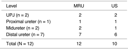
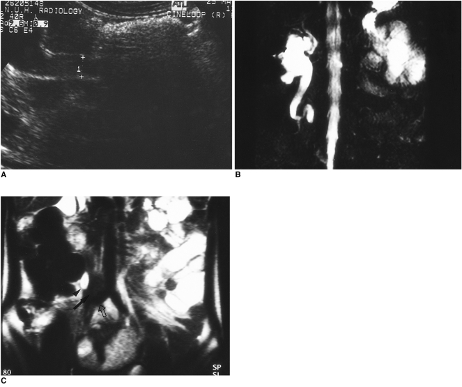
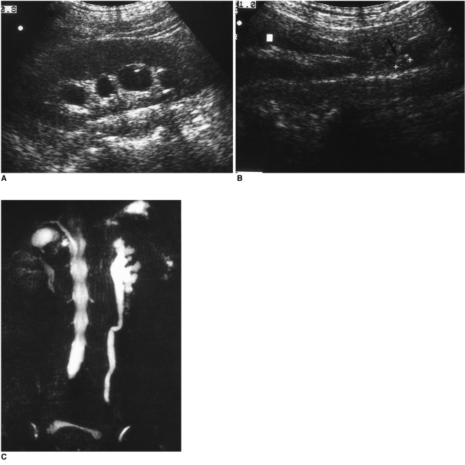
By demonstrating hydronephrosis and/or hydroureter, US revealed the presence of obstruction in all twelve cases, in ten of which (83.3%), the level of this could be determined (Table 1). Due to overlying bowel gas (Fig. 1) or misinterpretation of internal echo as stone (Fig. 2), US failed to demonstrate the level of obstruction in two cases.
Table 1.
Demonstration of the Level of Obstruction
Fig. 1.
Metastatic lymphadenopathy arising from uterine cervical carcinoma and causing hydronephrosis and hydroureter.
A. Due to bowel gas, the level of obstruction revealed by longitudinal US is not clear.
B. An MIP image acquired by HASTE MRU multi-slice scanning shows kinking and obstruction at the mid portion of the right ureter. Extensive high signal intensity in the left hemiabdomen is caused by dilated bowel.
C. A coronal source image obtained by multi-slice scanning shows an obstructive mass (arrow) located between the ureter (arrowhead) and iliac artery (open arrow), suggesting metastatic lymphadenopathy.
Fig. 2.
Obstruction of the distal ureter caused by transitional cell carcinoma.
A. US demonstrates dilated renal calyces.
B. Ureter is dilated at its mid portion, with echogenic lesion (arrow), falsely interpreted as midureter obstruction by ureter stone.
C. HASTE MRU does not reveal any filling defect in the left ureter. A dilated ureter at a more distal level is also demonstrated, but MRU failed to indicate a definite cause of obstruction.
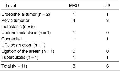
The cause of obstruction was demonstrated by US in six of eleven patients (54.5%) (Table 2). In one with bilateral obstruction, metastatic lymph nodes were seen on both sides.
Table 2.
Demonstration of the Cause of Obstruction
MRU
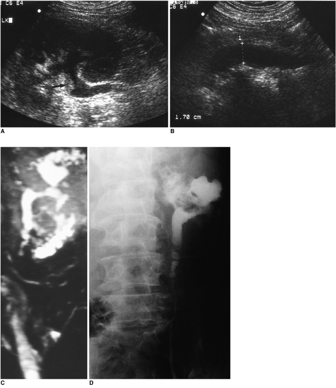
In all twelve cases (100%), MRU was able to detect urinary tract obstruction, and in all cases, the level of obstruction was also clearly demonstrated (Table 1), correlating closely with the findings of surgical exploration or other imaging studies (Fig. 3). The cause of obstruction was successfully demonstrated by MRU in eight of eleven patients (72.7%) (Table 2).
Fig. 3.
Midureter obstruction caused by transitional cell carcinoma.
A. US demonstrates an echogenic polypoid lesion involving the renal calyx and pelvis (arrow), with hydronephrosis.
B. Tapered ureter with luminal irregularity at its mid portion is also demonstrated.
C. An eccentric filling defect involving the pelvis and upper calyx of the left kidney, as well as tortousity of the ureter, are revealed by HASTE MRU.
D. MRU correlates closely with retrograde pyelogram.
MRU versus US
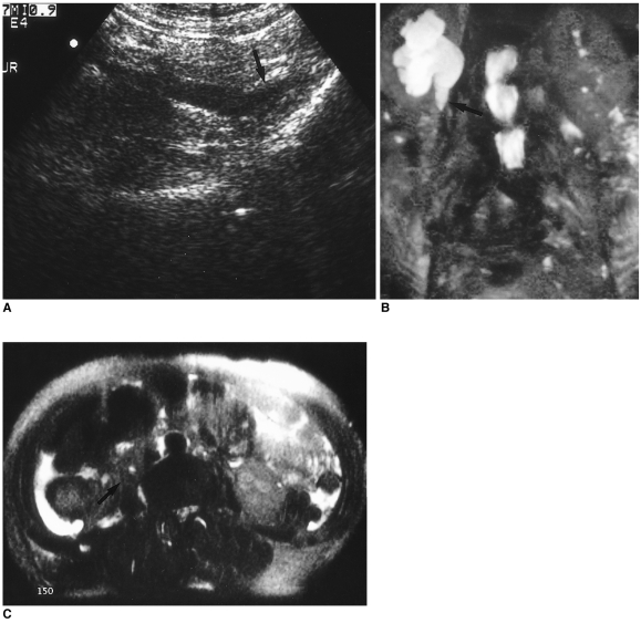
Among the ten cases in which the level of obstruction was revealed by US, MRU demonstrated the cause in seven, and US in six. In one case of ureteric metastasis, the cause of obstruction was revealed by MRU but not by US (Fig. 4). A case of metastatic lymphadenopathy, one of two in which the level of obstruction could not be determined by US, was demonstrated by MRU (Fig. 1). In three cases, MRU failed to reveal the definite cause of obstruction (Fig. 2).
Fig. 4.
Ureteric metastasis from uterine cervical carcinoma.
A. US demonstrates beaking appearance of the proximal ureter (arrow), with obstruction but without demonstrable intrinsic or extrinsic mass.
B. HASTE MRU also shows tapered ureter with beaking appearance (arrow).
C. Additional HASTE MR axial scan clearly demonstrates diffuse and evenly thickened right ureteric wall (arrow), suggesting ureteric metastasis.
DISCUSSION
HASTE (half-Fourier acquisition single-shot turbo spin-echo) imaging is an ultrafast heavily T2-weighted sequence with single breath hold. After a single excitation, using multiple 180°Δpulses, 128 echoes are generated, and all refocused echoes are sequentially phase-encoded by the application of a small phase-encoding gradient. HASTE imaging acquires only half of the lines in k spaces during a single echo train length, the other half being acquired by means of a half-Fourier reconstruction. Imaging time is thus markedly reduced, usually to less than two seconds per slice (3, 9, 10).
Several reports have described the application of MRU to the evaluation of hydronephrosis (3-5) or vesicoureteral reflux in children (11). Sigmund et al. (11) reported in his paper on MRU that using the RARE (rapid acquisition with relaxation enhancement) sequence, pelviureteric obstruction was demonstrated in all 19 of their patients. Differentiation of obstruction by means of vesicoureteral reflux was difficult, however. Regan et al. (3) applied HASTE MRU to ureteric obstruction and the condition was diagnosed correctly in all 41 urinary systems they studied. The presence of perirenal fluid was demonstrated in 20 of 23 acutely obstructed kidneys, and this sometimes obscured the level of obstruction.
Tang et al. compared MRU with EXU in patients with urinary obstruction (4), finding that in all their patients, HASTE MRU and EXU revealed the the level of obstruction equally well, but that the resolution of MRU was inferior to that of EXU. In six patients with severe hydronephrosis, EXU failed to adequately reveal the urinary tract, but HASTE MRU clearly demonstrated the dilated ureter.
The purpose of this study was practical. It was performed in order to determine the extent to which MRU is able to detect not only the presence, but also the level and cause of obstruction in cases in which opacification of the kidney and urinary system during EXU is insufficient. In all cases, MRU indicated the presence of obstruction, and the level of this was also demonstrated correctly. US, on the other hand, revealed the level of obstruction in ten of 12 urinary tracts; in the detection of hydronephrosis it has been reported to be sensitive but not specific (12-14). Most false-positive diagnoses are due to minimal dilatation in the absence of obstruction (13); most of the cases in this study, however, showed moderate to severe hydronephrosis, and it was thus relatively easy to trace the dilated ureter using US. In addition, only two of 12 ureters were obstructed at their mid portion, which due to bowel gas, is known to be an especially difficult area to trace with US. In our study protocol, moreover, US was performed after EXU, on the same day, and all patients had fasted overnight, as required for EXU. The bowel was therefore less distended, and this might have contributed to the better results of US.
Distinction between intrinsic and extrinsic obstruction was occasionally difficult, and in this respect, there were three cases in which MRU failed to reveal the etiology. This limitation of HASTE MRU was largely due to the fact that signal suppression from surrounding tissue provides strong contrast, a characteristic of HASTE imaging (3).
Because of a better signal-to-noise ratio, the images obtained using the multi-slice acquisition technique are usually better than those acquired using the single-slice technique (4), and for this reason we used the former procedure. MIP images, however, are projectional images that selectively display high signal intensity pixels, and the bright signal intensity from urine may thus entirely surround a small intraluminal lesion and obscure it. Periureteral lesions, which are of relatively lower signal intensity, were not clearly demonstrated by projection imaging, but source images, obtained by multi-slice scanning, helped to detect their presence (Fig. 2C).
One disadvantage of HASTE MRU is its inability to provide functional information about the kidney; however, a number of reports describing the findings of gadolinium-enhanced excretory MRU have been published (6, 15). With diuretic-enhanced excretory MRU, asymmetric excretion of the contrast agent suggested unilateral renal dysfunction. Its ability to assess the ureter and bladder was significantly superior to that of EXU, but the dilated urinary tract of a nonexcreting or very poorly functioning kidney could be accurately demonstrated and assessed only on HASTE images (6).
Several limitations of this study should be documented. First, patients with non-opacified kidney due to reasons other than obstruction, such as unilateral renal functional impairment due to longstanding vesicoureteral reflux or hypoplastic unilateral kidney were not included. Second, this was not a true double-blind study, since US preceded MRU and only obstruction revealed by the former was taken into consideration. In each image, interpretation of the findings of MRU involved an awareness of the presence of obstruction. Third, the number of selected patients is relatively small, so the statistical significance of differences between the findings of MRU and those of US is unknown. Further study involving a larger series is needed.
In conclusion, MRU is thought to be an effective modality for evaluation of the urinary tract when this fails to opacify during EXU, and in detecting the level and cause of obstruction, appears to be superior to US.
References
- 1.Cronan JJ. Contemporary concepts in imaging urinary tract obstruction. Radiol Clin North Am. 1991;29:527–542. [PubMed] [Google Scholar]
- 2.Webb JAW. Ultrasonography in the diagnosis of renal obstruction. Sensitive but not very specific. Br Med J. 1990;301:944–946. doi: 10.1136/bmj.301.6758.944. [DOI] [PMC free article] [PubMed] [Google Scholar]
- 3.Regan F, Bohlman ME, Khazan R, Rodriguez R, Schultze-Haakh H. MR urography using HASTE imaging in the assessment of ureteric obstruction. AJR. 1996;1678:1115–1120. doi: 10.2214/ajr.167.5.8911161. [DOI] [PubMed] [Google Scholar]
- 4.Tang Y, Yamashita Y, Namimoto T, et al. The value of MR urography that uses HASTE sequences to reveal urinary tract disorders. AJR. 1996;167:1497–1502. doi: 10.2214/ajr.167.6.8956584. [DOI] [PubMed] [Google Scholar]
- 5.O'Malley ME, Soto JA, Yucel EK, Hussain S. MR urography: evaluation of three-dimenstional fast spin-echo technique in patients with hydronephrosis. AJR. 1997;168:387–392. doi: 10.2214/ajr.168.2.9016213. [DOI] [PubMed] [Google Scholar]
- 6.Nolte-Ernsting CC, Bucker A, Adam GB, et al. Gadolinium-enhanced excretory MR urography after low-dose diuretic injection: comparison with conventional excretory urography. Radiology. 1998;209:147–157. doi: 10.1148/radiology.209.1.9769826. [DOI] [PubMed] [Google Scholar]
- 7.Rothpearl A, Frager D, Subramanian A, et al. MR urography: technique and application. Radiology. 1995;194:125–130. doi: 10.1148/radiology.194.1.7997538. [DOI] [PubMed] [Google Scholar]
- 8.Hattery RR, King BF. Technique and application of MR urography. Radiology. 1995;194:25–27. doi: 10.1148/radiology.194.1.7997563. [DOI] [PubMed] [Google Scholar]
- 9.Roy C, Saussine C, Guth S, et al. MR urography in the evaluation of urinary tract obstruction. Abdom Imaging. 1998;23:27–34. doi: 10.1007/s002619900279. [DOI] [PubMed] [Google Scholar]
- 10.Kiefer B, Grassner J, Hausman R. Image acquisition in a second with half-Fourier acquisition single shot turbo spin-echo. J Magn Reson Imaging. 1994;4(Suppl):86–87. [Google Scholar]
- 11.Sigmund G, Stoever B, Zimmerhackl LB, Frankenschmidt A, Nitzsche E, Leititis JU. RARE-MR urography in the diagnosis of upper urinary tract abnormalities in children. Pediatr Radiol. 1991;21:416–420. doi: 10.1007/BF02026676. [DOI] [PubMed] [Google Scholar]
- 12.Kamholtz RG, Cronan JJ, Dorfman GS. Obstruction and the minimally dilated renal collecting system: US evaluation. Radiology. 1989;170:51–53. doi: 10.1148/radiology.170.1.2642347. [DOI] [PubMed] [Google Scholar]
- 13.Hill MC, Rich JI, Mardiat JG, Finder CA. Sonography vs. excretory urography in acute flank pain. AJR. 1985;144:1235–1238. doi: 10.2214/ajr.144.6.1235. [DOI] [PubMed] [Google Scholar]
- 14.Malave SR, Neiman HL, Spies SM, Cisternino SJ, Adamo G. Diagnosis of hydronephrosis: comparision of radionuclide scanning and sonography. AJR. 1980;135:1179–1185. doi: 10.2214/ajr.135.6.1179. [DOI] [PubMed] [Google Scholar]
- 15.Li W, Chavez D, Edelman RR, Prasad PV. Magnetic resonance urography by breath-hold contrast-enhanced three-dimenstional FISP. J Magn Reson Imaging. 1997;7:309–311. doi: 10.1002/jmri.1880070209. [DOI] [PubMed] [Google Scholar]


