Abstract
Rationale: Persons with obstructive sleep apnea may have significant residual hypersomnolence, despite therapy. Long-term hypoxia/reoxygenation events in adult mice, simulating oxygenation patterns of moderate–severe sleep apnea, result in lasting hypersomnolence, oxidative injury, and proinflammatory responses in wake-active brain regions. We hypothesized that long-term intermittent hypoxia activates brain NADPH oxidase and that this enzyme serves as a critical source of superoxide in the oxidation injury and in hypersomnolence.
Objectives: We sought to determine whether long-term hypoxia/reoxygenation events in mice result in NADPH oxidase activation and whether NADPH oxidase is essential for the proinflammatory response and hypersomnolence.
Methods: NADPH oxidase gene and protein responses were measured in wake-active brain regions in wild-type mice exposed to long-term hypoxia/reoxygenation. Sleep and oxidative and proinflammatory responses were measured in adult mice either devoid of NADPH oxidase activity (gp91phox-null mice) or in which NADPH oxidase activity was systemically inhibited with apocynin osmotic pumps throughout hypoxia/reoxygenation.
Main Results: Long-term intermittent hypoxia increased NADPH oxidase gene and protein responses in wake-active brain regions. Both transgenic absence and pharmacologic inhibition of NADPH oxidase activity throughout long-term hypoxia/reoxygenation conferred resistance to not only long-term hypoxia/reoxygenation hypersomnolence but also to carbonylation, lipid peroxidation injury, and the proinflammatory response, including inducible nitric oxide synthase activity in wake-active brain regions.
Conclusions: Collectively, these findings strongly support a critical role for NADPH oxidase in the lasting hypersomnolence and oxidative and proinflammatory responses after hypoxia/reoxygenation patterns simulating severe obstructive sleep apnea oxygenation, highlighting the potential of inhibiting NADPH oxidase to prevent oxidation-mediated morbidities in obstructive sleep apnea.
Keywords: intermittent hypoxia, non-REM sleep, oxidation, peroxynitrite
Obstructive sleep apnea (OSA) with daytime hypersomnolence is present in at least 2 to 4% of adults in developed countries (1). This disorder manifests as repeated events of sleep state–dependent reductions in upper airway dilator motoneuronal activity, with consequent upper airway occlusions and oxyhemoglobin desaturations, each terminating with abrupt arousal and reoxygenation (2). The hypoxia and reoxygenation events may occur as frequently as once every minute of sleep. Despite therapy to alleviate OSA events, many individuals with OSA have residual sleepiness (3, 4). Mechanisms of the residual hypersomnolence in persons with OSA are not understood, but severity of hypoxemia in OSA predicts, in part, the severity of hypersomnolence (5, 6).
Long-term intermittent hypoxia (LTIH) in mice, modeling the patterns of hypoxia/reoxygenation observed in moderate to severe sleep apnea, results in protracted hypersomnolence (7, 8) and hippocampus-dependent memory impairments (9–11), with significant oxidative modifications in many brain regions, including wake-active regions (7, 8, 12) and the hippocampus (10, 11). The oxidative modifications observed after hypoxia/reoxygenation in wake-active neural groups that might contribute to impaired wakefulness and hypersomnolence include nitration, lipid peroxidation, and carbonylation (7, 8, 12). Inducible nitric oxide synthase (iNOS) contributes to nitration and lipid peroxidation injuries in the intermittent hypoxia model of sleep apnea (8, 11); however, transgenic absence of iNOS function does not confer resistance to intermittent hypoxia carbonylation injury and bestows only partial resistance on the proinflammatory gene response (8). A source of oxidation injury from long-term hypoxia/reoxygenation should be identified.
NADPH oxidase-dependent production of superoxide radical (O2−·) has been identified as a major contributor to oxidative injury in the brain under conditions of both inflammation and severe hypoxia/reperfusion injury (13–16). Moreover, NADPH oxidase has been implicated in oxidative neurodegeneration, including Alzheimer's disease (17, 18), and in dopaminergic neuronal injury in murine models of Parkinson's disease (19–21). Several recent reports have identified NADPH oxidase in select populations of neurons (22–24), raising the possibility that neuronal NADPH oxidase activation could contribute to enhanced neuronal vulnerability to oxidative injury. Presently, it is unknown whether NADPH oxidase is present in wake-active neurons, whether intermittent hypoxia that models sleep apnea increases NADPH oxidase in regions with wake-active neurons, or whether NADPH oxidase might mediate the intermittent hypoxia-induced hypersomnolence, oxidative injury, and/or proinflammatory responses.
In this series of studies, we first determined whether hypoxia/reoxygenation events in adult mice that model severe sleep apnea oxygenation increase NADPH oxidase gene and protein expression in wake-active brain regions, including the noradrenergic locus coeruleus, the lateral hypothalamic orexinergic group, the cholinergic lateral basal forebrain, and the posterior hypothalamic histaminergic group, and whether the changes in protein expression occur in wake-active neurons or surrounding cells. We then determined whether absence of NADPH oxidase activity (gp91phox−/− mice) or systemic NADPH inhibition throughout intermittent hypoxia exposure (via an apocynin osmotic pump) would prevent the constellation of hypersomnolence, carbonylation, lipid peroxidation, and proinflammatory gene expression in wake-active brain regions. We report for the first time that frequent hypoxia/reoxygenation events, which model oxygenation patterns in sleep apnea, induce NADPH oxidase and proinflammatory gene expression in select brain regions, including in wake-active neurons. Moreover, we have determined that lack of a functional NADPH oxidase and pharmacologic inhibition of NADPH oxidase confer resistance to intermittent hypoxia-induced neurobehavioral, redox, and proinflammatory changes, thereby highlighting a potential target to prevent oxidative morbidities in persons with OSA.
METHODS
Animals
Ten-week-old male C57BL/6J (B6) mice, gp91phox−/− mice (B6.129S6-Cybbtm1din) backcrossed 12 generations to C57BL/6J 000664, and gp91phox+/+ wild-type (WT) backcrossed substrain C57BL/6J 000664 mice (Jackson Laboratory, Bar Harbor, ME) were studied. Methods and study protocols were approved in full by the Institutional Animal Care and Use Committee of the University of Pennsylvania, conforming with the revised National Institutes of Health (NIH) Office of Laboratory Animal Welfare Policy. Food and water were provided ad libitum. Mice were confirmed pathogen free at the time of studies.
LTIH Protocol
A detailed description of the LTIH protocol was recently published (7, 8). An automated nitrogen/oxygen delivery profile system (Oxycycler model A84XOV; Biospherix, Redfield, NY) produced brief reductions in housing chamber ambient oxygen levels from 21 to 10% for 5 seconds every 90 seconds, resulting in arterial oxyhemoglobin saturation fluctuations between 95 to 98% and 83 to 86%. Sham LTIH, with ambient FiO2 fluctuations from 21 to 19% every 90 seconds, held arterial oxyhemoglobin values constant between 96 and 98%. Both conditions were produced for 10 hours of the lights-on period for a total of 8 weeks. Humidity, ambient CO2, and environmental temperature were held constant.
Measurement of Regional p67phox, Microglial, and Proinflammatory Gene Responses to LTIH
To determine if LTIH results in lasting increases in NADPH oxidase and microglial gene responses, real-time Taqman polymerase chain reaction (PCR) was performed on laser-captured microdissections or macropunches of selected brain regions in mice exposed to sham LTIH or LTIH, 2 weeks into recovery to parallel sleep studies, using our methods, as previously published (12, 25). To measure real-time transcriptase polymerase chain reaction, mice were perfused with phosphate-buffered saline (PBS) 2 weeks following completion of sham LTIH or LTIH. Brains were immediately frozen and sectioned (10 μm) for laser-capture microdissections of the following wake-active or hypoxia-sensitive regions: frontal cortex (layer V), nucleus basalis Meynert/substantia inominata/horizontal diagonal band, hippocampus CA1, striatum, lateral hypothalamus, dorsal raphe nucleus, and locus coeruleus, or the following sleep-active adjacent brain regions: median septal diagonal band, medial preoptic area, and ventrolateral preoptic area, collecting 100 captures (40-μm diameter) per region in each mouse (n = 6 mice/condition). RNA was purified and cDNA created for primer sets for primer/probe sets in Table 1, for Taqman real-time PCR (SDS-7900HT; Applied Biosystems, Inc., Foster City, CA). All primer probe sets showed excellent sensitivity and linearity (detection of ⩾ 100 copies/sample, r2 ⩾ 0.99).
TABLE 1.
Primer and probe gene identification and sequences
| Gene | GenBank | Sequences |
|---|---|---|
| p67phox | AB002664 | Sense = CGCTCTCGCCAGAACACA (1,244–1,261); antisense = TTGGTCACCCACCGTATGCT (1,392–1,373); probe = CTACCGGCGTCGGGACAGCC (1,272–1,291) |
| TNF-α | NM_013693 | Sense = CCAGGTTCTCTTCAAGGGACAA (573–594); antisense = CGGCAGAGAGGAGGTTGACTT (679–659); probe = CCTCACCCACACCGTCAGCCG (615–635) |
| iNOS | NM_010927 | Sense = CAGGAGATGGTCCGCAAGAG (3,254–3,273); antisense = AATTTCTGCAGCCATT TCCTTCT (1,509–1,530); probe = TGCACACAGGCTACTCCCGGC (3,288–3,308) |
| COX-2 | NM_011434 | Sense = TCCATTGACCAGAGCAGAGAGA (1,409–1,430); antisense = AATTTCTGCAGCCATT TCCTTCT (1,509–1,530); probe = TTCTCCCTGAAGCCGTACACATCA (1,409–1,430) |
Definition of abbreviations: COX-2 = cyclooxygenase 2; iNOS = inducible nitric oxide synthase; TNF-α = tumor necrosis factor α.
A second series of mice used for LTIH gene responses were gp91phox−/− and gp91phox+/+ mice exposed to either sham LTIH or LTIH for proinflammatory responses. To run real-time for four genes on the same sample, we used macrodissections (1-mm3 punches) of selected brain regions (n = 6–12 mice/strain/LTIH condition) from the following areas: frontal cortex, lateral basal forebrain, CA1 hippocampus, lateral hypothalamus, and medial septum, using the primer/probe sets for p67phox, cyclooxygenase 2 (COX-2), tumor necrosis factor α (TNF-α), and iNOS, as listed in Table 1.
To determine if NADPH oxidase inhibition would also prevent LTIH gene responses, a third series of mice examined for gene responses were WT mice treated systemically throughout LTIH or sham LTIH with 3 mg/kg/day apocynin (Sigma-Aldrich, St. Louis, MO) in dimethyl sulfoxide (DMSO), DMSO, or PBS delivered subcutaneously (n = 10/group) by way of microosmotic pump (1002; Durect, Cupertino, CA). Apocynin was selected over diphenylene iodinium, because the latter also inhibits NOS, xanthine oxidase, and other flavoenzymes (26, 27); and DMSO vehicle groups were included in the study and compared with saline pumps, because this hydrophobic diluent has hydroxyl scavenging activity and thus some neuroprotective effect in ischemic reperfusion studies (28).
Immunoreactivity to p47phox and p67phox in Wake-Active Regions
Cytosolic and membrane/organelle fractions in WT mice exposed to LTIH or sham LTIH but not allowed normoxia recovery (n = 5/condition) were separated using 100,000G × 60 minutes (19, 20). Polyclonal rabbit antimouse p47phox and p67phox (1:500; Upstate, Lake Placid, NY) added to homogenates were bound by a horseradish peroxidase–conjugated secondary anti-IgG (1:15,000, catalog #12-349; Upstate) and detected with chemiluminescence (SuperSignal Ultra; Pierce, Rockford, IL). Images were analyzed with NIH Image Analysis. Preliminary trials showed weak immunoreactivity 2 weeks into recovery.
Immunohistochemical Analysis
Mice were anesthetized (7, 8) and perfused transcardially with buffered aldehyde solution to preserve antigenicity of target proteins. Brains were postfixed and sectioned at 20 μm in a coronal plane, with a freezing microtome before storage in cryoprotectant at −20°C. Before immunohistochemical processing, brain sections were thoroughly washed in buffer to remove cryoprotectant. Sections were incubated with antibodies against NADPH oxidase components (p67phox and p47phox, 1:50; Upstate) (23) and tyrosine hydroxylase (1:1000 dilution; Chemicon, Temecula, CA) for 48 hours at 4°C. Tissue sections were then incubated with secondary antibodies (Alexa 488 and Alexa 594, 1:200; Molecular Probes, Eugene, OR) for 2 hours at room temperature. The tissues were subsequently washed in several changes of PBS, mounted on gelatin-coated slides, dehydrated in increasing concentrations of ethanol, cleared in xylene, and cover-slipped with Cytoseal 60 (VWR Scientific, West Chester, PA). Analysis was preformed using an LSM 510 Meta confocal microscope system (Zeiss, Gottingen, Germany).
Protein Carbonyl ELISA
In mice that had been exposed to LTIH or sham LTIH and then allowed 2-week normoxic recovery, concentrations of protein carbonyls in macrodissections of the lateral basal forebrain were determined using a commercially available ELISA kit (Zentec PC test; Zenith Technology, Dunedin, New Zealand) developed from established techniques (29), and modified as recently described (8).
Measurement of F2 Isoprostanes
Isoprostane, d4-8,12-iso-iPF2α-VI, (F2-iPs) analysis was also performed 2 weeks into recovery, as previously described (8, 12, 30), using macrodis-sections (0.5-mm3 bilaterally) from the lateral basal forebrain in mice after conditions of LTIH and sham LTIH mice for both gp91phox−/− and gp91phox+/+ strains. Thin-layer chromatography was used for purification of the eluate, and negative-ion, chemical ionization gas chromatography–mass spectrometry was used to assay F2-iPs (30, 31).
Sleep/Wake Protocol and Analysis of Sleepiness
After 8 weeks of LTIH or sham LTIH exposures, mice were returned to normoxic conditions for 1 week. Surgical implantation of electrodes and electrophysiologic recordings followed using previously described electrode implantation methods (32). After 3 days of postoperative recovery, mice were connected to recording cables in individual cages, and 4 days later, sleep recordings were initiated (beginning 2 weeks into recovery from LTIH). Baseline sleep was recorded for 5 days. On Recording Day 6, a baseline murine Multiple Sleep Latency Test was performed (four nap opportunities between 2:00 and 4:00 p.m.) to measure baseline sleep propensity (33). On Recording Day 7, sleep deprivation was performed for 6 hours of the light period (8:00 a.m. to 2:00 p.m.), followed by a second Multiple Sleep Latency Test, and then recovery sleep was recorded for 12 hours. The behavioral state acquisition and analysis program used for these studies was ACQ 3.4 (34), with modifications and behavioral state parameters as previously described (7, 9, 32). Primary variables were total sleep time/24 hours, total non-REM sleep time/24 hours, and REM sleep time/24 hours, and average sleep latency, before and after short-term sleep loss.
Statistical Analysis
Values reported represent mean ± SEM. Parameter differences were analyzed with one- and two-way analyses of variance, with LTIH conditions, brain region, strain, or drug treatment as the independent variables. When significant overall differences were observed, a priori within-group comparisons of means were made using Bonferroni's t posttests for preselected groups. The null hypothesis was rejected for probabilities less than 0.05.
RESULTS
Long-Term Hypoxia/Reoxygenation Increases NADPH Oxidase Gene Expression and Results in Increased NADPH Oxidase in Wake-Active Neurons
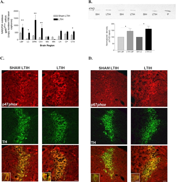
To determine whether LTIH increases NADPH oxidase gene expression in wake-active brain regions, we measured NADPH oxidase gene response in discrete brain regions by performing laser-captured microdissections in adult WT mice (n = 10 sham LTIH, n = 10 LTIH) for Taqman real-time PCR measurement of mRNA copies. LTIH increased NADPH oxidase subunit p67phox gene expression in most, but not all wake-active regions, as follows: the lateral basal forebrain (p < 0.001), the dorsal raphe nucleus (p < 0.001), and the locus coeruleus (p < 0.05). This expression was also increased, but not significantly, in the lateral hypothalamus, as illustrated in Figure 1A. To determine whether NADPH oxidase subunit proteins were increased in response to LTIH, Western blots were performed on homogenates from two representative wake-active regions: the locus coeruleus and the lateral basal forebrain. Micropunches from WT mice after sham LTIH and LTIH revealed increased p47phox in both regions in WT mice exposed to LTIH (Figure 1B). p47phox protein, as measured by relative density at the 47-kD band, increased by 50% (t = 2.9, p < 0.05) in the lateral basal forebrain and by 65% in the locus coeruleus (t = 2.8, p < 0.05). To determine whether NADPH oxidase protein was qualitatively increased within representative wake-active neurons, immunohistochemistry for two of the NADPH oxidase subunits was performed in WT mice exposed to 8 weeks' LTIH or sham LTIH for the noradrenergic locus coeruleus. We found increased p67phox and p47phox immunoreactivity in the noradrenergic locus coeruleus neurons (Figure 1C). Thus, LTIH results in increased NADPH oxidase subunit gene and protein expression in wake-active regions of the brain, and within the locus coeruleus, the increase is evident within noradrenergic neurons.
Figure 1.
Long-term intermittent hypoxia (LTIH), modeling oxygenation patterns in sleep apnea, results in increased NADPH oxidase gene and protein expression in wake-active brain regions. (A) NADPH oxidase subunit p67phox mRNA was measured in laser-captured neurons in brain regions selected because of behavioral state dependency or known hypoxia sensitivity. Taqman real-time reverse transcriptase polymerase chain reaction was performed on 50 laser-captured neurons each in the following brain regions: CA1 = hippocampal CA1 pyramidal cells; CTX = cortex; DRN = dorsal raphe nucleus; LBF = lateral basal forebrain (magnocellular preoptic, horizontal diagonal band, and substantia inominata); LC = locus coeruleus; LH = perifornicular lateral hypothalamus; MP = median preoptic area; MS = medial septum/vertical diagonal band; STR = striatum. Comparisons were drawn between wild-type mice exposed to LTIH (n = 10) and sham LTIH (n = 10). *p < 0.05; **p < 0.001. (B) NADPH oxidase sub-unit p47phox immunoreactivity (47 kD) in locus coeruleus and lateral basal forebrain micropunches in adult mice after LTIH (n = 8) and sham LTIH (n = 8) revealed increased p47phox in mice exposed to LTIH. *p < 0.01 in matched regions. P = positive control; SIH = sham intermittent hypoxia. (C, D) Immunohistochemical staining of noradrenergic locus coeruleus wake-active region shows enhanced NADPH oxidase subunit p47phox and p67phox expression (red) in locus coeruleus in mice exposed to LTIH. Double labeling with tyrosine hydroxylase localizes both subunits p47phox and p67phox to noradrenergic (tyrosine hydroxylase [TH], green) locus coeruleus wake-active neurons. Lower panel scale bar is 50 μm and inset bar is 20 μm.
Genetic Deletion of NADPH Oxidase Subunit gp91phox Confers Resistance to Long-Term Hypoxia Reoxygenation-induced Hypersomnolence and Sleepiness
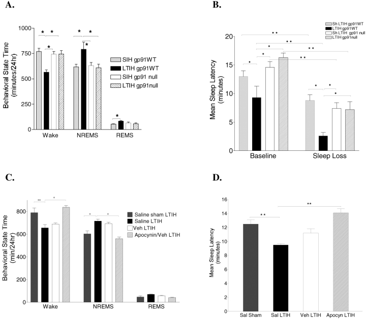
The LTIH effect on sleep latency also varied with genotype (F = 18, p < 0.0001; Figure 2B). Baseline sleep latency was reduced in WT mice exposed to LTIH (t = 2.9, p < 0.05) and markedly reduced relative to sham LTIH mice (t = 4.8, p < 0.0001), whereas no effect of LTIH (LTIH vs. sham LTIH) was observed in gp91phox-null mice. Sleep latency in gp91phox-null mice exposed to LTIH, however, was reduced by short-term (6 hours) enforced wakefulness in both the sham LTIH and LTIH (t = 4.8 and t = 5.1, respectively; both, p < 0.0001), suggesting that the homeostatic regulation is intact in the mutant mice. In summary, genetic deletion of gp91phox prevents LTIH-induced sleepiness without impairing sleep-loss sleepiness.
Figure 2.
NADPH oxidase is essential for the residual wake impairments after LTIH. (A) Wild-type (WT) control mice exposed to LTIH (LTIH gp91WT) show reduced wake time/24 hours and increased non-REM sleep (NREMS) at 2 weeks into normoxic recovery relative to sham LTIH (SIH WT). In contrast, mice lacking NADPH oxidase subunit gp91phox exposed to LTIH (LTIH gp91phox null) show no effect of LTIH on either wakefulness or NREMS. (B) The propensity to fall asleep, measured as the average sleep latency across four nap opportunities at the end of the lights-on period, is markedly reduced in WT mice exposed to LTIH (for both unperturbed baseline sleep and after 6 hours' sleep loss, p < 0.0001). In contrast, no effect of LTIH is observed for sleep latencies in gp91phox-null mice, despite a marked sleep loss effect on latencies for both sham LTIH and LTIH gp91-null mice, p < 0.0001. Shown are sleep latencies after unperturbed rest activity (Baseline) and after 6 hours of enforced wakefulness, same circadian time point (Sleep Loss). (C) Systemic apocynin treatment via osmotic pump (diagonally striped columns) prevented LTIH-induced impaired wakefulness and hypersomnolence in WT mice. In contrast, the apocynin vehicle (veh) dimethyl sulfoxide was without effect (white columns). (D) Apocynin therapy prevented LTIH-reduced sleep latency. Values presented are means ± SEM; asterisks denote statistical significance. For all panels: *p < 0.05; **p < 0.001.
Gp91phox-Null Mice Are Resistant to LTIH-induced Oxidative Protein Damage
Macrodissections of the lateral basal forebrain and the noradrenergic locus coeruleus were obtained from gp91phox-null and WT mice exposed to LTIH or sham LTIH (n = 10–11/strain and condition). The carbonyl content in the lateral basal forebrain in gp91phox-null mice relative to WT mice for sham LTIH appeared reduced by approximately 40%; however, this did not reach statistical significance (t = 2.9, p = 0.05; Figure 3A). WT mice exhibited an increase in carbonyl content in response to LTIH, relative to sham LTIH (t = 3.9, p < 0.05). In contrast, gp91phox-null mice did not show an increase in response to LTIH (p = not significant). Similar changes were observed in the locus coeruleus region. Thus, genetic deletion of gp91phox prevents the LTIH carbonyl response in representative brain regions contributing to wakefulness control.
Figure 3.
NADPH oxdase is essential for basal forebrain carbonyl and isoprostane responses to LTIH. (A) Carbonyl content as measured with ELISA in lateral basal forebrain macropunches in gp91phox-null mice and wild-type (WT) control mice after 8 weeks of LTIH (black bars) or sham LTIH exposures (gray bars). Carbonyl content (carbonyl protein/20 mg protein aliquot) is significantly increased in WT (gp91phox+/+) mice exposed to LTIH (n = 10), relative to sham LTIH exposure (n = 11); *p < 0.05. In contrast, gp91phox-null mice showed no effect of LTIH on carbonyl content, p = not significant. **p < 0.01, LTIH gp91phox+/+ versus LTIH gp91phox−/−. (B) Homogenized tissue samples were collected for measurement of isoprostane, 8,12-iso-iPF2-VI, from the lateral basal forebrain in mice after 8 weeks' exposure to either LTIH or sham LTIH. Using an internal standard, levels were assayed by negative-ion, chemical ionization gas chromatography and mass spectrometry. ***p < 0.001, WT sham LTIH versus WT LTIH; **p < 0.01, WT LTIH versus gp91phox-null LTIH.
Isoprostane levels were measured in similarly dissected lateral basal forebrain tissue blocks from mice (n = 5/intermittent hypoxic condition and genotype). gp91phox-null mice exposed to LTIH, relative to WT mice exposed to LTIH, had reduced isoprostanes in both the lateral basal forebrain and lateral hypothalamus (p < 0.01 for both regions); however, a trend toward increased isoprostanes in the LTIH-exposed gp91phox-null mice relative to sham LTIH gp91phox-null mice (25%, p = 0.06) suggests that the lipid peroxidation may not be completely blocked (Figure 3B).
Inhibition of NADPH Oxidase throughout LTIH Exposure Prevents Hypersomnolence and Sleepiness
To further investigate the role of NADPH oxidase in the LTIH-induced hypersomnolence and sleepiness, a series of WT mice were implanted with osmotic pumps to systemically deliver either a selective NADPH oxidase inhibitor, apocynin at 3 mg/kg/day, vehicle (DMSO), or buffered saline throughout LTIH exposure for 8 weeks. Pumps were removed and mice were implanted for sleep recordings as described above for studies in gp91phox-null mice. Apocynin therapy prevented both LTIH-reduced wake times and shortened sleep latency. WT mice treated throughout LTIH with apocynin, relative to saline treatment (t = 4.5, p < 0.001) and relative to DMSO vehicle (t = 2.9, p < 0.05) had increased wake times and reduced non-REM sleep time (t = 4.1, p < 0.001, and t = 2.7, p < 0.05, respectively, as shown in Figure 2C). In addition to completely preventing LTIH-reduced wake times for 24 hours, systemic delivery of apocynin also prevented LTIH-reduced sleep latencies. Specifically, sleep latencies in apocynin-treated mice were significantly higher than LTIH DMSO vehicle–treated mice (t = 3.4, p < 0.01) and were not significantly different from sleep latencies in WT mice without any intervention or after sham LTIH treatment (Figure 2D).
NADPH Oxidase Inhibition throughout LTIH Prevents the LTIH Proinflammatory Response
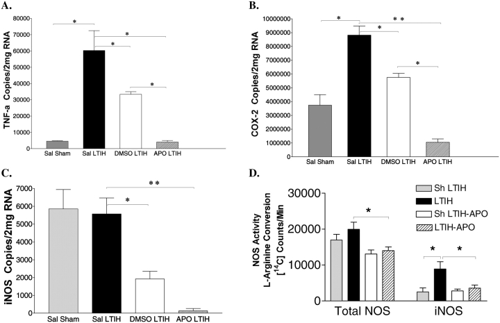
LTIH increases proinflammatory gene expression (TNF-α and COX-2) in the cortex, hippocampus, and wake-active regions, and this response is still present 2 weeks into recovery (8). To determine whether proinflammatory response is NADPH oxidase–dependent, we compared gene responses to LTIH in mice treated with apocynin, vehicle, or saline to sham LTIH–treated responses in a representative wake-active region, the lateral basal forebrain (Figure 4). Consistent with previous findings, the group treated with saline throughout LTIH (n = 6), relative to sham LTIH (n = 6), exhibited large increases in TNF-α (> 10-fold increase, t = 4.1, p < 0.01; Figure 4A) and in COX-2 mRNA (140% increase, t = 6.6, p < 0.001; Figure 4B). Treatment with apocynin throughout LTIH completely prevented the increases in TNF-α gene expression (Figure 4A). Specifically, LTIH TNF-α levels, in apocynin-treated mice did not differ from sham LTIH values (t = 0.7, p = not significant). Apocynin treatment during LTIH reduced the TNF-α response to 7% of the saline LTIH response (t = 4.8, p < 0.001). DMSO (vehicle alone) treatment during LTIH reduced the LTIH response to 35% of the expected (t = 2.8, p < 0.05); vehicle treatment, however, was less effective than apocynin in reducing the LTIH TNF-α response (apocynin vs. DMSO, 73% lower; t = 2.8, p < 0.05). Apocynin therapy had similar effects on the LTIH COX-2 gene response. Treatment with the apocynin in DMSO vehicle completely blocked the LTIH COX-2 response in the lateral basal forebrain (t = 10.2, p < 0.0001) to below the sham LTIH gene expression (65% reduction relative to sham LTIH, t = 3.5, p < 0.05; Figure 4B). Treatment with the DMSO vehicle also had some effect in reducing the LTIH gene response reduced the LTIH gene responses in the lateral basal forebrain, when compared with saline-treated LTIH (75% reduction, t = 8.6, p < 0.01). Thus, systemic apocynin was extremely effective in preventing the LTIH proinflammatory gene response. DMSO, the vehicle used for apocynin, had some effect on blunting the TNF-α and COX-2 increased expression, but was not as effective as apocynin.
Figure 4.
NADPH oxidase inhibition confers resistance to the long-term intermittent hypoxia (LTIH) proinflammatory response. Proinflammatory gene expression was measured in micropunches in the lateral basal forebrains in gp91phox WT control mice (n = 10) and gp91phox-null mice (n = 10) for the following: (A) tumor necrosis factor α (TNF-α), (B) cyclooxygenase 2 (COX-2), and (C) inducible nitric oxide synthase (iNOS) mRNA copies/2 μg total RNA. (D) Total NOS activity (left columns), measured as L-arginine conversion of [14C]L-arginine to L-citrulline, and iNOS activity (right columns) were measured using 5 mm S-ethyl-N-[4-trifluoromethyl)phenyl]isothiourea added to homogenates. Apocynin prevents the anticipated LTIH increase in iNOS activity. APO = apocynin in dimethylsulfoxide (DMSO) vehicle; sal = saline; sham = sham intermittent hypoxia control. *p < 0.05; **p < 0.01.
LTIH has been shown to have little effect on the iNOS gene response in wake-active regions but has a significant effect on iNOS activity (8). Thus, we sought to determine whether apocynin therapy affected iNOS gene response and whether inhibition of NADPH oxidase blocked the LTIH effect on iNOS activity. iNOS gene copies were reduced in both the DMSO (66% reduction in iNOS mRNA, p < 0.01) and apocynin groups (94% reduction in iNOS mRNA, p < 0.001; Figure 4C), with markedly reduced gene expression in the apocynin group relative to the vehicle group (t = 8.3, p < 0.001). Lateral basal forebrain iNOS activity was compared in mice treated with or without apocynin throughout LTIH (Figure 4D). Apocynin therapy, relative to saline therapy, had no effect on baseline (sham LTIH) iNOS activity. In contrast, apocynin prevented the LTIH increase in iNOS activity, resulting in a large reduction (60%) in iNOS activity in apocynin-treated versus untreated mice (t = 3.4, p < 0.05), so that apocynin-treated LTIH iNOS activity did not differ from sham LTIH iNOS activity levels in the lateral basal forebrain. Administration of the apocynin vehicle DMSO throughout LTIH resulted in higher iNOS activity than apocynin therapy (t = 2.8, p < 0.05). Thus, apocynin therapy throughout LTIH prevented the expected increase in iNOS activity.
DISCUSSION
NADPH oxidase is increasingly recognized for its dual-edge roles in health and disease, necessary for normal immunity and cell signaling, yet critical in the pathogenesis of diverse morbid conditions, including Alzheimer's disease, Parkinson's disease, and cardiovascular disease (13–21, 35–38). Here, we have demonstrated that the expression of several NADPH oxidase proteins is increased in a population of wake-active neurons under conditions of long-term hypoxia/reoxygenation events simulating the oxygenation patterns observed in persons with moderate to severe OSA. Moreover, we show that the NADPH oxidase response to long-term hypoxia/reoxygenation is detrimental to brain function. We have determined that this oxidase plays an essential role not only in the hypersomnolence and increased sleep propensity associated with the hypoxia/reoxygenation model of sleep apnea but also in the associated proinflammatory gene response, carbonylation, and lipid peroxidation injury to select brain regions—in particular, regions containing wake-active neurons. Collectively, these findings highlight the significance of long-term hypoxia/reoxygenation events as in sleep apnea and identify a novel pathway whereby the hypoxia/reoxygenation events induce NADPH oxidase activation in the brain, which then promotes oxidative injury, microglial activation, and proinflammatory gene expression. Therefore, these findings identify a potential target pathway for prevention of neurobehavioral morbidities commonly observed in persons treated for OSA.
Although oxidative injury in the brain has not been explored for OSA, persons with OSA have some evidence of oxidative stress peripherally (39–41). Specific isoprostanes are markers of in vivo lipid peroxidation that we have found is increased in LTIH and in exhaled breath condensate in patients with untreated OSA (39). The magnitude of isoprostane increase in persons with OSA is similar to the magnitude of increase observed in the brain in mice subjected to intermittent hypoxia. Further evidence that increased lipid peroxidation in persons with OSA relates directly to the sleep-disordered breathing is provided by the observation that effective treatment of sleep apnea with nasal continuous positive airway pressure results in a significant reduction in exhalate isoprostane levels (39). Peripheral polymorphonuclear neutrophils procured from humans with OSA manifest increased superoxide production in vitro relative to neutrophils from persons without OSA (41), and this increase in neutrophil superoxide in OSA also can be corrected with effective therapy for apneic events (41).
OSA is an independent risk factor not only for neurobehavioral morbidities but also for hypertension and ischemic heart disease (42–45), and NADPH oxidase has been implicated in the pathogenesis of these disorders (35, 36, 38). Having identified NADPH oxidase as an important source of intermittent hypoxia–induced injury in the brain, it will now be of significant interest to determine whether NADPH oxidase activation in persons with OSA contributes to the cardiovascular morbidities associated with this disease. The NADPH oxidase pathway may be a valuable pharmacotherapeutic target for both neurobehavioral and cardiovascular morbidities of this prevalent disorder.
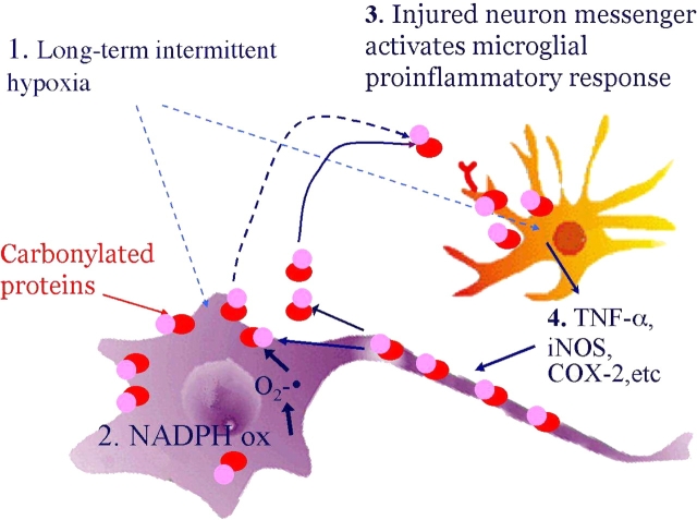
The present studies identify NADPH oxidase as essential for the proinflammatory response in intermittent hypoxia modeling sleep apnea. Peripheral TNF-α levels are elevated in persons with untreated OSA (46–48), whereas TNF-α levels improve with long-term continuous positive airway pressure therapy (46), supporting a direct link between proinflammatory response and OSA. The magnitude of gene expression increase we observed is surprisingly large, similar to TNF-α responses in mice subjected to severe ischemia with carotid ligation (49) and drug-induced dopaminergic neurotoxicity (50, 51). Local proinflammatory responses contribute to neuronal injury and may be an important factor in the microglially mediated injury to neurons (52–55). A next step will be to determine the role microglia play in hypoxia/reoxygenation injury to wake-active neurons. Both genetic deletion of gp91phox, which results in a functionally inactive NADPH oxidase, and pharmacologic inhibition of NADPH oxidase completely prevented the COX-2, TNF-α, and iNOS responses. Taken together, these findings strongly suggest that NADPH oxidase is upstream from the proinflammatory response. In Figure 5, we propose a model through which LTIH activates NADPH oxidase in select neural groups. This activation promotes the LTIH carbonyl protein injury previously shown (7). Redox-altered proteins or lipoproteins in the neural membrane activate adjacent microglia that, in turn, initiate a proinflammatory response, including iNOS activation as previously shown (8). The nitration and oxidation injury from the microglial proinflammatory response promotes a vicious cycle of continued oxidative neural injury and microglial activation. It may be through this vicious cycle that the responses persist for at least 2 weeks into recovery.
Figure 5.
Proposed model of LTIH-induced neural injury. Neurons with NADPH oxidase activation responses to LTIH are at increased risk of oxidative injury. The present study shows that LTIH activates NADPH oxidase in select neurons, including at least some groups of wake-active neurons. The mechanisms of LTIH activation of NADPH oxidase are unknown. However, NADPH oxidase activation and production of superoxide radical (O2−·) manifest, in part, as increased irreversible and progressive carbonylation injury to proteins. Redox alterations in surface proteins or secreted proteins activate adjacent microglia. Activation of microglia results in further increase of NADPH oxidase but also a proinflammatory response including iNOS, COX-2, and TNF-α. iNOS production of NO combined with adjacent O2−· molecules will result in peroxynitrite formation for lipid peroxidation of membranes. This lipid peroxidation of neural membranes and released COX-2 and TNF-α promote a vicious cycle of continued oxidative and inflammatory injury persisting after normalization of ambient oxygen tensions. NADPH oxidase blockade prevents all injuries described.
In conclusion, our findings have resulted in the identification of NADPH oxidase as an enzyme that is critically involved in all known components (behavioral, molecular, redox) of the intermittent hypoxia phenotype of brain injury. This indicates that NADPH oxidase could be a molecular target to further probe the mechanisms of neuronal injury in a highly prevalent form of intermittent hypoxia, OSA. Moreover, our findings have unveiled a selective vulnerability to hypoxia/reoxygenation neuronal proinflammatory injury, which can be predicted, at least in part, by whether NADPH oxidase is present in neurons. Finally, these studies raise the intriguing possibility that NADPH oxidase plays a critical role in sleep apnea–induced hypoxia/reoxygenation injury in other physiologic systems, including cardiovascular functions. Thus, NADPH oxidase and its downstream effector pathways should be considered molecular targets for pharmacologic and therapeutic intervention for the treatment and perhaps prevention of oxidative cardiovascular and neurobehavioral morbidities of OSA.
Supported in part by National Institutes of Health grants HL65225 and AG17628 (to S.C.V.) and AG 11542 (to D.P.).
Conflict of Interest Statement: G.Z. has no financial relationship with a commercial entity that has an interest in the subject of this manuscript. F.S. has no financial relationship with a commercial entity that has an interest in the subject of this manuscript. P.F. has no financial relationship with a commercial entity that has an interest in the subject of this manuscript. R.H. has no financial relationship with a commercial entity that has an interest in the subject of this manuscript. L.K. has no financial relationship with a commercial entity that has an interest in the subject of this manuscript. D.P. has no financial relationship with a commercial entity that has an interest in the subject of this manuscript. E.K. has no financial relationship with a commercial entity that has an interest in the subject of this manuscript. S.C.V. has received $5,000 for serving on a Sepracor advisory board in 2004 and served as a consultant for Organon in 2004 for $2,000.
References
- 1.Young T, Palta M, Dempsey J, Skatrud J, Weber S, Badr S. The occurrence of sleep-disordered breathing among middle-aged adults. N Engl J Med 1993;328:1230–1235. [DOI] [PubMed] [Google Scholar]
- 2.Remmers JE, deGroot WJ, Sauerland EK, Anch AM. Pathogenesis of upper airway occlusion during sleep. J Appl Physiol 1978;44:931–938. [DOI] [PubMed] [Google Scholar]
- 3.Patel SR, White DP, Malhotra A, Stanchina ML, Ayas NT. Continuous positive airway pressure therapy for treating sleepiness in a diverse population with obstructive sleep apnea: results of a meta-analysis. Arch Intern Med 2003;163:565–571. [DOI] [PubMed] [Google Scholar]
- 4.Douglas NJ, Engleman HM. Effects of CPAP on vigilance and related functions in patients with the sleep apnea/hypopnea syndrome. Sleep 2000;23:S147–S149. [PubMed] [Google Scholar]
- 5.Bedard MA, Montplaisir J, Richer F, Malo J. Nocturnal hypoxemia as a determinant of vigilance impairment in sleep apnea syndrome. Chest 1991;100:367–370. [DOI] [PubMed] [Google Scholar]
- 6.Tiihonen M, Partinen M. Polysomnography and maintenance of wakefulness test as predictors of CPAP effectiveness in obstructive sleep apnea. Electroencephalogr Clin Neurophysiol 1998;107:383–386. [DOI] [PubMed] [Google Scholar]
- 7.Veasey SC, Davis C, Zhan G, Hsu YJ, Fenik P, Pratico D, Gow AJ. Long-term intermittent hypoxia in mice: protracted hypersomnolence with oxidative injury to sleep-wake brain regions. Sleep 2004;27:194–201. [DOI] [PubMed] [Google Scholar]
- 8.Zhan G, Fenik P, Pratico D, Veasey SC. Inducible nitric oxide synthase in long-term intermittent hypoxia: hypersomnolence and brain injury. Am J Respir Crit Care Med 2005;171:1414–1420. [DOI] [PMC free article] [PubMed] [Google Scholar]
- 9.Gozal D, Daniel JM, Dohanich GP. Behavioral and anatomical correlates of chronic episodic hypoxia during sleep in the rat. J Neurosci 2001;21:2442–2450. [DOI] [PMC free article] [PubMed] [Google Scholar]
- 10.Row BW, Liu R, Wei X, Kheirandish L, Gozal D. Intermittent hypoxia is associated with oxidative stress and spatial learning deficits in the rat. Am J Respir Crit Care Med 2003;167:1540–1547. [DOI] [PubMed] [Google Scholar]
- 11.Row BW, Kheirandish L, Li RC, Guo SZ, Brittian KR, Hardy M, Bazan NG, Gozal D. Platelet-activating factor receptor-deficient mice are protected from experimental sleep apnea-induced learning deficits. J Neurochem 2004;89:189–196. [DOI] [PubMed] [Google Scholar]
- 12.Veasey SC, Zhan G, Fenik P, Pratico D. Long-term intermittent hypoxia: reduced hypoglossal nerve output. Am J Respir Crit Care Med 2004;170:665–672. [DOI] [PubMed] [Google Scholar]
- 13.Ozaki M, Deshpande SS, Angkeow P, Bellan J, Lowenstein CJ, Dinauer MC, Goldschmidt-Clermont PJ, Irani K. Inhibition of the Rac1 GTPase protects against nonlethal ischemia/reperfusion-induced necrosis and apoptosis in vivo. FASEB J 2000;14:418–429. [DOI] [PubMed] [Google Scholar]
- 14.Harada H, Hines IN, Flores S, Gao B, McCord J, Scheerens H, Grisham MB. Role of NADPH oxidase-derived superoxide in reduced size liver ischemia and reperfusion injury. Arch Biochem Biophys 2004;423:103–108. [DOI] [PubMed] [Google Scholar]
- 15.Kusaka I, Kusaka G, Zhou C, Ishikawa M, Nanda A, Granger DN, Zhang JH, Tang J. Role of AT1 receptors and NAD(P)H oxidase in diabetes-aggravated ischemic brain injury. Am J Physiol Heart Circ Physiol 2004;286:H2442–H2451. [DOI] [PubMed] [Google Scholar]
- 16.Rupin A, Paysant J, Sansilvestri-Morel P, Lembrez N, Lacoste JM, Cordi A, Verbeuren TJ. Role of NADPH oxidase-mediated superoxide production in the regulation of E-selectin expression by endothelial cells subjected to anoxia/reoxygenation. Cardiovasc Res 2004;63:323–330. [DOI] [PubMed] [Google Scholar]
- 17.Abramov AY, Canevari L, Duchen MR. Beta-amyloid peptides induce mitochondrial dysfunction and oxidative stress in astrocytes and death of neurons through activation of NADPH oxidase. J Neurosci 2004;24:565–575. [DOI] [PMC free article] [PubMed] [Google Scholar]
- 18.Jana A, Pahan K. Fibrillar amyloid-beta peptides kill human primary neurons via NADPH oxidase-mediated activation of neutral sphingomyelinase. Implications for Alzheimer's disease. J Biochem (Tokyo) 2004;279:51451–51459. [DOI] [PMC free article] [PubMed] [Google Scholar]
- 19.Gao HM, Liu B, Zhang W, Hong JS. Critical role of microglial NADPH oxidase-derived free radicals in the in vitro MPTP model of Parkinson's disease. FASEB J 2003;17:1954–1956. [DOI] [PubMed] [Google Scholar]
- 20.Wu DC, Teismann P, Tieu K, Vila M, Jackson-Lewis V, Ischiropoulos H, Przedborski S. NADPH oxidase mediates oxidative stress in the 1-methyl-4-phenyl-1,2,3,6-tetrahydropyridine model of Parkinson's disease. Proc Natl Acad Sci USA 2003;100:6145–6150. [DOI] [PMC free article] [PubMed] [Google Scholar]
- 21.Zhang W, Wang T, Qin L, Gao HM, Wilson B, Ali SF, Zhang W, Hong JS, Liu B. Neuroprotective effect of dextromethorphan in the MPTP Parkinson's disease model: role of NADPH oxidase. FASEB J 2004;18:589–591. [DOI] [PubMed] [Google Scholar]
- 22.Noh KM, Koh JY. Induction and activation by zinc of NADPH oxidase in cultured cortical neurons and astrocytes. J Neurosci 2000;20:RC111. [DOI] [PMC free article] [PubMed] [Google Scholar]
- 23.Serrano F, Kolluri NS, Wientjes FB, Card JP, Klann E. NADPH oxidase immunoreactivity in the mouse brain. Brain Res 2003;988:193–198. [DOI] [PubMed] [Google Scholar]
- 24.Wang G, Anrather J, Huang J, Speth RC, Pickel VM, Iadecola C. NADPH oxidase contributes to angiotensin II signaling in the nucleus tractus solitarius. J Neurosci 2004;24:5516–5524. [DOI] [PMC free article] [PubMed] [Google Scholar]
- 25.Zhan G, Shaheen F, Mackiewicz M, Fenik P, Veasey SC. Single cell laser dissection with molecular beacon polymerase chain reaction identifies 2A as the predominant serotonin receptor subtype in hypoglossal motoneurons. Neuroscience 2002;113:145–154. [DOI] [PubMed] [Google Scholar]
- 26.Kachadourian R, Johnson CA, Min E, Spasojevic I, Day BJ. Flavin-dependent antioxidant properties of a new series of meso-N,N'-dialkyl-imidazolium substituted manganese(III) porphyrins. Biochem Pharmacol 2004;67:77–85. [DOI] [PubMed] [Google Scholar]
- 27.Vasquez-Vivar J, Martasek P, Hogg N, Masters BS, Pritchard KA Jr, Kalyanaraman B. Endothelial nitric oxide synthase-dependent superoxide generation from adriamycin. Biochemistry 1997;36:11293–11297. [DOI] [PubMed] [Google Scholar]
- 28.Wu DC, Jackson-Lewis V, Vila M, Tieu K, Teismann P, Vadseth C, Choi DK, Ischiropoulos H, Przedborski S. Blockade of microglial activation is neuroprotective in the 1-methyl-4-phenyl-1,2,3,6-tetrahydropyridine mouse model of Parkinson disease. J Neurosci 2002;22:1763–1771. [DOI] [PMC free article] [PubMed] [Google Scholar]
- 29.Buss IH, Winterbourn CC. Protein carbonyl measurement by ELISA. Methods Mol Biol 2002;186:123–128. [DOI] [PubMed] [Google Scholar]
- 30.Pratico D, Uryu K, Leight S, Trojanoswki JQ, Lee VM. Increased lipid peroxidation precedes amyloid plaque formation in an animal model of Alzheimer amyloidosis. J Neurosci 2001;21:4183–4187. [DOI] [PMC free article] [PubMed] [Google Scholar]
- 31.Uryu K, Laurer H, McIntosh T, Pratico D, Martinez D, Leight S, Lee VM, Trojanowski JQ. Repetitive mild brain trauma accelerates Abeta deposition, lipid peroxidation, and cognitive impairment in a transgenic mouse model of Alzheimer amyloidosis. J Neurosci 2002;22:446–454. [DOI] [PMC free article] [PubMed] [Google Scholar]
- 32.Veasey SC, Valladares O, Fenik P, Kapfhamer D, Sanford L, Benington J, Bucan M. An automated system for recording and analysis of sleep in mice. Sleep 2000;23:1025–1040. [PubMed] [Google Scholar]
- 33.Veasey SC, Hsu Y-J, Thayer P, Fenik P. Murine Multiple Sleep Latency Test: phenotyping sleep propensity in mice. Sleep 2004;27:388–393. [DOI] [PubMed] [Google Scholar]
- 34.Benington JH. Sleep homeostasis and the function of sleep. Sleep 2000;23:959–966. [PubMed] [Google Scholar]
- 35.Cai H, Griendling KK, Harrison DG. The vascular NAD(P)H oxidases as therapeutic targets in cardiovascular diseases. Trends Pharmacol Sci 2003;24:471–478. [DOI] [PubMed] [Google Scholar]
- 36.Agarwal R, Campbell RC, Warnock DG. Oxidative stress in hypertension and chronic kidney disease: role of angiotensin II. Semin Nephrol 2004;24:101–114. [DOI] [PubMed] [Google Scholar]
- 37.Furukawa S, Fujita T, Shimabukuro M, Iwaki M, Yamada Y, Nakajima Y, Nakayama O, Makishima M, Matsuda M, Shimomura I. Increased oxidative stress in obesity and its impact on metabolic syndrome. J Clin Invest 2004;114:1752–1761. [DOI] [PMC free article] [PubMed] [Google Scholar]
- 38.Jung O, Schreiber JG, Geiger H, Pedrazzini T, Busse R, Brandes RP. gp91phox-containing NADPH oxidase mediates endothelial dysfunction in renovascular hypertension. Circulation 2004;109:1795–1801. [DOI] [PubMed] [Google Scholar]
- 39.Carpagnano GE, Kharitonov SA, Resta O, Foschino-Barbaro MP, Gramiccioni E, Barnes PJ. 8-Isoprostane, a marker of oxidative stress, is increased in exhaled breath condensate of patients with obstructive sleep apnea after night and is reduced by continuous positive airway pressure therapy. Chest 2003;124:1386–1392. [DOI] [PubMed] [Google Scholar]
- 40.Lavie L, Vishnevsky A, Lavie P. Evidence for lipid peroxidation in obstructive sleep apnea. Sleep 2004;27:123–128. [PubMed] [Google Scholar]
- 41.Schulz R, Mahmoudi S, Hattar K, Sibelius U, Olschewski H, Mayer K, Seeger W, Grimminger F. Enhanced release of superoxide from polymorphonuclear neutrophils in obstructive sleep apnea: impact of continuous positive airway pressure therapy. Am J Respir Crit Care Med 2000;162:566–570. [DOI] [PubMed] [Google Scholar]
- 42.Peker Y, Hedner J, Kraiczi H, Loth S. Respiratory disturbance index: an independent predictor of mortality in coronary artery disease. Am J Respir Crit Care Med 2000;162:81–86. [DOI] [PubMed] [Google Scholar]
- 43.Nieto FJ, Young TB, Lind BK, Shahar E, Samet JM, Redline S, D'Agostino RB, Newman AB, Lebowitz MD, Pickering TG. Association of sleep-disordered breathing, sleep apnea, and hypertension in a large community-based study. Sleep Heart Health Study. JAMA 2000;283:1829–1836. [DOI] [PubMed] [Google Scholar]
- 44.Shahar E, Whitney CW, Redline S, Lee ET, Newman AB, Javier Nieto F, O'Connor GT, Boland LL, Schwartz JE, Samet JM. Sleep-disordered breathing and cardiovascular disease: cross-sectional results of the Sleep Heart Health Study. Am J Respir Crit Care Med 2001;163:19–25. [DOI] [PubMed] [Google Scholar]
- 45.Parra O, Arboix A, Montserrat JM, Quinto L, Bechich S, Garcia-Eroles L. Sleep-related breathing disorders: impact on mortality of cerebrovascular disease. Eur Respir J 2004;24:267–272. [DOI] [PubMed] [Google Scholar]
- 46.Alberti A, Sarchielli P, Gallinella E, Floridi A, Floridi A, Mazzotta G, Gallai V. Plasma cytokine levels in patients with obstructive sleep apnea syndrome: a preliminary study. J Sleep Res 2003;12:305–311. [DOI] [PubMed] [Google Scholar]
- 47.Dyugovskaya L, Lavie P, Lavie L. Phenotypic and functional characterization of blood γδ T cells in sleep apnea. Am J Respir Crit Care Med 2003;168:242–249. [DOI] [PubMed] [Google Scholar]
- 48.Minoguchi K, Tazaki T, Yokoe T, Minoguchi H, Watanabe Y, Yamamoto M, Adachi M. Elevated production of tumor necrosis factor-alpha by monocytes in patients with obstructive sleep apnea syndrome. Chest 2004;126:1473–1479. [DOI] [PubMed] [Google Scholar]
- 49.Schroeter M, Kury P, Jander S. Inflammatory gene expression in focal cortical brain ischemia: differences between rats and mice. Brain Res Mol Brain Res 2003;117:1–7. [DOI] [PubMed] [Google Scholar]
- 50.Hebert G, Arsaut J, Dantzer R, Demotes-Mainard J. Time-course of the expression of inflammatory cytokines and matrix metalloproteinases in the striatum and mesencephalon of mice injected with 1-methyl-4-phenyl-1,2,3,6-tetrahydropyridine, a dopaminergic neurotoxin. Neurosci Lett 2003;349:191–195. [DOI] [PubMed] [Google Scholar]
- 51.Nakajima A, Yamada K, Nagai T, Uchiyama T, Miyamoto Y, Mamiya T, He J, Nitta A, Mizuno M, Tran MH, et al. Role of tumor necrosis factor-alpha in methamphetamine-induced drug dependence and neurotoxicity. J Neurosci 2004;24:2212–2225. [DOI] [PMC free article] [PubMed] [Google Scholar]
- 52.Robertson J, Beaulieu JM, Doroudchi MM, Durham HD, Julien JP, Mushynski WE. Apoptotic death of neurons exhibiting peripherin aggregates is mediated by the proinflammatory cytokine tumor necrosis factor-alpha. J Cell Biol 2001;155:217–226. [DOI] [PMC free article] [PubMed] [Google Scholar]
- 53.Del Villar K, Miller CA. Down-regulation of DENN/MADD, a TNF receptor binding protein, correlates with neuronal cell death in Alzheimer's disease brain and hippocampal neurons. Proc Natl Acad Sci USA 2004;101:4210–4215. [DOI] [PMC free article] [PubMed] [Google Scholar]
- 54.Li R, Yang L, Lindholm K, Konishi Y, Yue X, Hampel H, Zhang D, Shen Y. Tumor necrosis factor death receptor signaling cascade is required for amyloid-beta protein-induced neuron death. J Neurosci 2004;24:1760–1771. [DOI] [PMC free article] [PubMed] [Google Scholar]
- 55.Li RC, Row BW, Gozal E, Kheirandish L, Fan Q, Brittian KR, Guo SZ, Sachleben LR Jr, Gozal D. Cyclooxygenase 2 and intermittent hypoxia-induced spatial deficits in the rat. Am J Respir Crit Care Med 2003;168:469–475. [DOI] [PubMed] [Google Scholar]


