Abstract
A relativistic plasma shutter technique is proposed and tested to remove the sub-100 ps pedestal of a high-intensity laser pulse. The shutter is an ultrathin foil placed before the target of interest. As the leading edge of the laser ionizes the shutter material it will expand into a relativistically underdense plasma allowing for the peak pulse to propagate through while rejecting the low intensity pedestal. An increase in the laser temporal contrast is demonstrated by measuring characteristic signatures in the accelerated proton spectra and directionality from the interaction of 30 TW pulses with ultrathin foils along with supporting hydrodynamic and particle-in-cell simulations.
The development of ultrashort, high-power lasers has produced record pulse intensities up to 1022W∕cm2.1, 2 However, as the peak intensity increases the required contrast ratio between: (i) a temporal (nanosecond scale) pedestal caused by the amplified spontaneous emission (ASE); (ii) (sub-100 ps) prepulses, caused by the residual aberrations of the spectral phase, and the main pulse needs to be proportionally improved to allow for preplasma free interaction with solid targets. Several techniques have been proposed and tested to improve temporal contrast of high-intensity lasers. Among these techniques are the use of saturable absorbers,3 polarization rotation in a hollow fiber,4 cross-polarized wave generation (XPW)5 before pulse stretching, double chirped pulse amplification,6 plasma mirrors,7, 8 and second harmonic generation after pulse compression. The use of high contrast, prepulse free pulses for interaction of ultrahigh intensity lasers with solids is of primary importance for a number of high field experiments such as high harmonic generation and proton acceleration. Specifically, experimental data and particle-in-cell (PIC) simulations suggest the maximum proton energy increases, in some cases, dramatically, as the target thickness is reduced for sufficiently high laser intensity and contrast.9, 10, 11
In this letter, a relativistic, transmissive plasma shutter is proposed and tested in order to improve the sub-100 ps contrast of a high-intensity laser pulse and allow a clean interaction with ultrathin targets. The relativistic plasma shutter is a thin foil placed in front of the main target so that the leading edge of the laser pulse fully ionizes the foil. In order for the plasma shutter to be effective, the ablated foil must expand such that the peak electron density ne is greater than the nonrelativistic critical density and less than the relativistic critical density . This plasma will transmit the high-intensity peak pulse while rejecting the low intensity pedestal resulting in an increase in temporal contrast. The use of relativistic transparency to shape short laser pulses was first proposed by Vshivkov et al.12 Here, the improved contrast is confirmed by measuring high energy protons at three different angles from the target rear with and without a plasma shutter inserted. Hydrodynamic and PIC are used to explain the target deformation and proton acceleration direction.
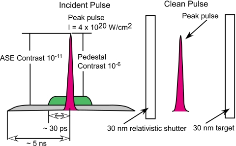
The experiment was performed using the Hercules laser system at the University of Michigan. The target consisted of two silicon nitride (Si3N4) foils of varying thickness (30 or 50 nm) separated by 20 μm. A schematic illustration is shown in Fig. 1. An f∕2 off axis parabolic mirror focused the 800 nm, p-polarized, 30 fs duration, laser pulse into a 3 μm full width at half maximum (FWHM) focal spot diameter irradiating the rear foil at a 20° angle of incidence. The peak intensity on target was 4×10²⁰ W∕cm2 and approximately 2×10²⁰ W∕cm2 on the shutter surface. The laser system had a 10% shot-to-shot energy fluctuation. The ASE contrast, as measured by a third order autocorrelator, was 10−11 by using XPW technique,13 allowing for the ASE intensity (∼109 W∕cm2) to be below the damage threshold for dielectric targets. However, the contrast measured from 30 to ∼1 ps before the laser peak begins to degrade quickly from 10−11 to approximately 10−6.
Figure 1.
Schematic of the relativistic plasma shutter for ultraintense laser pulses.
The thickness of the shutter was chosen based on experimental laser transmission data. For example, if 0% of the laser light was transmitted through a 100 nm shutter, then the target was opaque and still above the relativistic critical density when the peak pulse arrived. If a 100 nm shutter transmits ∼100% of the laser energy, then the target was below the relativistic critical density allowing the laser to transmit through. The transmission data was measured by placing targets of varying thickness 20 μm before the laser focus. For thicknesses of 200 nm and above, approximately 0% of the laser energy was transmitted. The transmission rises for 100 and 75 nm foils and was on average of 6% and 40% of the laser energy, respectively. 50 nm thick targets varied widely in transmitted energy, ranging from ∼5% to 95% transmission because the expanded plasma cloud was at the threshold of the relativistic critical density. However, 30 nm targets consistently transmitted 70% to 99% of the laser energy, effectively removing the sub-100 ps laser prepulse. Therefore, 30 nm foils were chosen and used as a shutter throughout the experiment.
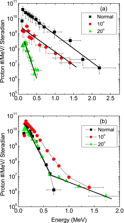
Figure 2 shows the proton spectrum simultaneously measured along three different angles, 0°, 10°, and 20° (laser axis) to the normal for a 50 nm stand alone target and a shuttered 50 nm target. This was done using 3 cm long, 0.45 T magnetic spectrometers with a 120 μm diameter pinhole in front of CR-39 track detectors. The proton spectrum from the 50 nm Si3N4 target shows the highest energy protons are along the target normal direction, and fewer low energy protons are accelerated along the laser axis [Fig. 2a]. The data clearly shows in Fig. 2b that when the shutter is inserted, the highest energy protons lie along the laser axis (20°), while the protons along the target normal have decreased in energy. Therefore, as consequence of the 30 nm plasma shutter, the protons move away from the target normal direction toward the laser axis. To further investigate this pulse cleaning technique, 30 nm targets were irradiated with and without a shutter. Because of the sub-100 ps laser prepulse no single 30 nm thick Si3N4 target generated any detectable protons. However, by inserting the plasma shutter, 1.8 MeV protons were observed.
Figure 2.
Proton beam energy spectrum from CR-39 detectors (a) 50 nm Si3N4 stand alone target and (b) with a 30 nm Si3N4 shutter placed in front of a 50 nm Si3N4 target. By inserting the shutter, the highest energy protons shift away from the target normal toward the laser axis (20°) which is characteristic of a high contrast laser solid interaction.
To understand this data it is convenient to separate the laser interaction into two stages. First, the laser prepulse ionizes the shutter creating a cloud of plasma expanding in front of the main interaction target. This is simulated using the one-dimensional hydrodynamics code HYADES.14 The second stage, which is studied using two-dimensional (2D) PIC simulations, tracks the high-intensity pulse interacting with the main target.
The hydrodynamic code simulated Si3N4 targets irradiated with an 800 nm, 1 mJ pulse ramping from 109 to 1015 W∕cm2 over 30 ps, coming in at a 20° angle of incidence. The simulations show that 30 nm shutters are transformed into a ∼10 μm thick, ∼3nc peak density cloud of plasma. This is above the nonrelativistic critical density but below the relativistic critical density of .
The PIC simulations were performed for two cases. First, the case of the laser pulse interacting with a 50 nm thick, 400nc density target to illustrate what is expected during a high contrast interaction. Here target deformation is observed causing the protons to shift toward the laser axis. The second case describes a 50 nm Si3N4 target without a shutter which has expanded several microns due to the laser prepulse. For this case, the PIC simulations show the protons are accelerated in the target normal direction. The hydro simulations show the laser prepulse transforms the 50 nm target into a 5 μm thick, 10nc electron density cloud and is used as the target in the PIC simulations.
The 2D PIC simulations are based on the code REMP.15 Particle acceleration from the high-intensity laser interactions was studied by using a two-layer silicon-hydrogen target. The incident target surface is assumed to be Si ions, and the rear target surface is the proton contamination layer. The two-layer setup allows for particles to be independently tracked from both locations. The temporal and spatial profile of the pulse are Gaussian with a laser power of 30 TW, pulse duration of 30 fs, 20° incidence angle, and a spot size of 3 μm (FWHM).
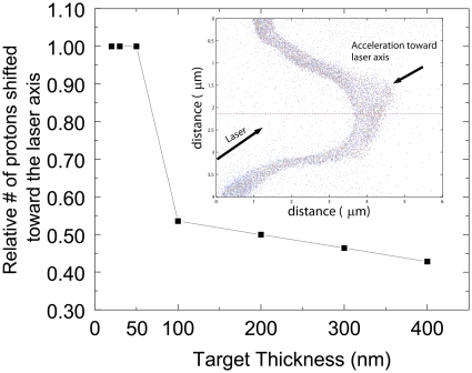
In the first case, representing high contrast, the following interaction scenario is observed for the 50 nm thick, 400nc density Si–H target. As the peak intensity of the laser pulse enters the plasma, the laser’s Gaussian spatial profile is imprinted upon the accelerated electrons which quickly propagate through the thin target. PIC simulations show the laser ponderomotive force on the electrons accelerates them forward creating a charge separation sheath which then accelerates the ions with a similar spatial profile. The Si ion and H density show the target deforming with a structure matching that of the laser pulse and shows an asymmetry in the density profile due to the particles being accelerated toward the laser axis. However, target deformation is observed only for sufficiently thin targets. Figure 3 shows PIC simulation results of the top 20% highest energy protons in the simulation which are accelerated toward the laser axis direction as a function of target thickness for a 20° off-normal incident laser. As the target thickness decreases, it becomes easier for the laser pressure to deform the rear surface which in turn directs the protons toward the laser axis. As shown in Fig. 3 the number of proton deflected quickly saturates because the once the target rear surface matches the laser’s spatial profile the thinner targets will do the same. The insert shows for a 20° incident laser pulse the ion and proton shift toward the laser axis.
Figure 3.
PIC simulation results showing the relative number of high energy protons directed toward the laser axis as a function of target thickness. The simulations were performed using a 30 TW laser incident at 20° off-normal. The insert shows the resulting asymmetry of the ion and proton density acceleration directed toward the laser axis for a 50 nm thick target.
In the second case, without the plasma shutter, the laser prepulse expands the 50 nm target several microns, however the targets’ rear surface expands symmetrically. This causes the accelerated protons to be directed along the target normal direction, which is consistent with the experimental results.
In conclusion, after the insertion of the 30 nm shutter two important results were experimentally observed. First, the proton beam’s direction shifted from the normal axis toward the laser axis signifying target deformation due to the laser light pressure. If the target experiences a deformation due to the laser pulse, the proton beam will replicate the structure. As the PIC simulations showed, the laser pressure will deform a thin target, but this can only happen if the contrast is sufficiently high. The second result was the production of 1.8 MeV protons from a shuttered 30 nm target, while no protons were detected for a 30 nm stand alone target. The relativistic plasma shutter technique is simple to implement and scaleable as the laser power is increased. Use of this technique requires a detailed knowledge of the laser prepulse parameters in order to optimize the laser contrast.
Acknowledgments
This study was supported by the NSF through the FOCUS Center (Grant No. PHY-0114336) and in part by the NIH (Grant No. R21 CA120262-01).
References
- Bahk S. W., Rousseau P., Planchon T. A., Chvykov V., Kalintchenko G., Maksimchuk A., Mourou G. A., and Yanovsky V., Opt. Lett. 29, 2837 (2004). 10.1364/OL.29.002837 [DOI] [PubMed] [Google Scholar]
- Yanovsky V., Chvykov V., Kalinchenko G., Rousseau P., Planchon T., Matsuoka T., Maksimchuk A., Nees J., Cheriaux G., Mourou G., and Krushelnick K., Opt. Express 16, 2109 (2008). 10.1364/OE.16.002109 [DOI] [PubMed] [Google Scholar]
- Itatani J., Faure J., Nantel M., Mourou G., and Watanabe S., Opt. Commun. 148, 70 (1998). 10.1016/S0030-4018(97)00638-X [DOI] [Google Scholar]
- Homoelle D., Gaeta A. L., Yanovsky V., and Mourou G., Opt. Lett. 27, 1646 (2002). 10.1364/OL.27.001646 [DOI] [PubMed] [Google Scholar]
- Jullien A., Augé-Rochereau F., Chériaux G., Chambaret J. -P., d’Oliveira P., Auguste T., and Falcoz F., Opt. Lett. 29, 2184 (2004). 10.1364/OL.29.002184 [DOI] [PubMed] [Google Scholar]
- Kalashnikov M. P., Risse E., Schonnagel H., and Sandner W., Opt. Lett. 30, 923 (2005). 10.1364/OL.30.000923 [DOI] [PubMed] [Google Scholar]
- Kapteyn H. C., Murnane M. M., Szoke A., and Falcone R. W., Opt. Lett. 16, 490 (1991). 10.1364/OL.16.000490 [DOI] [PubMed] [Google Scholar]
- Thaury C., Quere F., Geindre J. -P., Levy A., Ceccotti T., Monot P., Bougeard M., Reau F., D’Oliviera P., Audebert P., Marjoribanks R., and Martin Ph., Nat. Phys. 3, 424 (2007). 10.1038/nphys595 [DOI] [Google Scholar]
- Mackinnon A. J., Sentoku Y., Patel P. K., Price D. W., Hatchett S., Key M. H., Andersen C., Snavely R., and Freeman R. R., Phys. Rev. Lett. 88, 215006 (2002). 10.1103/PhysRevLett.88.215006 [DOI] [PubMed] [Google Scholar]
- Neely D., Foster P., Robinson A., Lindau F., Lundh O., Persson A., Wahlström C. G., and McKenna P., Appl. Phys. Lett. 89, 021502 (2006). 10.1063/1.2220011 [DOI] [Google Scholar]
- Bulanov S. S., Brantov A., Bychenkov V. Yu., Chvykov V., Kalinchenko G., Matsuoka T., Rousseau P., Reed S., Yanovsky V., Krushelnick K., Litzenberg D. W., and Maksimchuk A., Med. Phys. 35, 1770 (2008). 10.1118/1.2900112 [DOI] [PMC free article] [PubMed] [Google Scholar]
- Vshivkov V. A., Naumova N. M., Pegoraro F., and Bulanov S. V., Phys. Plasmas 5, 2727 (1998). 10.1063/1.872961 [DOI] [Google Scholar]
- Chvykov V., Rousseau P., Reed S., Kalinchenko G., and Yanovsky V., Opt. Lett. 31, 2993 (2006). 10.1364/OL.31.002993 [DOI] [PubMed] [Google Scholar]
- Larsen J. T. and Lane S. M., J. Quant. Spectrosc. Radiat. Transf. 51, 179 (1994). 10.1016/0022-4073(94)90078-7 [DOI] [Google Scholar]
- Esirkepov T. Zh., Comput. Phys. Commun. 135, 144 (2001). 10.1016/S0010-4655(00)00228-9 [DOI] [Google Scholar]


