

Abstract
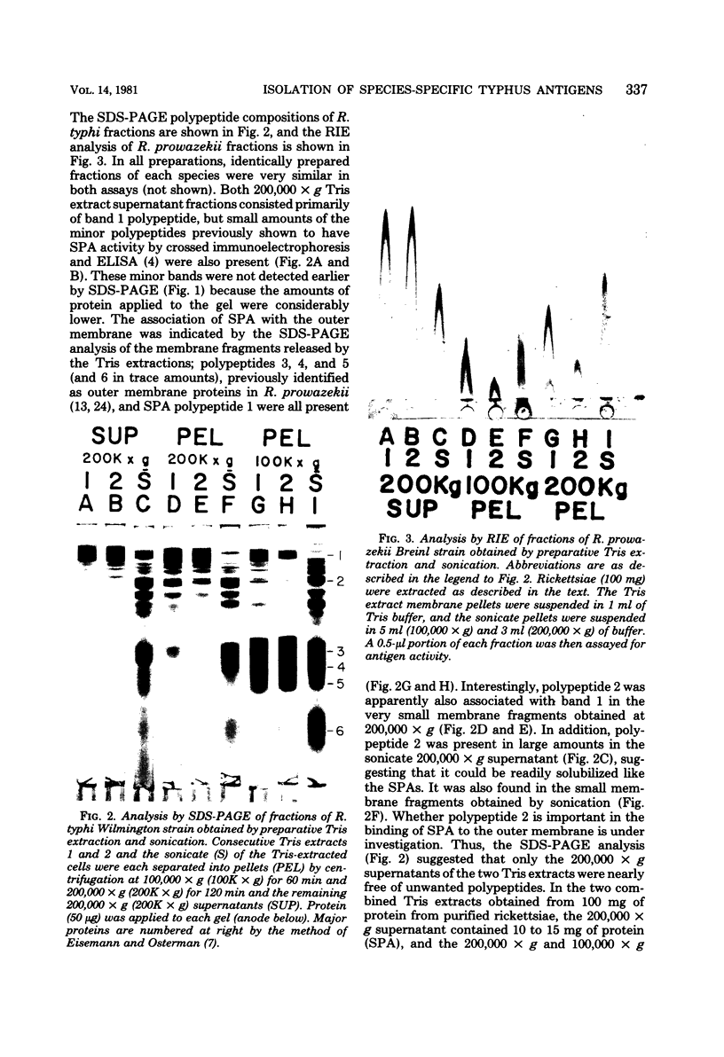
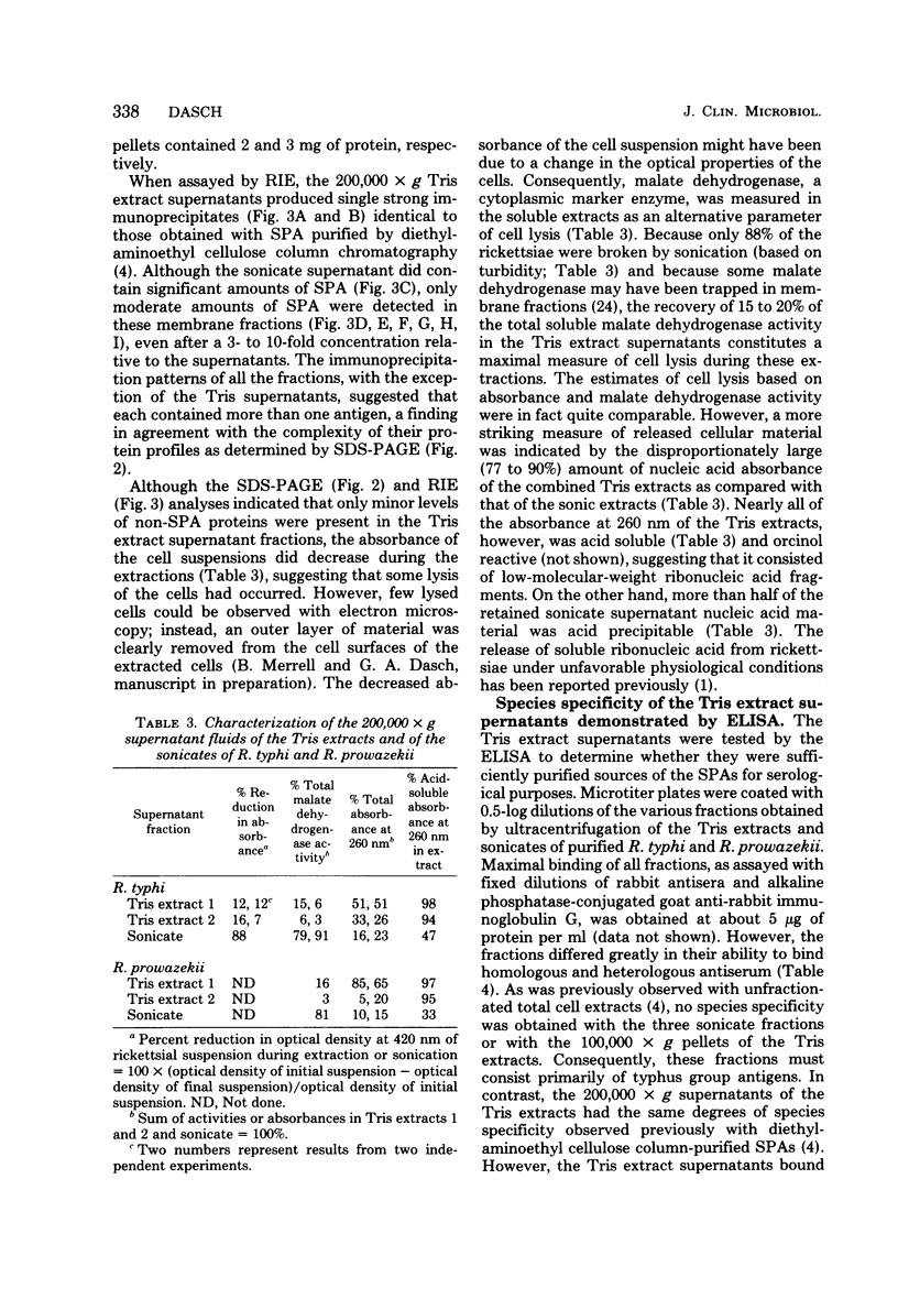
A simple procedure for the selective isolation of the protective species-specific protein antigens (SPAs) of Rickettsia typhi and Rickettsia prowazekii was developed to permit use of the SPAs in the immunodiagnosis and immunoprophylaxis of typhus infections. Although the SPAs were readily extracted from lysozyme- or detergent-treated rickettsiae, as measured by rocket immunoelectrophoresis, other polypeptides were also present, as shown by sodium dodecyl sulfate-polyacrylamide gel electrophoresis. In contrast, both water and seven buffers, each at a 10 mM concentration and pH 7.6, were nearly equally effective in the selective release of the SPAs from whole cells by extraction for 30 min at 45 degrees C. High-ionic-strength buffers and MgCl2 abolished this SPA release, thus suggesting that divalent cations were important in the binding of the SPAs to the cell envelope. The efficacy of the dilute buffer extraction procedure for isolation of large amounts of SPAs was tested by further characterization of the supernatants obtained by centrifugation (200,000 x g) of two successive tris-(hydroxymethyl)aminomethane-hydrochloride buffer (Tris) extracts. With this procedure, between 10 and 15 mg of SPA was obtained from 100 mg of purified rickettsiae. Although low-molecular-weight ribonucleic acid fragments were released into the Tris extracts in significant amounts, only the SPAs were detected, in significant quantities, as measured by polyacrylamide gel electrophoresis and rocket immunoelectrophoresis. The Tris extracts contained the same major and minor SPA polypeptides as those observed previously in SPA preparations obtained by extensive diethylaminoethyl-cellulose column chromatography, but the Tris SPAs were more satisfactory antigens in an enzyme-linked immunosorbent assay.
Full text
PDF
Images in this article
Selected References
These references are in PubMed. This may not be the complete list of references from this article.
- COHN Z. A., HAHN F. E., CEGLOWSKI W., BOZEMAN F. M. Unstable nucleic acids of Rickettsia mooseri. Science. 1958 Feb 7;127(3293):282–283. doi: 10.1126/science.127.3293.282. [DOI] [PubMed] [Google Scholar]
- Dasch G. A., Samms J. R., Weiss E. Biochemical characteristics of typhus group rickettsiae with special attention to the Rickettsia prowazekii strains isolated from flying squirrels. Infect Immun. 1978 Feb;19(2):676–685. doi: 10.1128/iai.19.2.676-685.1978. [DOI] [PMC free article] [PubMed] [Google Scholar]
- Dasch G. A., Samms J. R., Williams J. C. Partial purification and characterization of the major species-specific protein antigens of Rickettsia typhi and Rickettsia prowazekii identified by rocket immunoelectrophoresis. Infect Immun. 1981 Jan;31(1):276–288. doi: 10.1128/iai.31.1.276-288.1981. [DOI] [PMC free article] [PubMed] [Google Scholar]
- Dasch G. A., Weiss E. Characterization of the Madrid E strain of Rickettsia prowazekii purified by renografin density gradient centrifugation. Infect Immun. 1977 Jan;15(1):280–286. doi: 10.1128/iai.15.1.280-286.1977. [DOI] [PMC free article] [PubMed] [Google Scholar]
- Eisemann C. S., Osterman J. V. Proteins of typhus and spotted fever group rickettsiae. Infect Immun. 1976 Jul;14(1):155–162. doi: 10.1128/iai.14.1.155-162.1976. [DOI] [PMC free article] [PubMed] [Google Scholar]
- Golinevitch H. M., Voronova Z. A. The superficial protective antigen of R. prowazeki. J Hyg Epidemiol Microbiol Immunol. 1968;12(4):413–419. [PubMed] [Google Scholar]
- Golinevitch H., Voronova Z., Friazinova I., Doutova G., Dolgov H., Goudima O., Nikolskaya V., Botcharova T. L'antigène soluble purifié de R. prowazeki (le vaccine chimique antityphique) Rev Immunol Ther Antimicrob. 1969 Jul-Sep;33(4):229–258. [PubMed] [Google Scholar]
- Hollingdale M. R., Vinson J. W., Herrmann J. E. Immunochemical and biological properties of the outer membrane-associated lipopolysaccharide and protein of Rochalimaea quintana. J Infect Dis. 1980 May;141(5):672–679. doi: 10.1093/infdis/141.5.672. [DOI] [PubMed] [Google Scholar]
- Osterman J. V., Eisemann C. S. Surface proteins of typhus and spotted fever group rickettsiae. Infect Immun. 1978 Sep;21(3):866–873. doi: 10.1128/iai.21.3.866-873.1978. [DOI] [PMC free article] [PubMed] [Google Scholar]
- Palmer E. L., Mallavia L. P., Tzianabos T., Obijeski J. F. Electron microscopy of the cell wall of Rickettsia prowazeki. J Bacteriol. 1974 Jun;118(3):1158–1166. doi: 10.1128/jb.118.3.1158-1166.1974. [DOI] [PMC free article] [PubMed] [Google Scholar]
- Palmer E. L., Martin M. L., Mallavia L. Ultrastucture of the surface of Rickettsia prowazeki and Rickettsia akari. Appl Microbiol. 1974 Oct;28(4):713–716. doi: 10.1128/am.28.4.713-716.1974. [DOI] [PMC free article] [PubMed] [Google Scholar]
- Popov V. L., Ignatovich V. F. Electron microscopy of surface structures of Rickettsia prowazeki stained with ruthenium red. Acta Virol. 1976 Oct;20(5):424–428. [PubMed] [Google Scholar]
- Reiss-Gutfreund R. J., Cappucinelli P., Cavallo G. The soluble antigens of Rickettsia prowazeki, R. typhi and R. canada. Investigation of their interrelationship by various serological methods. Z Immunitatsforsch Exp Klin Immunol. 1975 Feb;148(4):315–329. [PubMed] [Google Scholar]
- Silverman D. J., Wisseman C. L., Jr Comparative ultrastructural study on the cell envelopes of Rickettsia prowazekii, Rickettsia rickettsii, and Rickettsia tsutsugamushi. Infect Immun. 1978 Sep;21(3):1020–1023. doi: 10.1128/iai.21.3.1020-1023.1978. [DOI] [PMC free article] [PubMed] [Google Scholar]
- Silverman D. J., Wisseman C. L., Jr, Waddell A. D., Jones M. External layers of Rickettsia prowazekii and Rickettsia rickettsii: occurrence of a slime layer. Infect Immun. 1978 Oct;22(1):233–246. doi: 10.1128/iai.22.1.233-246.1978. [DOI] [PMC free article] [PubMed] [Google Scholar]
- Sleytr U. B. Regular arrays of macromolecules on bacterial cell walls: structure, chemistry, assembly, and function. Int Rev Cytol. 1978;53:1–62. doi: 10.1016/s0074-7696(08)62240-8. [DOI] [PubMed] [Google Scholar]
- Smith D. K., Winkler H. H. Radioiodination of an outer membrane protein in intact Rickettsia prowazekii. Infect Immun. 1980 Aug;29(2):831–834. doi: 10.1128/iai.29.2.831-834.1980. [DOI] [PMC free article] [PubMed] [Google Scholar]
- Smith D. K., Winkler H. H. Separation of inner and outer membranes of Rickettsia prowazeki and characterization of their polypeptide compositions. J Bacteriol. 1979 Feb;137(2):963–971. doi: 10.1128/jb.137.2.963-971.1979. [DOI] [PMC free article] [PubMed] [Google Scholar]
- Thorne K. J. Regularly arranged protein on the surfaces of Gram-negative bacteria. Biol Rev Camb Philos Soc. 1977 May;52(2):219–234. doi: 10.1111/j.1469-185x.1977.tb01351.x. [DOI] [PubMed] [Google Scholar]
- Weber K., Osborn M. The reliability of molecular weight determinations by dodecyl sulfate-polyacrylamide gel electrophoresis. J Biol Chem. 1969 Aug 25;244(16):4406–4412. [PubMed] [Google Scholar]
- Weiss E., Coolbaugh J. C., Williams J. C. Separation of viable Rickettsia typhi from yolk sac and L cell host components by renografin density gradient centrifugation. Appl Microbiol. 1975 Sep;30(3):456–463. doi: 10.1128/am.30.3.456-463.1975. [DOI] [PMC free article] [PubMed] [Google Scholar]
- Woodman D. R., Weiss E., Dasch G. A., Bozeman F. M. Biological properties of Rickettsia prowazekii strains isolated from flying squirrels. Infect Immun. 1977 Jun;16(3):853–860. doi: 10.1128/iai.16.3.853-860.1977. [DOI] [PMC free article] [PubMed] [Google Scholar]