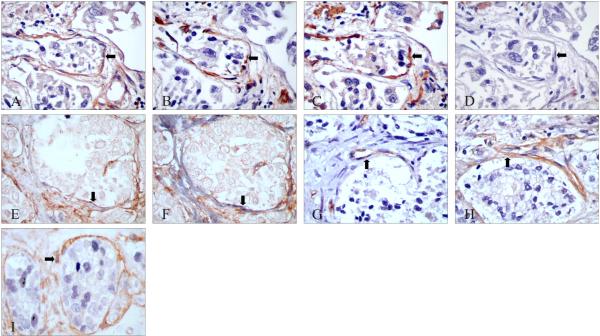
Figure 2. Immunohistochemical characterization of PAR-1 localization in reactive stroma.
A-D: Serial sections stained with PAR-1, vimentin, α-SMA and desmin antibodies, respectively. The peritumoral stromal cells were PAR-1 positive (A, black arrow), vimentin positive (B), α-SMA positive (C) but desmin negative (D). E*, F*: Serial sections with double staining of PAR-1 (brown) & desmin (gray) (E), and PAR-1 (brown) & α-SMA (gray) (F). The peritumoral stromal cells were PAR-1 and α-SMA positive, but desmin negative (black arrows). G, H: Serial sections stained with CD34 (G, black arrow) or PAR-1 (H, black arrow). I*: Co-expression of PAR-1 (in brown) and PCNA (in gray) in reactive stromal cells (black arrow).
Original magnification: A to I ×400
*No counterstaining was performed in double-label IHC.

