

Abstract
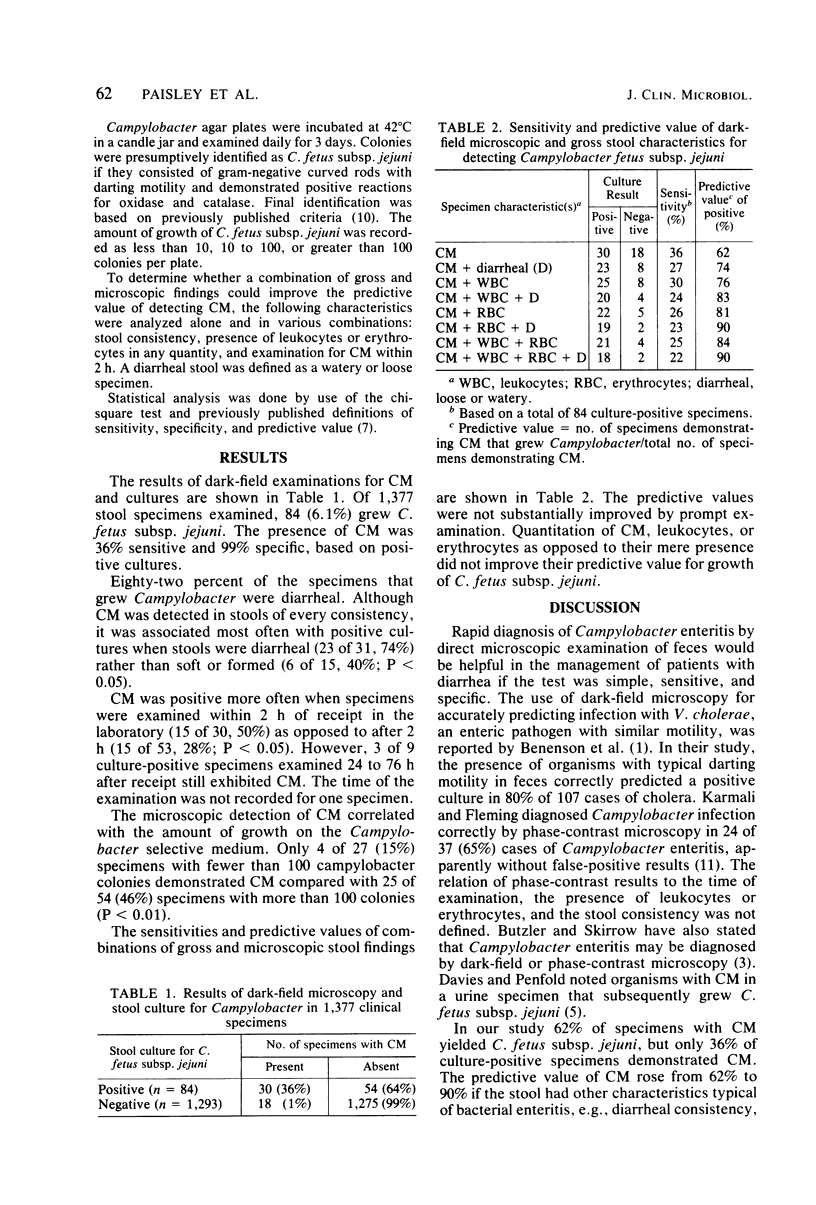
To determine the value of direct dark-field microscopy for diagnosing enteritis due to Campylobacter fetus subsp. jejuni, we examined 1,377 human fecal specimens for bacteria with typical Campylobacter darting motility, leukocytes, and erythrocytes. Eighty-four specimens (6.1%) grew C.fetus subsp. jejuni. Of the 48 specimens showing Campylobacter motility, 30 (62%) grew C. fetus subsp. jejuni. The sensitivity, specificity, and predictive value of observing Campylobacter motility were 36%, 99%, and 62%, respectively. The predictive value of detecting Campylobacter motility was improved if the specimens were diarrheal (23 of 31, 74%), leukocytes were present (25 of 33, 76%), erythrocytes were present (22 of 27, 81%), or if all of the above findings were present (18 of 20, 90%). The sensitivity of detecting Campylobacter darting motility was highest if specimens were examined within 2 h of arrival in the laboratory (15 of 30, 50%) as opposed to after 2 h (15 of 53, 28%; P less than 0.01). Prompt dark-field microscopic examination of diarrheal stool specimens is valuable for the presumptive diagnosis of Campylobacter enteritis.
Full text
PDF
Selected References
These references are in PubMed. This may not be the complete list of references from this article.
- BENENSON A. S., ISLAM M. R., GREENOUGH W. B., 3rd RAPID IDENTIFICATION OF VIBRIO CHOLERAE BY DARKFIELD MICROSCOPY. Bull World Health Organ. 1964;30:827–831. [PMC free article] [PubMed] [Google Scholar]
- Blaser M. J., Berkowitz I. D., LaForce F. M., Cravens J., Reller L. B., Wang W. L. Campylobacter enteritis: clinical and epidemiologic features. Ann Intern Med. 1979 Aug;91(2):179–185. doi: 10.7326/0003-4819-91-2-179. [DOI] [PubMed] [Google Scholar]
- Butzler J. P., Skirrow M. B. Campylobacter enteritis. Clin Gastroenterol. 1979 Sep;8(3):737–765. [PubMed] [Google Scholar]
- Chester B., Poulos E. G. Rapid presumptive identification of vibrios by immobilization in distilled water. J Clin Microbiol. 1980 May;11(5):537–539. doi: 10.1128/jcm.11.5.537-539.1980. [DOI] [PMC free article] [PubMed] [Google Scholar]
- Davies J. S., Penfold J. B. Campylobacter urinary infection. Lancet. 1979 May 19;1(8125):1091–1092. doi: 10.1016/s0140-6736(79)92995-7. [DOI] [PubMed] [Google Scholar]
- DuPont H. L., Hornick R. B. Clinical approach to infectious diarrheas. Medicine (Baltimore) 1973 Jul;52(4):265–270. [PubMed] [Google Scholar]
- George H. A., Hoffman P. S., Smibert R. M., Krieg N. R. Improved media for growth and aerotolerance of Campylobacter fetus. J Clin Microbiol. 1978 Jul;8(1):36–41. doi: 10.1128/jcm.8.1.36-41.1978. [DOI] [PMC free article] [PubMed] [Google Scholar]
- Harris J. C., Dupont H. L., Hornick R. B. Fecal leukocytes in diarrheal illness. Ann Intern Med. 1972 May;76(5):697–703. doi: 10.7326/0003-4819-76-5-697. [DOI] [PubMed] [Google Scholar]
- Karmali M. A., Fleming P. C. Campylobacter enteritis in children. J Pediatr. 1979 Apr;94(4):527–533. doi: 10.1016/s0022-3476(79)80004-9. [DOI] [PubMed] [Google Scholar]
- Nelson J. D., Haltalin K. C. Accuracy of diagnosis of bacterial diarrheal disease by clinical features. J Pediatr. 1971 Mar;78(3):519–522. doi: 10.1016/s0022-3476(71)80240-8. [DOI] [PubMed] [Google Scholar]
- Pickering L. K., DuPont H. L., Olarte J., Conklin R., Ericsson C. Fecal leukocytes in enteric infections. Am J Clin Pathol. 1977 Nov;68(5):562–565. doi: 10.1093/ajcp/68.5.562. [DOI] [PubMed] [Google Scholar]