A 46-year-old woman with a history of hypercholesterolemia, hypothyroidism, and cigarette smoking presented to the emergency department because of recurrent episodes of substernal chest pain. Her family history included early-onset myocardial infarction. Electrocardiography revealed sinus rhythm without significant ST-T changes. Recent exercise-stress echocardiography had shown preserved left ventricular function (ejection fraction, 0.60) and a normal stress test result.
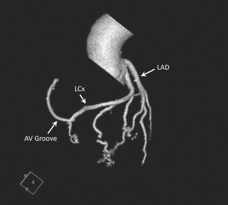
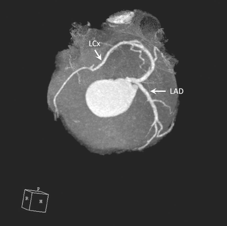
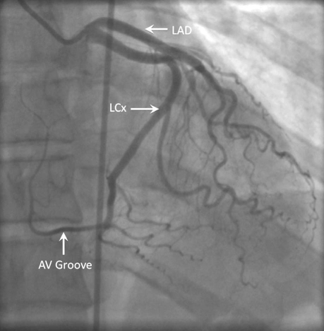
The patient was next evaluated by 64-slice multidetector computed tomography of the coronary arteries, which revealed an anomalous single coronary artery arising from the left sinus of Valsalva, together with an absence of the right coronary artery (RCA). The left circumflex coronary artery (LCx) was the dominant vessel; it appeared to continue, without significant stenosis, beyond the atrioventricular groove up to the level normally occupied by the RCA (Fig. 1). The left main coronary artery bifurcated into the left anterior descending coronary artery (LAD) and the LCx. Only a single coronary ostium (that of the left main) was seen to arise from the aorta (Fig. 2). The patient's calcium score was zero. Subsequently, her coronary anatomy was confirmed by cardiac catheterization (Fig. 3). She was treated medically with aspirin, metoprolol, and rovastatin.
Fig. 1 Computed tomographic angiogram. Three-dimensional volume-rendering view (anterior) shows an anomalous single coronary artery from the left sinus of Valsalva, together with an absence of the right coronary artery. The left circumflex artery (LCx) continues beyond the atrioventricular (AV) groove as right coronary artery distribution.
LAD = left anterior descending coronary artery
Fig. 2 Computed tomographic angiogram. Three-dimensional volume-rendering view (superior) shows an anomalous single coronary artery arising from the left sinus of Valsalva and the left circumflex coronary artery (LCx) continuing as right coronary artery distribution. There is no evidence of a dual ostium.
LAD = left anterior descending coronary artery
Fig. 3 Coronary angiogram of the left coronary artery in the posteroanterior projection confirms that the left circumflex coronary artery (LCx) continues as right coronary artery distribution.
AV = atrioventricular; LAD = left anterior descending coronary artery
Comment
Isolated single coronary artery is extremely rare, with an incidence of 0.024% to 0.066% in the general population.1 Most patients are asymptomatic, and prognoses vary. Group I anomalies, defined as solitary dominant vessels that follow the course of either a normal right or a normal left coronary artery (in accordance with the modification of Lipton's classification2,3), are extremely rare and generally have a benign clinical course.4 Our images show an anomalous coronary artery (L-1 subtype) that originates from the left sinus of Valsalva, gives off the LCx in normal fashion, and then continues in the atrioventricular groove up to the level of the RCA. The blood supply to the right ventricular free wall is similar to that provided by a native RCA that arises from the right coronary cusp.
Most coronary anomalies are found incidentally during coronary angiography. A recent study5 demonstrated that multidetector computed tomography can be a noninvasive alternative to conventional coronary angiography for imaging anomalous coronary arteries. Medical treatment is recommended in patients who present with single coronary artery in the absence of ischemia.6
Footnotes
Address for reprints: Tanyanan Tanawuttiwat, MD, 4440 West 95th Street, Oak Lawn, IL 60453
E-mail: ttanawuttiwat@gmail.com
References
- 1.Desmet W, Vanhaecke J, Vrolix M, Van de Werf F, Piessens J, Willems J, de Geest H. Isolated single coronary artery: a review of 50,000 consecutive coronary angiographies. Eur Heart J 1992;13(12):1637–40. [DOI] [PubMed]
- 2.Lipton MJ, Barry WH, Obrez I, Silverman JF, Wexler L. Isolated single coronary artery: diagnosis, angiographic classification, and clinical significance. Radiology 1979;130(1):39–47. [DOI] [PubMed]
- 3.Yamanaka O, Hobbs RE. Coronary artery anomalies in 126,595 patients undergoing coronary arteriography. Cathet Cardiovasc Diagn 1990;21(1):28–40. [DOI] [PubMed]
- 4.Sheth M, Dovnarsky M, Cha SD, Kini P, Maranhao V. Single coronary artery: right coronary artery originating from distal left circumflex. Cathet Cardiovasc Diagn 1988;14(3):180–1. [DOI] [PubMed]
- 5.Kacmaz F, Ozbulbul NI, Alyan O, Maden O, Demir AD, Balbay Y, et al. Imaging of coronary artery anomalies: the role of multidetector computed tomography. Coron Artery Dis 2008;19(3):203–9. [DOI] [PubMed]
- 6.Akcay A, Tuncer C, Batyraliev T, Gokce M, Eryonucu B, Koroglu S, Yilmaz R. Isolated single coronary artery: a series of 10 cases. Circ J 2008;72(8):1254–8. [DOI] [PubMed]


