Abstract
We report a case of gastric pseudoterranoviasis proven by gastrofiberscopy on Dec. 13, 1994. The 34-year-old male patient, residing in Chungju-shi, was admitted to Konkuk University Hospital complaining of prickling epigastric pain. The symptoms suddenly attacked him two days after eating raw marine fish at Chonan-shi. By the gastrofiberscopic examination, a long white-yellowish nematode was found from the fundus region of stomach. The worm was 34.50 × 0.84 mm in size, and was identified as a 3rd stage larva of Pseudoterranova decipiens judging from the position of the intestinal cecum. This is the 12th confirmed case of human pseudoterranoviasis in Korea.
Keywords: pseudoterranoviasis, gastrofiberscopy, Korea
Pseudoterranova decipiens, the seal nematode, has been found in many seas around the world (Bristow and Berland, 1992), including the Antarctic Ocean (Chai et al., 1995). While numerous cases of anisakiasis have been presented by many workers after the first human case report (Kim et al., 1971), only 11 cases among them were identified as Pseudoterranova type A larva (Seo et al., 1984; Lee et al., 1985; Im et al., 1990, 1995; Im and Shin, 1991; Sohn and Seol, 1994a; Lee et al., 1998; Koh et al., 1999). Recently, we have seen a case of gastric disorder caused by a larval P. decipiens. This paper deals with the 12th human case of pseudoterranoviasis in Korea.
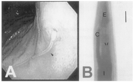
The patient, residing in Chungju-shi, Chungchungbuk-do, was a 34-year-old man. He was admitted to the Department of Internal Medicine in Konkuk University Hospital on Dec 13, 1994. The chief complaint of the patient was prickling epigastric pain and nausea, which developed two days after eating raw marine fish at Chonan-shi, Chungchungnam-do. He was diagnosed as having gastroenteritis. On physical examination, rebound tenderness was positive at the epigastrium, and no specific abnormal findings were revealed from laboratory examinations. Gastrofiberscopic exmaination was performed under the impression of anisakiasis. A long white, yellowish nematode larva invading the gastric mucosa was found in the fundus region and removed (Fig. 1A).
Fig. 1.
The larva of Pseudoterranova decipiens found in the stomach of 34-year old patient. A, Gastrofundoscopic view of the present case showing a long white-yellowish larva penetrating the gastric mucosa in the fundus (arrowhead). B, Ventricular level of the worm showing the esophagus (E), ventriculus (V), intestine (I) and intestinal cecum (C). Bar = 300 µm.
The recovered worm was fixed in 10% formalin, cleared in alcohol glycerine and mounted in glycerin jelly. The mounted specimen was observed and measured under a light microscope. The nematode larva is slender and the mouth is surrounded by three lips without a boring tooth. The intestinal cecum is stretched anteriorly to the level of one-third portion of the ventriculus (Fig. 1B). The tail is conical shaped, and reproductive organs are not developed.
The worm measured 34.50 mm long (L) and 0.84 mm wide (W). The total length of the esophagus (E) was 3.38 mm. The ventriculus (V), 1.25 mm long, is directly connected to the muscular esophagus (M), 2.13 mm long, without any appendage. The cecum (C) 1.13 mm, tail 0.12 mm. The morphological indices of the worm are as follows; α(L/W) = 27.60; β1(L/E) = 10.21; β2(L/M) = 15.81; β3(L/V) = 27.60; γ(L/C) = 30.53. From the morphological characteristics and indices, the worm is identified as the 3rd stage larva of P. decipiens.
The majority of human gastric anisakiasis reported in the literature is Anisakis simplex type A larvae (Sohn and Seol, 1994b). Therefore, most clinicians might be under the false impression that all anisakiasis are due to the infection with Anisakis type I larvae. In fact, many reports on anisakiasis overlooked the species identification. Sohn and Seol (1994b) mentioned that species identification had been done only in 38 out of 155 cases of anisakiasis reported in Korea. Presumably, more cases of pseudoterranoviasis might have existed in Korea.
The present worm was diagnosed as P. decipiens based on the absence of ventricular appendage but the presence of an intestinal cecum. In addition, the worm has a cecum reaching beyond the one third anterior level of ventriculus and a mucron at posterior end, suggesting the 3rd stage.
The larva from this case, 34.50 mm long, was found two days after infection. The longest larva of P. decipiens reported in Korea was that of Lee et al. (1998), 42.6 mm long, and the duration of infection was three days. The next was that of Koh et al. (1999), 38.27 mm when it was found 16 days after ingesting raw marine fish. The length was 29.73 mm in the report of Sohn and Seol (1994a) where the duration of infection was two days, and 25.76 mm in the report of Seo et al. (1984), with a 6 hr duration. Except for the case of Lee et al. (1998), the length of P. decipiens larvae were roughly proportional to the duration of infection.
On the other hand, the length of Anisakis type I larvae did not exceed 25 mm in analyzing six cases of type I anisakiasis (Kim et al., 1991; Sohn and Seol, 1994b; Jeong and Song, 1995). Seol et al. (1994) reported that the length of Anisakis type I larvae were within the range of 13.3-28.9 mm (mean: 19.5). Additionally, the length of Anisakis type I larvae extracted from the yellow corvina was ranged from 13.4 to 25.0 mm (mean: 20.7) (Chai et al., 1986). Considering these results, P. decipiens larvae seemed to be somewhat longer than those of Anisakis type I.
Third stage larvae of P. decipiens are frequently found in the muscle of cod (Gadus morhua) in areas where seal are present (Brattey et al., 1990). However, there is still no report on the fish intermediate host of P. decipiens in Korea. The previous cases suggested the possible infection source as Sebasters inermis (Sohn and Seol, 1994a), bleekeri or Bothidae sp. (Koh et al., 1999), squid and yellow corvina (Im and Shin, 1991). Chai et al. (1986) investigated the yellow corvina (Pseudosciaena manchurica) from a local market in Seoul for the presence of P. decipiens larvae, but only Anisakis type I (80.4%) and Contracaecum (19.6%) were extracted. From Anago anago (Astroconger myriaster) purchased in Noryangin Market, the larvae of P. decipiens were not found either (Chai et al., 1992).
In Japan, marine fish such as halibut, cod (Alaska pollack), sailfin sand fish, nurt smelt and arctic smelt were reported to be fish host of P. decipiens (Nagano, 1989). In addition, a coalfish had been reported as a source of P. decipiens infection in France (Pinel et al., 1996). It is apparent that more research is needed to determine the infection status of market fish with marine nematodes, with special reference to P. decipiens in Korea.
References
- 1.Brattey J, Bishop CA, Myers RA. Geographic distribution and abundance of Pseudoterranova decipiens (Nematoda: Ascaridoidea) in the musculature of Atlantic cod (Gadus morhua) from Newfoundland and Labrador. Can Bull Fisheries Aquatic Sci. 1990;222:67–82. [Google Scholar]
- 2.Bristow GA, Berland B. On the ecology and distribution of Pseudoterranova decipiens (Nematoda: Anisakidae) in an intermediate host, Hippoglassoides platessoides, in Northern norwegian waters. Intern J Parasitol. 1992;22:203–208. doi: 10.1016/0020-7519(92)90102-q. [DOI] [PubMed] [Google Scholar]
- 3.Chai JY, Chu YM, Sohn WM, Lee SH. Larval anisakids collected from the yellow corvina in Korea. Korean J Parasitol. 1986;24:1–11. doi: 10.3347/kjp.1986.24.1.1. [DOI] [PubMed] [Google Scholar]
- 4.Chai JY, Cho SR, Kook J, Lee SH. Infection status of the sea eel (Astroconger myriaster) purchased from the Noryangjin fish market with anisakid larvae. Korean J Parasitol. 1992;30:157–162. doi: 10.3347/kjp.1992.30.3.157. [DOI] [PubMed] [Google Scholar]
- 5.Chai JY, Guk SM, Sung JJ, et al. Recovery of Pseudoterranova decipiens (Anisakidae) larvae from codfish of the Antarctic Ocean. Korean J Parasitol. 1995;33:231–234. doi: 10.3347/kjp.1995.33.3.231. [DOI] [PubMed] [Google Scholar]
- 6.Im KI, Shin HJ, Kim BH, Moon S. Gastric anisakiasis cases in Cheju-do, Korea. Korean J Parasitol. 1995;33:179–186. doi: 10.3347/kjp.1995.33.3.179. [DOI] [PubMed] [Google Scholar]
- 7.Im KI, Shin HJ. Morphological observation of Terranova sp. larva found in humanstomach wall. Yonsei Rep Trop Med. 1991;22:35–41. [Google Scholar]
- 8.Im IK, Yong TS, Shin HJ, et al. Gastric anisakiasis in Korea with review of 47 cases. Yonsei Rep Trop Med. 1990;21:1–7. [Google Scholar]
- 9.Jeong H, Song SB. Gastric anisakiasis. Report of four cases and Review of literature. Proceed Doctor's Assoc Pusan. 1995;31:1–5. [Google Scholar]
- 10.Kim CH, Chung BS, Moon YI, Cun SH. A case report on human infection with Anisakis sp. in Korea. Korean J Parasitol. 1971;9:39–43. doi: 10.3347/kjp.1971.9.1.39. [DOI] [PubMed] [Google Scholar]
- 11.Kim SE, Kim SL, Lee KS, et al. Nine cases of acute gastric anisakiasis. Korean J Gastroenterol. 1991;23:866–872. [Google Scholar]
- 12.Koh MS, Huh S, Sohn WM. A case of gastric pseudoterranoviasis in a 43-year old man in Korea. Korean J Parasitol. 1999;37:47–49. doi: 10.3347/kjp.1999.37.1.47. [DOI] [PMC free article] [PubMed] [Google Scholar]
- 13.Lee AH, Kim SM, Choi KY. A case of human infection with the larva of Terranova type A. Korean J Pathol. 1985;19:463–467. [Google Scholar]
- 14.Lee IH, Jang S, Lee CY, et al. A case of human infection of the larvae from Pseudoterranova decipiens. Korean J Gastrointest Endoscopy. 1998;18:732–736. [Google Scholar]
- 15.Nagano K. In: In gastric anisakiasis in Japan-Epidemiology, diganosis, treatment. Ishikura H, Namiki M, editors. Tokyo, Japan: Springerverlag; 1989. pp. 133–140. [Google Scholar]
- 16.Pinel C, Beaudevin M, Chermette R, et al. Gastric anisakidosis due to Pseudoterranova decipiens larva. Lancet. 1996;347:1829. doi: 10.1016/s0140-6736(96)91648-7. [DOI] [PubMed] [Google Scholar]
- 17.Seo BS, Chai JY, Lee SH, et al. A human case infected by the larva of Terranova type A in Korea. Korean J Parasitol. 1984;22:248–252. doi: 10.3347/kjp.1984.22.2.248. [DOI] [PubMed] [Google Scholar]
- 18.Seol SY, Ok SC, Pyo JS, et al. Twenty cases of gastric anisakiasis caused by Anisakis type I larva. Korean J Gastroenterol. 1994;26:17–24. [Google Scholar]
- 19.Sohn WM, Seol SY. A human case of gastric anisakiasis by Pseudoterranova decipiens larva. Korean J Parasitol. 1994a;32:53–56. doi: 10.3347/kjp.1994.32.1.53. [DOI] [PubMed] [Google Scholar]
- 20.Sohn WM, Seol SY. A case of acute intestinal anisakiasis caused by Anisakis type I larva. Inje Med J. 1994b;15:355–359. [Google Scholar]


