Abstract
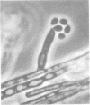
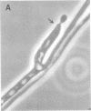
In an epidemiological study, nine isolates of dematiaceous fungi were recovered from the interior of a local feed and seed warehouse. Sample sites include brick walls and floors. Air samples also were included. Samples were collected in saline and plated on Mycobiotic and Sabouraud agar. The nine dematiaceous fungi recovered from these samples were identified with microscopic morphology, thermotolerance, biochemical reactions, and animal virulence test. Four isolates were identified as nonpathogens on the basis of positive gelatin tests. The identified pathogens included Fonsecaea pedrosoi, Cladosporium bantianum (C trichoides), Wangiella dermatitidis (Dixon et al., Mycopathologia 70:153-161, 1980), and Exophiala jeanselmei. These five organisms were injected into NCI/ALB mice. Only the isolate of C. bantianum was neurotropic, as demonstrated histopathologically and by the recovery of the organism from brain tissue. None of the remaining four isolates were seen or cultured from any of the mouse tissues analyzed. The recovery of pathogenic dematiaceous fungi from environmental sites is not uncommon. However, this study is noteworthy in that it represents only the second reported isolation of C. bantianum and the first isolation of F. pedrosoi from the environment in North America and suggests that these fungi may be more ubiquitous in this region that previously believed.
Full text
PDF


Images in this article
Selected References
These references are in PubMed. This may not be the complete list of references from this article.
- Dixon D. M., Shadomy H. J., Shadomy S. Dematiaceous fungal pathogens isolated from nature. Mycopathologia. 1980 Mar 31;70(3):153–161. doi: 10.1007/BF00443026. [DOI] [PubMed] [Google Scholar]
- Dixon D. M., Shadomy H. J., Shadomy S. Isolation of cladosporium trichoides from nature. Mycopathologia. 1977 Dec 16;62(2):125–127. doi: 10.1007/BF01259404. [DOI] [PubMed] [Google Scholar]
- Gezuele E., Mackinnon J. E., Conti-Díaz I. A. The frequent isolation of Phialophora verrucosa and Phialophora pedrosoi from natural sources. Sabouraudia. 1972 Nov;10(3):266–273. [PubMed] [Google Scholar]
- Jang S. S., Biberstein E. L., Rinaldi M. G., Henness A. M., Boorman G. A., Taylor R. F. Feline brain abscesses due to Cladosporium trichoides. Sabouraudia. 1977 Jul;15(2):115–123. [PubMed] [Google Scholar]
- KLITE P. D., KELLEY H. B., Jr, DIERCKS F. H. A NEW SOIL SAMPLING TECHNIQUE FOR PATHOGENIC FUNGI. Am J Epidemiol. 1965 Jan;81:124–130. doi: 10.1093/oxfordjournals.aje.a120491. [DOI] [PubMed] [Google Scholar]
- McGinnis M. R. Recent taxonomic developments and changes in medical mycology. Annu Rev Microbiol. 1980;34:109–135. doi: 10.1146/annurev.mi.34.100180.000545. [DOI] [PubMed] [Google Scholar]
- Newsholme S. J., Tyrer M. J. Cerebral mycosis in a dog caused by Cladosporium trichoides Emmons 1952. Onderstepoort J Vet Res. 1980 Mar;47(1):47–49. [PubMed] [Google Scholar]
- Padhye A. A., McGinnis M. R., Ajello L. Thermotolerance of Wangiella dermatitidis. J Clin Microbiol. 1978 Oct;8(4):424–426. doi: 10.1128/jcm.8.4.424-426.1978. [DOI] [PMC free article] [PubMed] [Google Scholar]
- Salfelder K., Schwarz J., Romero A., De Liscano T. R., Zambrano Z., Diaz I. Habitat de Nocardia asteroides, Phialophora pedrosoi y Cryptococcus neoformans en Venezuela. Mycopathol Mycol Appl. 1968 Mar 22;34(2):144–154. doi: 10.1007/BF02051423. [DOI] [PubMed] [Google Scholar]
- Velasquez L. F., Restrepo A. Chromomycosis in the toad (Bufo marinus) and a comparison of the etiologic agent with fungi causing human chromomycosis. Sabouraudia. 1975 Mar;13(Pt 1):1–9. doi: 10.1080/00362177585190021. [DOI] [PubMed] [Google Scholar]


