Abstract
ATP, acting on P2X7 receptors, stimulates changes in intracellular calcium concentrations, maturation, and release of interleukin-1β (IL-1β), and following prolonged agonist exposure, cell death. The functional effects of P2X7 receptor activation facilitate several proinflammatory processes associated with arthritis. Within the nervous system, these proinflammatory processes may also contribute to the development and maintenance of chronic pain. Emerging data from genetic knockout studies have indicated specific roles for P2X7 receptors in inflammatory and neuropathic pain states. The discovery of multiple distinct chemical series of potent and highly selective P2X7 receptor antagonists have enhanced our understanding of P2X7 receptor pharmacology and the diverse array of P2X7 receptor signaling mechanisms. These antagonists have provided mechanistic insight into the role(s) P2X7 receptors play under pathophysiological conditions. In this review, we integrate the recent discoveries of novel P2X7 receptor-selective antagonists with a brief update on P2X7 receptor pharmacology and its therapeutic potential.
Keywords: Calcium flux, Pore formation, Pain, Inflammation, Interleukin-1, Nerve injury, Microglia, Hyperalgesia, Neural-glial interactions
Introduction
P2X7 receptors belong to the family of ATP-sensitive ionotropic P2X receptors that are composed of seven homomeric receptor subtypes (P2X1–P2X7) [1]. P2X7 receptors are unique among the P2X receptor family since they are activated by high concentrations of ATP (> 100 μM) [2]. The P2X7 subunit, previously termed the P2Z receptor [3], was initially cloned from rat [4] and human brain [5], and from human macrophages [6]. P2X7 receptors are selectively expressed on cells of hematopoietic lineage including mast cells, lymphocytes, erythrocytes, fibroblasts, and peripheral macrophages [4]. Within the CNS, functional P2X7 receptors are localized on microglia and Schwann cells, as well as on astrocytes [5, 7]. The existence of functional P2X7 receptors on peripheral or central neurons remains controversial due to the poor specificity of both antibodies and ligands targeting the rat P2X7 receptor [8]. In rat peripheral sensory ganglia (dorsal root), P2X7 receptors appear to be selectively localized on glial cells, but not neurons [9].
P2X7 receptor-mediated changes in intracellular potassium concentrations lead to the activation of caspase-1 and the rapid maturation and release of the proinflammatory cytokine, interleukin-1β (IL-1β) [10–13]. Increased IL-1β concentrations, in turn, trigger the induction of procaspase-I [14], nitric oxide synthase, cycloxygenase-2, and tumor necrosis factor-α (TNF-α) [15–18]. Activation of caspase-3 has also been linked to P2X7 receptor activation and may underlie receptor-associated cytolytic mechanisms including pore formation [19]. Inhibitors of caspase-1 and caspase-3 effectively block P2X7 receptor-mediated IL-1β release and Yo-Pro uptake, respectively [20]. Activation of P2X7 receptors has also been associated with other downstream signaling pathways including phospholipase D (PLD) [21], phospholipase A2 (PLA2), nuclear factor kappa B (NF-κB) [22, 23], and mitogen-activated protein kinases (MAPKs) [22, 24–27].
Unlike other members of the P2 receptor superfamily, homomeric P2X7 receptors are activated by high concentrations of ATP (> 100 μM) and 2′, 3′-O-(4-benzoylbenzoyl)-ATP (BzATP) which has significantly greater potency (EC50 = 20 μM) than ATP (EC50 > 100 μM) [2]. It has been demonstrated that the human receptor has a lower sensitivity to agonists than the rat receptor [6]. Originally, functional P2X7 receptors were thought to exist as homomers; however, recent evidence has emerged indicating that P2X7 receptors may form functional heteromeric combinations with P2X4 receptors [28, 29].
There is growing evidence to indicate a pathophysiological role for P2X7 receptors in inflammatory responses [13]. For example, detailed analysis of an independently generated P2X7 receptor knockout (KO) mouse revealed that P2X7 (-/-) mice show a disruption of ATP-induced processing of pro-IL-1β in macrophages [12]. Using a monoclonal antibody-induced model of collagen arthritis, these investigators also demonstrated that P2X7 (-/-) mice show a decreased incidence and severity of arthritis in this model as compared to wild-type control mice [30]. Although published data are lacking, a P2X7 antagonist from Astra-Zeneca, AZ9056 (structure not disclosed), is reportedly under clinical development for the treatment of rheumatoid arthritis and inflammatory bowel disease [31].
A more recent study has demonstrated that P2X7 knockout mice show reduced pain sensitivity following both complete Freund’s adjuvant-induced inflammation and partial injury of the sciatic nerve [32]. These data were consistent with the mechanistic role of P2X7 receptors in modulating IL-1β release and the ability of IL-1β to alter pain sensitivity in experimental models. It has been shown, for example, that increased IL-1 levels are associated with enhanced nociceptive signaling in a concentration-related fashion [33, 34]. The P2X7 receptor has also been linked with release of the excitatory neurotransmitter glutamate, well-known for its involvement in pain transmission. In hippocampal slices from P2X7 KO mice, ATP-evoked release of [3H]glutamate was virtually absent, in contrast to the robust neurotransmitter release observed with wild-type controls [35]. Under electrical stimulation, [3H]glutamate release was also significantly attenuated in the KO animals, although some level of residual release was present. These observations are consistent with results implicating the participation of P2X7 in excitatory amino acid release from murine astrocytes [36]. The potential role of P2X7 in modulating glutamate release thus provides an additional rationale for the reduced nociceptive responses of the P2X7 KO mice under chronic inflammation or nerve injury conditions. In view of the participation of P2X7 in the release of IL-1β and glutamate, the localization of P2X7 receptors on glial cells, and the growing appreciation for the role of activated glia in promoting the development and maintenance of pathological pain [37–39], it is tempting to regard the P2X7 receptor as a central player in this neuroimmune interface.
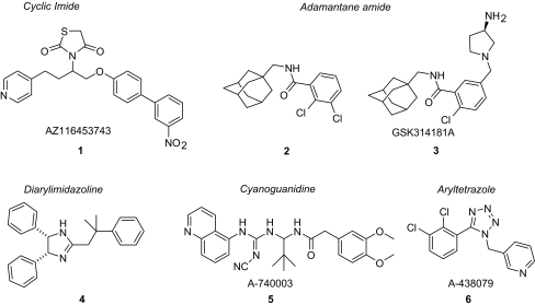
Complementing the genetic data above are recent studies using receptor-selective antagonists indicating a specific role for P2X7 receptor activation in pain signaling [40–42]. Similar to the nociceptive phenotype of mice lacking P2X7 receptors [32] or lacking both isoforms of IL-1 [43], systemic administration of P2X7 receptor-selective antagonists (e.g., A-740003 (5), A-438079 (6), Fig. 2) produced dose-dependent antinociceptive effects in models of neuropathic [40, 41] and inflammatory pain [41]. These data are also consistent with an independent study of the Astra-Zeneca adamantane P2X7 antagonist (3), renamed GSK314181A, that showed dose-dependent antinociception in an inflammatory pain model [42]. Collectively, the foregoing results illustrate the potential role of the P2X7 receptor in modulating IL-1β, and perhaps glutamate, to reduce nociception in neuropathic pain models.
Fig. 2.
Classes of small molecule P2X7 antagonist
Medicinal chemistry advances
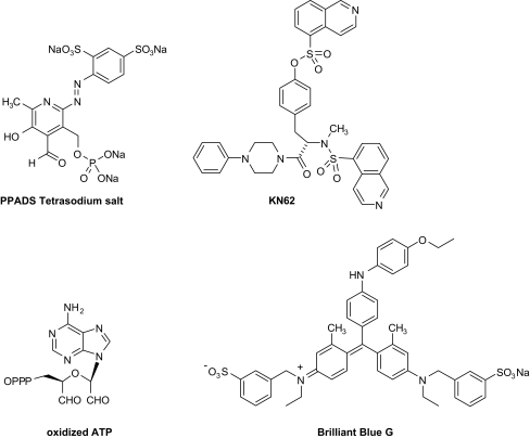
The landscape of P2X7 medicinal chemistry has evolved considerably over the last 5 years. The historically known P2X7 antagonists such as KN-62, PPADS, o-ATP and Brilliant Blue G (see Fig. 1) have gradually given way to a plethora of new molecules that encompass a diversity of structural types (Fig. 2). The cyclic imides (1), adamantane amides (2 and 3), diarylimidazolines (4), cyanoguanidines (5), and substituted aryltetrazoles (6) exemplify those that have been the subject of recent publications [44–46]. Many other recent advances have only been described in the patent literature [44] or are the subject of forthcoming reports (vide infra). All of these new entries in the field appear to possess better drug-like characteristics in terms of the “rule of 5” and may offer more opportunity for the development of viable therapeutic agents.
Fig. 1.
Historical P2X7 antagonists
A common characteristic of many P2X7 antagonists is that of disparate potency between the human and rat P2X7 receptors. The absence of activity at a rodent P2X7 receptor is, from a preclinical standpoint, an obvious impediment to determining the potential therapeutic applications of P2X7 antagonism. KN-62 is a prime example of this phenomenon, having potent activity at the human receptor but lacking activity at the rat receptor (rP2X7 pIC50 < 4; hP2X7 pIC50 ~ 6.7) [46]. The more recently disclosed AZ116453743 (1) [47] similarly shows a significant reduction in potency at the rat receptor relative to human (rP2X7 pIC50 < 5; hP2X7 pIC50 ~ 8). As will be described below, however, strides are being made in identifying structural types that possess similar potencies across species. These studies have led to the novel finding that selective, competitive P2X7 antagonists are efficacious in models of neuropathic and inflammatory pain as well as inflammatory peritonitis.
Adamantane carboxamides
There has been a significant amount of patent activity around the adamantane amide pharmacophore [44] in addition to the early literature report that provided some initial insight into the structure-activity relationships (SAR) of this series at the human P2X7 receptor [48]. Very recently, a second study was published that expanded upon the previous findings and also revealed some SAR trends for this series at the rat P2X7 receptor [49]. Reflecting a common trend in the field of P2X7 ligands, the potency of 2 at rat P2X7 (pIC50 = 6.1) was found to be approximately tenfold less than that at the human receptor (pIC50 = 7.2), as measured by Ca2+ flux in a FLIPR instrument [50]. On the other hand, GSK314181A (3) was reported to be potent at the rat P2X7 receptor (pIC50 = 9.0) as measured using a whole cell patch clamp technique [42] (human data not reported). In a pattern that will be seen repeated across different series of P2X7 antagonists, it was observed that the presence of an ortho substituent on the aromatic ring of the benzamide portion gives rise to substantially increased potency. The absence of an ortho substituent such as a halogen or a methyl group is accompanied by an attendant drop-off in activity. Compound 2 was among the most potent compounds reported (P2X7 pA2 = 8.8). Although chain extension from methylene to ethylene was tolerated between the amide and the adamantane, excision of the methylene resulted in a large decrease in activity. Similarly, replacement of the amide NHCO group with NMeCO (N-methylamide) and NHCH2 (aminomethyl) was also not tolerated. Further investigation of the linker portion between the phenyl and adamantane revealed that reversing the connectivity of the amide also provided analogs with potent P2X7 activity such as 7 (P2X7 pA2 = 8.3) and 8 (P2X7 pA2 = 7.4) shown in Fig. 3. Many of the published adamantane amide analogs were found to possess poor metabolic stability and pharmacokinetic characteristics; however, the indazole-containing compound 8 (P2X7 pA2 = 7.4) was improved in this regard, albeit with a corresponding reduction in potency.
Fig. 3.
Adamantane carboxamide P2X7 antagonists
Numerous patent applications have disclosed additional structural variations on the adamantane amides with the apparent thrust of activities being to improve the solubility and pharmacokinetic characteristics of this highly lipophilic class of molecules [44]. The limited data available from these sources suggest that this pharmacophore is amenable to incorporation of solubilizing amine-containing functionality with retention of robust P2X7 activity. GSK314181A (3) represents a prime example of this structural type as evidenced by the presence of the substituted pyrrolidine appended to the aromatic ring. As mentioned, this molecule displays potent activity at rat P2X7 receptors and has additionally been found to possess activity in the rat CFA model of inflammatory hyperalgesia as assessed using a weight bearing measurement [42].
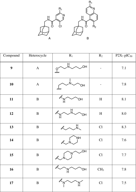
Further expanding on the theme of heterocyclic amides such as the indazole analog 8, both substituted pyridines (A) and quinolines (B) (Table 1) were reported in two recent patent applications to possess potent P2X7 activity [51, 52]. Most interestingly, these heterocycles are amenable to substitution with a variety of amine and alcohol groups that would be expected to significantly improve the solubility characteristics in this series. As can be seen in Table 1, both secondary and tertiary amines, as well as diamino (piperazine) and alcohol moieties are tolerated in the pendant chain. From the limited data, it is not possible to discern any significant differences between the pyridine- and quinoline-based analogs.
Table 1.
Pyridine and quinoline adamantane amides
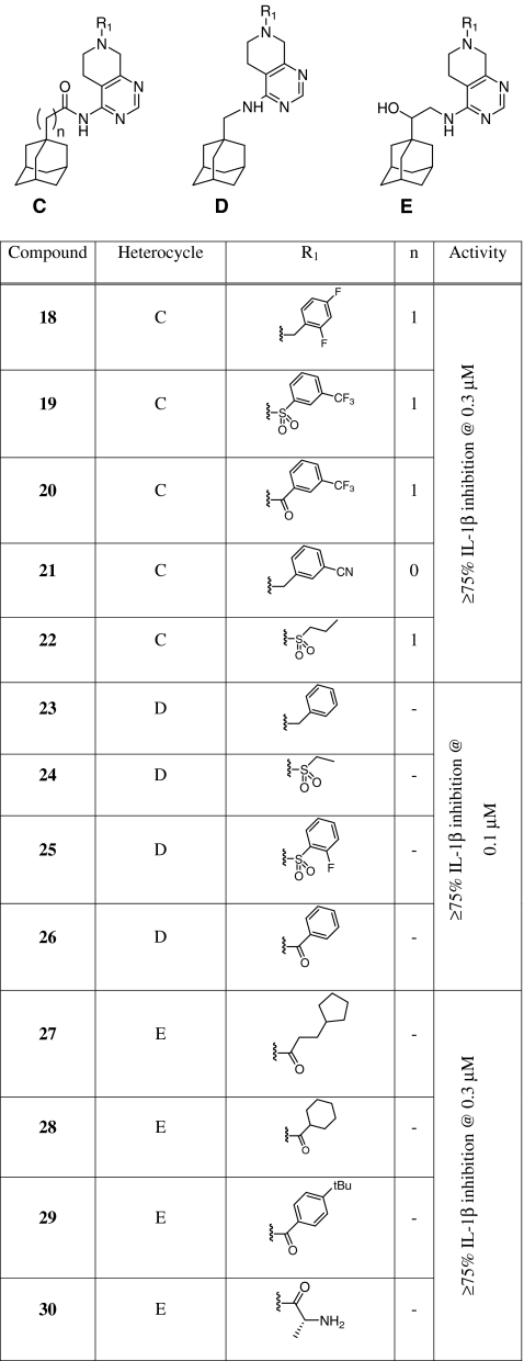
Additional adamantane-derived structural variations that have appeared in the patent literature are shown in Table 2. These analogs are based upon a 5,6,7,8-tetrahydropyrido[3,4-d]pyrimidine unit connected to the adamantane via an amide (C), aminomethyl (D), or aminoethanol (E) linker [53, 54]. The connectivity of the amides 18–22 is the same as for 7 and 8, reversed from compounds 9–17 in Table 1. The amide may be directly attached to the adamantane as in 21 or connected through a methylene group (18–20, 22). Although only single concentration data are provided it is possible to gain a sense of some general SAR trends with these pyridopyrimidines. In all three variations C-E, there appears to be a significant degree of tolerance for structural variation at the R1 position. Arylamides (20, 26, 29), alkylamides (27, 28), arylsulfonamides (19, 25), alkylsulfonamides (22, 24), and benzylamines (18, 21, 23) all show activity at less than 0.3 μM. The presence of an electron-withdrawing group attached to the nitrogen is not required as evidenced by the activity of the benzylamine examples. The primary amine example 30 showed appreciable activity but, interestingly, the analog that is epimeric at the alanine-derived carbon did not [54].
Table 2.
Tetrahydropyridopyrimidine adamantane amides
Aryl carbohydrazides
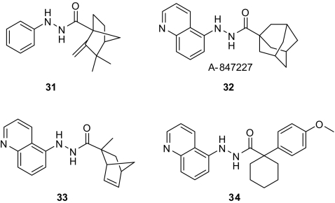
A series of hydrazide-containing P2X7 antagonists (Fig. 4) has recently appeared in the literature which bear a resemblance to some adamantane amide analogs described above [55]. Starting with the high throughput screening lead 31 (hP2X7 pIC50 = 7.3), SAR studies led to the discovery of the highly potent antagonist A-847227 (32: hP2X7 pIC50 = 8.0). Adamantane substitution and variations thereof provided the most potent analogs in this series but other groups also gave rise to potent, if somewhat attenuated, activity. For example, cycloalkyl (33: hP2X7 pIC50 = 7.7), alkyl (not shown), and aryl spirocyclohexyl (34: hP2X7 pIC50 = 7.3) groups all showed potent activity as P2X7 antagonists. On the left hand aryl group, an ortho substituent was essential for potency as has been observed in other series. The absence of an ortho substituent is not compensated for by substitution elsewhere on the aryl group. Tying the ortho substituent back in the form of a quinoline ring (32) retained the P2X7 potency while significantly improving the physiochemical properties relative to similar substituted phenyl analogs. The isoquinoline moiety in this region also showed appreciable potency (not shown).
Fig. 4.
Arylhydrazide P2X7 antagonists
Several hydrazide analogs were assayed for the ability to inhibit IL-1β release in vitro and in a zymosan peritonitis model in vivo. As observed for other classes of P2X7 antagonists, activity to inhibit Ca2+ flux translated into comparable potency to inhibit IL-1β release in vitro (A-847227 (32; IL-1β pIC50 = 8.3). In addition, these molecules demonstrate significant activity to attenuate IL-1β release in the zymosan model in mice. For example, A-847227 (32) was found to reduce IL-1β release by 63% when dosed at 20 μmol/kg (i.p.). Consistent with the effects seen with compounds 5 and 6, A-847227 (32) also displayed antiallodynic activity in the Chung model of neuropathic pain with an ED50 of 92 μmol/kg (i.p.).
Cyanoguanidines
A novel class of cyanoguanidines, represented by A-740003 (5), has recently been discovered to possess potent and selective P2X7 antagonist properties. The relatively good pharmacokinetic (PK) properties and clean pharmacology of A-740003 made it an attractive tool compound with which to probe the potential therapeutic consequences of selective P2X7 receptor antagonism. In contrast to some earlier classes of P2X7 receptor antagonists reported in the literature, A-740003 was found to possess potent activity at the rat P2X7 receptor, making it suitable for in vivo efficacy studies in rats. A-740003, a potent, competitive P2X7 antagonist, was efficacious in models of neuropathic and inflammatory pain upon i.p. administration in rats. In vitro, A-740003 potently inhibited Ca2+ flux, Yo-Pro uptake, and IL-1β release (pIC50 = 7.0–7.3).
The cyanoguanidine-containing P2X7 antagonist A-740003 bears a remarkable similarity to compound 35 (Fig. 5), a structure that was previously discovered to possess activity as an ATP-sensitive potassium channel (KATP) opener [56, 57]. This is an interesting finding, given that the KATP channel and P2X7 are each a ligand-gated ion channel wherein the endogenous ligand is ATP. Another parallel between compounds 5 and 35 is that they both were derived from thiourea screening hits, which themselves were not pursued due to toxicological concerns associated with this functional group. A number of cyanoguanidine KATP openers were assayed at P2X7 and generally found to possess very weak activity (Donnelly-Roberts, unpublished results), providing some indication that the SAR trends for KATP and P2X7 within this pharmacophore do not substantially overlap. The human P2X7 pIC50 for the KATP opener 35 was approximately 5.7, considerably weaker than 5. In general, direct attachment of the aryl group to the amide on the right hand side (RHS) of the structure was a requirement for potent activity at KATP, whereas the presence of an additional 1 carbon spacer conferred more potent P2X7 activity. On the left hand side (LHS), a substituent in the ortho position increased activity at P2X7 (Donnelly-Roberts, unpublished observations) but decreased KATP activity [62, 63]. Although substitution patterns that were preferred for KATP tended to erode activity at P2X7, there were some exceptions. Compound 36, for example, a hybrid of a preferred LHS for P2X7 and RHS for KATP possesses potent activity at hP2X7 (pIC50 = 7.2) but weak activity at KATP (pIC50 ~ 5) [57].
Fig. 5.
Cyanoguanidine KATP opener 35 and P2X7 antagonist 36
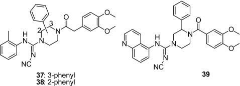
The preceding discussion concerns observations that are confined to the aromatic rings on either end of the molecule. Additional SAR studies around A-740003 led to a novel modification wherein the central t-butyl-substituted carbon linker has been replaced with a substituted piperazine unit (Fig. 6) [58]. In the case of 37 and 38, the piperazine is substituted with a phenyl group in either of the two available positions. Both of these compounds retain good potency as P2X7 antagonists (37: hP2X7 pIC50 = 7.2; 38: hP2X7 pIC50 = 7.3), seemingly indifferent to the location of the phenyl substituent. Replacement of the 3,4-dimethoxyphenyl on the RHS was tolerated with various heterocycles such as pyridine, isoxazole, and thiophene. The carbon chain connecting the piperazine amide with the aromatic group on the RHS could be varied from zero to three carbons with little effect on hP2X7 potency. On the left hand side, the ortho-tolyl group could be replaced with the 5-quinolinyl present in A-740003 (Fig. 6; compound 39). Although most compounds from this series showed three- to fivefold greater potency at hP2X7 than rP2X7, 39 was nearly equipotent at rat (pIC50 = 7.5) and human (pIC50 = 7.2) P2X7.
Fig. 6.
Cyanoguanidine-piperazine P2X7 antagonists
Aryltetrazoles/aryltriazoles
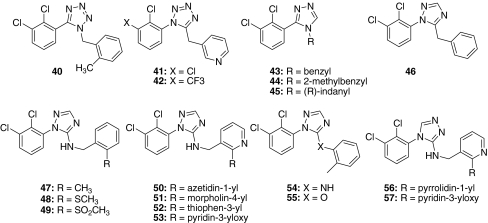
The growing diversity of small molecule P2X7 antagonists is exemplified by the recent reports detailing SAR studies on aryltetrazoles [40] and aryltriazoles [50] represented respectively by A-438079 (6) (hP2X7 pIC50 = 6.3; Fig. 2) and 44 (hP2X7 pIC50 = 7.1; Fig. 7) (see also Table 3). The nature of the heterocyclic core in this pharmacophore was found to be critical for activity as potency decreased in the following order: tetrazole > triazole > pyrazole > imidazole [50]. The connectivity of the aryl and benzyl groups to the core via either nitrogen or carbon appears to be of little consequence as potency for the N-aryl compounds 41 and 46 is similar to the C-aryl connected counterparts 6 and 43 (Table 3). In the case of the triazole core, three additional regioisomers were evaluated beyond those shown in Fig. 7, for which the potencies were comparable to 43 and 46. As will be described below in greater detail, the aryltetrazoles and aryltriazoles display the same tendency observed with other chemotypes in requiring the presence of an ortho-substituted aromatic group in the molecule for potency to be maintained.
Fig. 7.
Aryltetrazole and aryltriazole P2X7 antagonists
Table 3.
Aryltetrazole and aryltriazole P2X7 antagonists
| Compound | hP2X7 pIC50 | rP2X7 pIC50 | |
|---|---|---|---|
| Tetrazoles | 6 | 6.9 | 6.5 |
| 40 | 7.8 | 7.2 | |
| 41 | 7.0 | 6.6 | |
| 42 | 7.9 | 7.3 | |
| Triazoles | 43 | 6.3 | 6.4 |
| 44 | 7.1 | 6.7 | |
| 45 | 7.7 | 7.1 | |
| 46 | 6.7 | 6.4 | |
| Aminotriazoles | 47 | 7.8 | 6.7 |
| 48 | 7.3 | 6.5 | |
| 49 | 7.7 | 7.4 | |
| 50 | 8.0 | 7.6 | |
| 51 | 7.6 | 6.7 | |
| 52 | 7.8 | 6.9 | |
| 53 | 7.9 | 6.9 | |
| 54 | 7.8 | 6.6 | |
| 55 | 5.8 | 5.6 | |
| 56 | 7.8 | 7.9 | |
| 57 | 7.5 | 7.5 |
SAR exploration of the RHS of the tetrazole pharmacophore began with an evaluation of preferred substitutions on the phenyl ring. Although highly potent compounds such as 40 were discovered, the poor physiochemical properties of these molecules led to an investigation of heterocyclic attachments that might confer improved properties in this regard. The 3-pyridyl group was found to be an acceptable moiety on the RHS, balancing potency with improved solubility characteristics (e.g., A-438079). With a 3-pyridyl group in place, it was possible to explore variations on the LHS of A-438079. On the left hand aromatic ring of the structure, substitution in both the ortho and meta positions with either halogen or CF3 groups was a definite requirement for potency. In the meta position, CF3 proved superior to chloro in terms of P2X7 potency (42 vs 41). Additional placement of fluorine or chlorine in the para position was tolerated although typically no further improvement in potency was realized. Other arrangements of halogens on the left side aromatic were substantially less active as were modifications that replaced the halogens with alkyl or alkoxy groups. Replacement of the ortho-substituted phenyl with a 5-quinolinyl group, present in A-740003, on the LHS of the tetrazole resulted in a completely inactive analog [40].
In the case of the aryltriazoles, even the weak basicity of the core was sufficient to allow extensive study of the preferred substitution patterns on the RHS without the solubility limitations associated with the tetrazole. Thus, complementary SAR studies were conducted on the left and right hand sides of the tetrazoles and triazoles, respectively. A more extensive study on the right hand aromatic of the triazoles around 43 revealed a fair degree of tolerance for the interchange of groups in the ortho position. Substitution of relatively small electron-donating (OCH3) and electron-withdrawing groups (CF3, CHF2) similarly increased potency in comparison to the simple phenyl [50]. The best potencies, however, were achieved with the more weakly electron-donating methyl (compound 44) (same trend observed for the tetrazole 40) and methylthio groups (not shown). Both 2,3 and 2,5 disubstitution was found to retain the potency of the simple ortho-substituted analogs; however, the presence of a lone substituent in the meta or para position markedly decreased potency regardless of the particular moiety present. Connecting the ortho substituent to the benzylic position by forming an indane ring provided the most potent triazole reported (45), with the (R) enantiomer being several times more potent than the (S) enantiomer. Interestingly, this same modification on the tetrazole core did not result in a similar increase in potency (Donnelly-Roberts, unpublished observations). In both the tetrazoles and triazoles, increasing the length of the spacer between the core and the right hand side aromatic group to two carbons significantly decreased P2X7 potency.
In animal pain models, A-438079 and 44 were found to inhibit mechanical allodynia in the Chung model of neuropathic pain upon i.p. dosing with ED50s of 76 and 125 μmol/kg, respectively. Additionally, A-438079 displayed activity against vincristine-induced neuropathy and was effective in the formalin model of persistent pain [59]. Up to a dose of 300 μmol/kg (i.p.), A-438079 did not attenuate rotarod performance. A-438079 has been further shown from in vivo electrophysiological experiments to inhibit both evoked and spontaneous firing of different classes of spinal cord neurons following intravenous administration [59].
A noteworthy extension of the scope of the SAR in this class of P2X7 antagonist has recently appeared showing the potential for nitrogen incorporation into the linker between the core and the RHS aryl group [60] (Fig. 7, compounds 47–57). Although an oxygen linker led to weak activity (55), amino (54) and aminomethyl (47) linkers were well tolerated with the latter variation giving rise to a number of highly potent antagonists, particularly at hP2X7. The activity seen with the two atom aminomethyl linker is especially surprising in light of the poor activity reported with a two carbon linker [40, 50].
Some important similarities and differences are observed between the SAR trends in the methylene-linked (43–46) and amino/aminomethyl-linked (47–54, 56–57) analogs. Like 43–46, compounds 50–53 and 54 showed ~tenfold greater potency relative to their counterparts lacking a substituent in the ortho position. The requirement of an ortho substituent was less pronounced, however, where the right hand aromatic was phenyl as in 47–49. Another illustration of the differences encountered with the aminomethyl-linked triazoles can be seen by comparing the 2-SO2Me and 2-SMe substitutions with their counterparts in the carbon-linked class. Whereas the 2-SO2Me group lost potency relative to 2-SMe in the carbon-linked series [50], it instead shows relatively enhanced potency with the aminomethyl linker (49 vs 48), particularly at the rat receptor. Reduced potency with monosubstitution in the meta or para position is also observed in the aminomethyl-linked series.
The diversity of substitution at the ortho position has been greatly expanded in the aminomethyl series, with heterocyclic (50–52, 56) and heteroaryloxy (53, 57) groups all exhibiting potent activity. This tolerance for structural variation was confined to the aminomethyl-linked analogs, however, as replacement of the ortho-methyl on compound 54 with ortho-morpholinyl (structure not shown) resulted in a 100-fold reduction in activity compared with compound 51 [60].
Regarding regioisomers, similar to observations made in the methylene-linked triazoles, the regioisomeric aminotriazole 57 possessed essentially the same potency as 53 at the human P2X7 receptor. Although no data have been published, it is worth noting that aminopyrazole and aminotetrazole variations on this series have appeared in the patent literature [61, 62].
Reflecting a common theme in the P2X7 field, different potencies at the rat and human P2X7 receptors were often noted for both the tetrazoles and triazoles (Table 3). Generally, these structural types were three- to tenfold more potent at human P2X7 compared to rat, although a few analogs possessed essentially equivalent potency across species such as 56 and 57. Although the SAR trends summarized above for the tetrazole and triazole analogs are based upon data measuring the inhibition of Ca2+ flux at the recombinant human and rat P2X7 receptors, very similar results were obtained using native THP-1 cells measuring Yo-Pro uptake [40]. Likewise, the rank order potencies for inhibition of IL-1β release paralleled those from the Ca2+ flux experiments, with the caveat that potencies were somewhat attenuated for cytokine release [40].
Conclusion
The discovery of potent and receptor-selective P2X7 antagonists has significantly expanded the field of P2X receptor pharmacology. The plethora of emerging new chemotypes have greatly enhanced our appreciation for the important structural features that impart P2X7 receptor activity while also serving to illustrate that this target remains fertile ground for future exploration. A noteworthy commonality that has been observed across the diversity of P2X7 receptor antagonist structural types is the preference or even requirement for the presence of an ortho-substituted aromatic group in the molecule. Additionally, substantial progress has been made toward the discovery of potent antagonists at both rodent and human P2X7 receptors that possess improved drug-like properties (e.g., solubility/pharmacokinetics). In this regard, the P2X7 receptor appears to be both more drugable, as well as susceptible to pharmacological modulation in comparison to other P2X receptors like P2X3 and P2X4 [2].
While it is clear that P2X7 receptors play an important mechanistic role in chronic ongoing inflammation, multiple P2 receptor-based mechanisms likely contribute to the ability of ATP to alter nociceptive sensitivity following tissue injury [64]. Activation of P2X3, P2X2/3, P2X4, P2X7, and P2Y (e.g. P2Y2) receptors can modulate pain via direct (neuronal excitability) and non-direct (neural-glial cell interactions) mechanisms [63]. The recent report of functional heteromeric P2X4/7 receptors [29] may also help bring some mechanistic clarity to the similar functional roles of these receptors in chronic inflammation and pain. While both P2X4 and P2X7 receptors have been implicated in glial-based modulatory mechanisms involved in sensory neurotransmission [9, 65], the discovery of selective P2X4 antagonists are needed to differentiate the respective roles of these receptors in pain signaling. The identification of novel P2X7 receptor-selective antagonists has provided new research tools for both in vitro and in vivo studies of P2X7 receptor pharmacology and led to the generation of new data that indicate an expanded role for this receptor in pain signaling associated with nerve injury and inflammation.
References
- 1.North AR (2002) Molecular physiology of P2X receptors. Physiol Rev 82:1013–1067 [DOI] [PubMed]
- 2.Jacobson KA, Jarvis MF, Williams M (2002) Purine and pyrimidine (P2) receptors as drug targets. J Med Chem 45:4057–4093 [DOI] [PMC free article] [PubMed]
- 3.Di Virgilio FD, Vishawanath V, Ferrari D (2001) On the role of the P2X7 receptor in the immune system. In: Abbracchio MP, Williams M (eds) Purinergic and pyrimidinergic signalling. Handbook of experimental pharmacology, vol 151. Springer, Heidelberg, pp 355–373
- 4.Surprenant A, Rassendren F, Kawashima E, North RA, Buell G (1996) The cytolytic P2Z receptor for extracellular ATP identified as a P2X receptor (P2X7). Science 272:735–738 [DOI] [PubMed]
- 5.Collo G, Neidhart S, Kawashima E, Kosco-Vibois M, North RA, Buell G (1997) Tissue distribution of the P2X7 receptor. Neuropharmacology 36:1277–1283 [DOI] [PubMed]
- 6.Rassendren F, Buell GN, Virginio C, Collo G, North RA, Surprenant A (1997) The permeabilizing ATP receptor, P2X7. Cloning and expression of a human cDNA. J Biol Chem 272:5482–5486 [DOI] [PubMed]
- 7.Sim JA, Young MT, Sung HY, North RA, Surprenant A (2004) Reanalysis of P2X7 receptor expression in rodent brain. J Neurosci 24:6307–6314 [DOI] [PMC free article] [PubMed]
- 8.Anderson CM, Nedergaard M (2006) Emerging challenges of assigning P2X(7) receptor function and immunoreactivity in neurons. Trends Neurosci 29:257–262 [DOI] [PubMed]
- 9.Zhang X-F, Han P, Faltynek CR, Jarvis MF, Shieh C-C (2005) Functional expression of P2X7 receptors in non-neuronal cells of rat dorsal root ganglion. Brain Res 1052:63–70 [DOI] [PubMed]
- 10.Kahlenberg J, Dubyak GW (2004) Mechanisms of caspase-1 activation by P2X7 receptor-mediated K+ release. Am J Physiol Cell Physiol 286:C1100–C1108 [DOI] [PubMed]
- 11.Perregaux DG, Gabel CA (1994) Interleukin-1 beta maturation and release in response to ATP and nigericin. Evidence that potassium depletion mediated by these agents is a necessary and common feature of their activity. J Biol Chem 269:15195–15203 [PubMed]
- 12.Solle M, Labasi J, Perregaux DG, Stam E, Petrushova N, Koller BH, Griffiths RJ, Gabel CA (2001) Altered cytokine production in mice lacking P2X7 receptors. J Biol Chem 276:125–132 [DOI] [PubMed]
- 13.Ferrari D, Pizzirani C, Adinolfi E, Lemoli RM, Curti A, Idzko M, Panther E, Di Virgilio F (2006) The P2X7 receptor: a key player in IL-1 processing and release. J Immunol 176:3877–3883 [DOI] [PubMed]
- 14.Verhoef PA, Estacion M, Schilling W, Dubyak GR (2003) P2X7 receptor-dependent blebbing and the activation of Rho-effector kinases, caspases, and IL-1 beta release. J Immunol 170:5728–5738 [DOI] [PubMed]
- 15.Woolf CJ, Allchorne A, Safieh-Garabedian B, Poole S (1997) Cytokines, nerve growth factor and inflammatory hyperalgesia: the contribution of tumour necrosis factor alpha. Br J Pharmacol 121:417–424 [DOI] [PMC free article] [PubMed]
- 16.Samad TA, Moore KA, Sapirstein A, Billet S, Allchorne A, Poole S, Bonventre JV, Woolf CJ (2001) Interleukin-1beta-mediated induction of Cox-2 in the CNS contributes to inflammatory pain hypersensitivity. Nature 410:471–475 [DOI] [PubMed]
- 17.Parvathenani LK, Svetlana T, Greco CR, Roberts SB, Robertson B, Posmantur R (2003) P2X7 mediates superoxide production in primary microglia and is up-regulated in a transgenic mouse model of Alzheimer’s disease. J Biol Chem 278:13309–13317 [DOI] [PubMed]
- 18.Burnstock G (2006) Pathophysiology and therapeutic potential of purinergic signaling. Pharmacol Rev 58:58–78 [DOI] [PubMed]
- 19.Perregaux DG, Labasi J, Laliberte R, Stam E, Solle M, Koller B, Griffiths R, Gabel CA (2001) Interleukin-1b posttranslational processing-exploration of P2X7 receptor involvement. Drug Dev Res 53:83–90 [DOI]
- 20.Donnelly-Roberts DL, Namovic M, Faltynek CR, Jarvis MF (2004) Mitogen-activated protein kinase and caspase signaling pathways are required for P2X7 receptor (P2X7R)-induced pore formation in human THP-1 cells. J Pharmacol Exp Ther 308:1053–1061 [DOI] [PubMed]
- 21.Humphreys BD, Dubyak GR (1996) Induction of the P2z/P2X7 nucleotide receptor and associated phospholipase D activity by lipopolysaccharide and IFN-gamma in the human THP-1 monocytic cell line. J Immunol 157:5627–5637 [PubMed]
- 22.Aga M, Johnson CJ, Hart AP, Guadarrama AG, Suresh M, Svaren J, Bertics PJ, Darien BJ (2002) Modulation of monocyte signaling and pore formation in response to agonists of the nucleotide receptor P2X7. J Leukoc Biol 72:222–232 [PubMed]
- 23.Ferrari D, Wesselborg S, Bauer M, Schulze-Osthoff K (1997) Extracellular ATP activates transcription factor NF-kappaB through the P2Z purinoreceptor by selectively targeting NF-kappab p65. J Cell Biol 139:1635–1643 [DOI] [PMC free article] [PubMed]
- 24.Armstrong JN, Brust TB, Lewis RG, MacViar BA (2002) Activation of presynaptic P2X7-like receptors depresses mossy fiber-CA3 synaptic transmission through p38 mitogen-activated protein kinase. J Neurosci 22:5938–5945 [DOI] [PMC free article] [PubMed]
- 25.Bradford MD, Soltoff SP (2002) P2X7 receptors activate protein kinase D and p42/p44 mitogen-activated protein kinase (MAPK) downstream of protein kinase C. Biochem J 366:745–755 [DOI] [PMC free article] [PubMed]
- 26.Gendron F-P, Neary JT, Theiss PM, Sun GY, Gonzalez FA, Weismann GA (2003) Mechanisms of P2X7 receptor-mediated ERK1/2 phosphorylation in human astrocytoma cells. Am J Physiol Cell Physiol 284:C571–C581 [DOI] [PubMed]
- 27.Monterio da Cruz C, Ventura ALM, Schachter J, Costa-Junior HM, da Silva Souza HA, Gomes FR, Coutinho-Silva R, Ojcius DM, Persechini PM (2006) Activation of ERK1/2 by extracellular nucleotides in macrophages is mediated by multiple P2 receptors independently of P2X7-associated pore or channel formation. Br J Pharmacol 147:324–334 [DOI] [PMC free article] [PubMed]
- 28.Dubyak GR (2007) Go it alone no more—P2X7 joins the society of heteromeric ATP-gated receptor channels. Mol Pharmacol 72:1402–1405 [DOI] [PubMed]
- 29.Guo C, Masin M, Qureshi OS, Murrel-Lagnado RD (2007) Evidence for functional P2X4/P2X7 heteromeric receptors. Mol Pharmacol 72:1447–1456 [DOI] [PubMed]
- 30.Labasi JM, Petrushova N, Donovan C, McCurdy S, Lira P, Payette MM, Brissette W, Wicks JR, Audoly L, Gabel CA (2002) Absence of the P2X7 receptor alters leukocyte function and attenuates an inflammatory response. J Immunol 168:6436–6445 [DOI] [PubMed]
- 31.Adis Data Information (2007) http://pipeline.dialog.com/abb_lab/adrd.htm. Cited Sept 2007
- 32.Chessell IP, Hatcher J, Bountra C, Michel AD, Hughes JP, Green P, Egerton J, Murfin M, Richardson J, Peck WL, Grahames CBA, Casula MA, Yiangou Y, Birch R, Anand P, Buell GN (2005) Disruption of the P2X7 purinoceptor gene abolishes chronic inflammatory and neuropathic pain. Pain 114:386–396 [DOI] [PubMed]
- 33.Bianchi M, Dib B, Panerai AE (1998) Interleukin-1 and nociception in the rat. J Neurosci Res 53:645–650 [DOI] [PubMed]
- 34.Horai R, Saijo S, Tanioka H, Nakae S, Sudo K, Okahara A, Ikuse T, Asano M, Iwakura Y (2000) Development of chronic inflammatory arthropathy resembling rheumatoid arthritis in interleukin 1 receptor antagonist-deficient mice. J Exp Med 191:313–320 [DOI] [PMC free article] [PubMed]
- 35.Papp L, Vizi ES, Sperlágh B (2004) Lack of ATP-evoked GABA and glutamate release in the hippocampus of P2X7 receptor -/- mice. Neuroreport 15:2387–2391 [DOI] [PubMed]
- 36.Duan S, Anderson CM, Keung EC, Chen Y, Chen Y, Swanson RA (2003) P2X7 Receptor-mediated release of excitatory amino acids from astrocytes. J Neurosci 23:1320–1328 [DOI] [PMC free article] [PubMed]
- 37.Milligan ED, Maier SF, Watkins LR (2003) Review: neuronal-glial interactions in central sensitization. Semin Pain Med 1:171–183 [DOI]
- 38.Wieseler-Frank J, Maier SF, Watkins LR (2004) Glial activation and pathological pain. Neurochem Int 45:389–395 [DOI] [PubMed]
- 39.Raghavendra V, DeLeo JA (2004) The role of astrocytes and microglia in persistent pain. Adv Mol Cell Biol 31:951–966 [DOI]
- 40.Nelson DW, Gregg RJ, Kort ME, Perez-Medrano A, Voight EA, Wang Y, Namovic MT, Grayson G, Donnelly-Roberts DL, Niforatos W, Honore P, Jarvis M, Faltynek CR, Carroll WA (2006) Structure-activity relationship studies on a series of novel, substituted 1-benzyl-5-phenyltetrazole P2X7 antagonists. J Med Chem 49:3659–3666 [DOI] [PubMed]
- 41.Honore PM, Donnelly-Roberts D, Namovic M, Hsieh G, Zhu C, Mikusa J, Hernandez G, Zhong C, Gauvin D, Chandran P, Harris R, Perez-Medrano A, Carroll W, Marsh K, Sullivan J, Faltynek C, Jarvis MF (2006) A-740003 (N-(1-{[(cyanoimino)(5-quinolinylamino) methyl]amino}-2,2-dimethylpropyl)-2-(3,4-dimethoxyphenyl)acetamide, a novel and selective P2X7 receptor antagonist dose-dependently reduces neuropathic pain in the rat. J Pharmacol Exp Ther 319:1376–1385 [DOI] [PubMed]
- 42.Lappin SC, Winyard LA, Clayton N, Chambers LJ, Demont EH, Chessell IP, Richardson JC, Gunthorpe MJ (2005) Reversal of mechanical hyperalgesia in a rat model of inflammatory pain by a potent and selective P2X7 antagonist (abstract 958.2). Society for Neuroscience
- 43.Honore P, Wade CL, Zhong C, Harris RR, Wu C, Ghayur T, Iwakura Y, Decker MW, Faltynek C, Sullivan J, Jarvis MF (2006) Interleukin-1alphabeta gene-deficient mice show reduced nociceptive sensitivity in models of inflammatory and neuropathic pain but not post-operative pain. Behav Brain Res 167:355–364 [DOI] [PubMed]
- 44.Romagnoli R, Baraldi PG, DiVirgilio F (2005) Recent progress in the discovery of antagonists acting at P2X7 receptor. Expert Opin Ther Patents 15:271–287 [DOI]
- 45.Gunosewoyo H, Coster MJ, Kassiou M (2007) Molecular probes for P2X7 receptor studies. Curr Med Chem 14:1505–1523 [DOI] [PubMed]
- 46.Donnelly-Roberts DL, Jarvis MF (2007) Discovery of P2X7 receptor-selective antagonists offers new insights into P2X7 receptor function and indicates a role in chronic pain states. Br J Pharmacol 151:571–579 [DOI] [PMC free article] [PubMed]
- 47.Stokes L, Jiang LH, Alcaraz L, Bent J, Bowers K, Fagura M, Furber M, Mortimore M, Lawson M, Theaker J, Laurent C, Braddock M, Surprenant A (2006) Characterization of a selective and potent antagonist of human P2X7 receptors, AZ11645373. Br J Pharmacol 149:880–887 [DOI] [PMC free article] [PubMed]
- 48.Baxter A, Bent J, Bowers K, Braddock M, Brough S, Fagura M, Lawson M, McInally T, Mortimore M, Robertson M, Weaver R, Webborn P (2003) Hit-to-lead studies: the discovery of potent adamantane amide P2X7 receptor antagonists. Bioorg Med Chem Lett 13:4047–4050 [DOI] [PubMed]
- 49.Furber M, Alcaraz L, Bent JE, Beyerbach A, Bowers K, Braddock M, Caffrey MV, Cladingboel D, Collington J, Donald DK, Fagura M, Ince F, Kinchin EC, Laurent C, Lawson M, Luker TJ, Mortimore MMP, Pimm AD, Riley RJ, Roberts N, Robertson M, Theaker J, Thorne PV, Weaver R, Webborn P, Willis P (2007) Discovery of potent and selective adamantane-based small-molecule P2X7 receptor antagonists/interleukin-1beta inhibitors. J Med Chem 50:5882–5885 [DOI] [PubMed]
- 50.Carroll WA, Kalvin DM, Perez Medrano A, Florjancic AS, Wang Y, Donnelly-Roberts DL, Namovic MT, Grayson G, Honoré P, Jarvis MF (2007) Novel and potent 3-(2,3-dichlorophenyl)-4-(benzyl)-4H-1,2,4-triazole P2X7 antagonists. Bioorg Med Chem Lett 17:4044–4048 [DOI] [PubMed]
- 51.WO/2003/041707
- 52.WO/2003/080579
- 53.WO/2006/102610
- 54.WO/2007/028022
- 55.Nelson DW, Sarris K, Kalvin DM, Namovic MT, Grayson G, Donnelly-Roberts, DL, Harris R, Honore P, Jarvis MF, Faltynek CR, Carroll WA (2008) Structure-activity studies on N’-aryl carbohydrazide P2X7 antagonists. J Med Chem Apr 26 [Epub ahead of print] [DOI] [PubMed]
- 56.Perez-Medrano A, Buckner SA, Coghlan MJ, Gregg RJ, Gopalakrishnan M, Kort ME, Lynch JK, Scott VE, Sullivan JP, Whiteaker KL, Carroll WA (2004) Design and synthesis of novel cyanoguanidine ATP-sensitive potassium channel openers for the treatment of overactive bladder. Bioorg Med Chem Lett 14:397–400 [DOI] [PubMed]
- 57.Perez-Medrano A, Brune ME, Buckner SA, Coghlan MJ, Fey TA, Gopalakrishnan M, Gregg RJ, Kort ME, Scott VE, Sullivan JP, Whiteaker KL, Carroll WA (2007) Structure-activity studies of novel cyanoguanidine ATP-sensitive potassium channel openers for the treatment of overactive bladder. J Med Chem 50:6265–6273 [DOI] [PubMed]
- 58.Morytko MJ, Betschmann P, Woller K, Ericsson A, Chen H, Donnelly-Roberts DL, Namovic MT, Jarvis MF, Carroll WA, Rafferty P (2008) Synthesis and in vitro activity of N’-cyano-4-(2-phenylacetyl)-N-o-tolylpiperazine-1-carboximidamide P2X7 antagonists. Bioorg Med Chem Lett 18:2093–2096 [DOI] [PubMed]
- 59.McGaraughty S, Chu KL, Namovic MT, Donnelly-Roberts DL, Harris RR, Zhang XF, Shieh CC, Wismer CT, Zhu CZ, Gauvin DM, Fabiyi AC, Honore P, Gregg RJ, Kort ME, Nelson DW, Carroll WA, Marsh K, Faltynek CR, Jarvis MF (2007) P2X7-related modulation of pathological nociception in rats. Neuroscience 146:1817−1818 [DOI] [PubMed]
- 60.Florjancic AS, Peddi S, Perez-Medrano A, Namovic MT, Grayson G, Donnelly-Roberts DL, Jarvis MF, Carroll WA (2008) Synthesis and in vitro activity of 1-(2,3-dichlorophenyl)-N-(pyridin-3-ylmethyl)-1H-1,2,4-triazol-5-amine and 4-(2,3-dichlorophenyl)-N-(pyridin-3-ylmethyl)-4H-1,2,4-triazol-3-amine P2X7 antagonists. Bioorg Med Chem Lett 18:2089–2092 [DOI] [PubMed]
- 61.WO/2007/056091
- 62.WO/2005/111003
- 63.Donnelly-Roberts DL, McGaraughty S, Shieh C-C, Honore P, Jarvis MF (2008) Painful purinergic receptors. J Pharmacol Exp Ther 324:409–415 [DOI] [PubMed]
- 64.Fields RD, Burnstock G (2006) Purinergic signalling in neuron-glia interactions. Nat Rev Neurosci 7:423–436 [DOI] [PMC free article] [PubMed]
- 65.Tsuda M, Shigemoto-Mogami Y, Koizumi S, Mizokoshi A, Kohsaka S, Salter M, Inoue K (2003) P2X4 receptors induced in spinal microglia gate tactile allodynia after nerve injury. Nature 424:778–783 [DOI] [PubMed]


