Fig. 3.

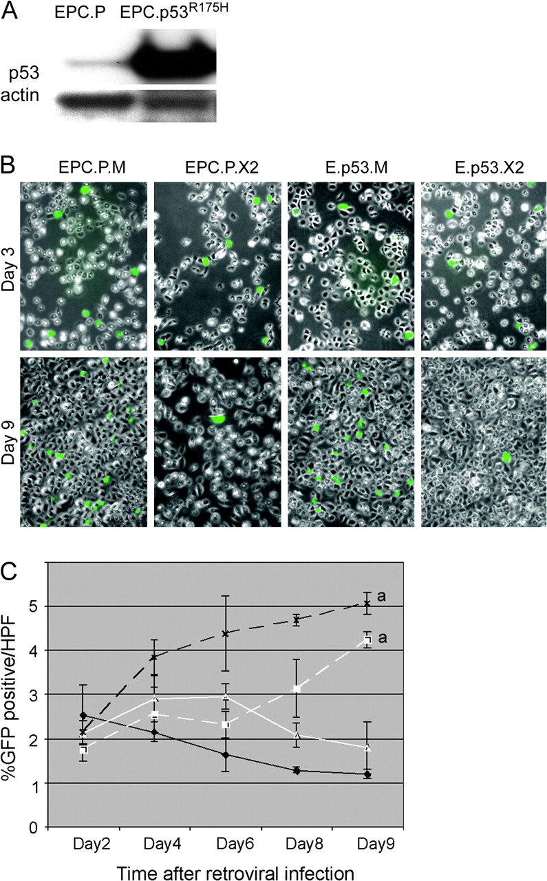
A dominant-negative p53 does not rescue Cdx2 expression in EPC-hTERT keratinocytes. EPC-hTERT keratinocytes expressing a dominant-negative p53 were established using the pBabe-puro-p53R175H vector to direct expression of the p53R175H mutant. (A) Western blot of p53 protein levels in EPC-hTERT cells receiving the empty pBabe control vector (EPC.P) or the pBabe-puro-p53R175H (EPC.p53R175H). The blot was reprobed for actin levels as loading control. (B) As before, fluorescent image for GFP+ cells overlaid with phase contrast image. EPC.P or EPC.p53R175H cells were subsequently infected with the MIGR-Cdx2 virus to induce Cdx2 expression (EPC.P.X2 and E.p53.X2) or the MIGR1 empty control vector (EPC.P.M and E.p53.M). Photodocumentation was obtained at 3 and 9 days post-infection with the MIGR-based vectors. At 3 days, nearly equivalent numbers of GFP+ cells with both vectors. By day 9, there is a highly significant reduction in GFP+ expression in those cells infected with the MIGR-Cdx2 virus. (C) The number of GFP+ cells per total cells in six high-power fields (HPFs) was determined on days 2, 4, 6, 8 and 9 post-retroviral infection. Averages and standard deviations were calculated and graphed. Black X, dashed line: EPC.P.M. White triangle, solid line: EPC.P.X2. White square, dashed line: E.p53.M. Black diamond, solid line: E.p53.X2. a, significantly differs from day 9 MIGR controls using analysis of variance and Tukey Rank Mean testing, P < 0.05.
