Abstract
Studies show that sex plays a role in stress-related depression, with women experiencing a higher vulnerability to its effect. Two major targets of antidepressants are brain-derived neurotrophic factor (BDNF) and cyclic adenosine monophosphate response element–binding protein (CREB). The aim of this study was to investigate the levels of CREB, phosphorylation of CREB (pCREB), and BDNF in stress-related brain regions of male and female rats after stress and recovery. CREB and pCREB levels were examined in CA1, CA2, CA3, paraventricular nucleus of the thalamus (PVT), amygdala, anterior cingulate area, dorsal part (ACAd), and infralimbic area of prefrontal cortex (PFC), whereas dentate gyrus (DG) and prelimbic area (PL) of PFC were examined for BDNF levels. Our results demonstrate that levels of CREB and pCREB in male CA1, CA2 and CA3, PVT, amygdala, and ACAd were reduced by stress, whereas the same brain regions of female rats exhibited no change. BDNF levels were decreased by chronic stress in female PL but were increased by acute stress in female DG. BDNF levels in male DG and PL were found not to undergo change in response to stress. Abnormalities in morphology occurred after chronic stress in males but not in females. In all cases, the levels of CREB, pCREB, and BDNF in recovery animals were comparable to the levels of these proteins in control animals. These findings demonstrate a sexual dimorphism in the molecular response to stress and suggest that these differences may have important implications for potential therapeutic treatment of depression.
Keywords: depression, neuroplasticity, recovery, sex differences, stress response
Introduction
Depression is one of the leading causes of disability in the world when measured by the number of years lived with a disabling condition (Blendy 2006). Although depression is highly heritable, the onset of depression appears to be triggered by environmental factors such as stress (Bale 2006; Berton and Nestler 2006). Epidemiological studies demonstrate that women are more vulnerable than men to stress-related psychopathologies and that depression occurs twice as frequently in women as in men (Kessler et al. 1993; Carter-Snell and Hegadoren 2003; Kessler 2003; Sun and Alkon 2006). However, few studies have been conducted in females, and the mechanisms underlying these sex differences have not been clarified.
Results from studies in males indicate that brain-derived neurotrophic factor (BDNF) and cyclic adenosine monophosphate response element–binding protein (CREB) are key mediators of the therapeutic response to antidepressants (D'Sa and Duman 2002). CREB and CREB-dependent gene expression are activated in response to many signal transduction cascades implicated in neuronal plasticity (Lonze and Ginty 2002). In male rats, CREB overexpression causes a behavioral phenotype similar to that seen with antidepressant therapy (Chen et al. 2001). Furthermore, enhancing cortical and hippocampal phosphorylation of CREB (pCREB) has been proposed as a common antidepressant mechanism, based largely on studies of antidepressant action in the naive male rodent (Thome et al. 2000; Blendy 2006). pCREB at serine 133 leads to transcription of genes including BDNF (Carlezon et al. 2005) that is required for activity-dependent survival of neurons (Ghosh et al. 1994; Shieh et al. 1998) and plays a pivotal role in the action of antidepressants (Wang et al. 2008). In contrast to the effects of antidepressant treatment, stress decreases BDNF levels (Smith et al. 1995; Nibuya et al. 1999) and suppresses local CREB phosphorylation (Laifenfeld et al. 2005). Therefore, CREB, pCREB, and CREB-regulated genes such as BDNF are major targets of antidepressant drug action. Our previous study showed that stress decreases pCREB in dentate gyrus (DG) and prelimbic area (PL) of prefrontal cortex (PFC) in male rats whereas not in female rats. However, open-field tests showed no behavioral difference between male and female rats following stress (Lin et al. 2008). Clearly, these 2 findings are contradictory and fail to explain the sex differences seen in human depression. As such, other proteins like BDNF should be further investigated. More brain regions related to stress besides DG and PL should also be examined.
Multiple brain regions are likely involved in the organization of responses to stressful stimuli, including regions of prefrontal and cingulate cortex, hippocampus, amygdala, and thalamus (Drevets 2001; Liotti and Mayberg 2001). Until now, studies have primarily focused on PFC and hippocampus because these are the key regions responsible for glucocorticoid feedback in the hypothalamic–pituitary–adrenocortical axis (Popoli et al. 2002; Sheline 2003; Sairanen et al. 2007). For example, shortly after a stressful event, corticosteroids increase cellular excitability in subfields of the hippocampus (Joëls 2008). However, stress responses are not limited to PFC and hippocampus. For example, the paraventricular nucleus of the thalamus (PVT) may be an important target for antidepressant therapy because this midline thalamic nucleus responds strongly to various stressors (Beck and Fibiger 1995). Also, many of the projection targets of PVT neurons, including PFC and amygdala, show strong stress responses (Bubser and Deutch 1999). Certain nuclei of the amygdala are critical in responses to rewarding and aversive stimuli, and these nuclei show abnormalities in depressed human subjects (Talarovicova et al. 2007). The amygdala is also considered central in mediating stress-related changes in hippocampal functions (Yang et al. 2008), and this observation is consistent with studies reporting abnormalities in hippocampus and PFC in depressed human subjects (Drevets 2001; Bissette et al. 2003; Rajkowska 2003). Finally, recent compelling evidence suggests that estrogen is involved in depression, a factor that may help to explain sex differences observed in human depression. Estrogen receptors are found in multiple brain regions, including amygdala where estrogen receptor alpha (ERα) dominates and thalamus where ERβ dominates (Ostlund et al. 2003), so these brain regions may be important in the study of sex differences in stress-related psychopathologies.
To further explore the mechanisms underlying the sex differences in stress-related depression, we exposed male and female rats to inescapable footshock to mimic depression. Chronic footshock exposure has been proposed as a valid animal model for affective disorders (Westenbroek, Ter Horst, et al. 2003). Using this model, we investigated the effects of footshock (acute and chronic) and recovery (chronic footshock for 3 weeks followed by 3 weeks of recovery) on CREB, pCREB, and BDNF levels. CREB and pCREB levels were examined in CA1, CA2, CA3, PVT, amygdala, anterior cingulate area, dorsal part (ACAd), and infralimbic area (ILA), whereas DG and PL were examined for BDNF levels. Prior to stress induction, males and females showed no differences in protein levels or brain morphology. During the stress response, male rats showed significant changes in pCREB levels and in brain morphology, whereas females showed no such response. However, females showed changes in BDNF levels during the stress response, whereas males did not. Both sexes returned to baseline protein levels and morphology following recovery. These results suggest that sex may play a role in the response to stress and that future targets for clinical antidepressants may be sex dependent.
Materials and Methods
Animals
Each experimental group consisted of (8) 6- to 7-week-old male or female Wistar rats, individually housed with ad libitum access to food and water. A plastic tube (diameter 8 × 17 cm) was placed in each cage as a shelter. The light–dark cycle was reversed (lights on 19:00–7:00). All experiments were designed to minimize the number of animals required, and all procedures were approved by the Animals Ethics Committee of the University of Groningen. The effect of the estrous cycle on the stress response was not specifically investigated in the current study.
Male and female rats were randomly assigned to 4 experimental groups: 1) control group: subjected to no footshock throughout the experiment; 2) acute stress group: received 6 footshocks on day 42 and exposed to footshock box with light stimulus only on day 43; 3) recovery group: received footshocks daily for 3 weeks followed by a 3-week period with no footshock and on day 43 exposure to footshock box with only light but no shock; 4) chronic stress group: received footshocks daily for 3 weeks followed by 3 weeks of alternating days of exposure to footshock box with footshocks and without receiving footshock and on day 43 exposure to footshock box only with light.
Stress Procedure
The “footshock chamber” consists of a box containing an animal space positioned on a metallic grid floor connected to a shock generator and scrambler. Rats in the stress group were placed in a box and received variable (2–6) inescapable footshocks with randomized starting time (between 9:00 AM and 5:00 PM) and intervals during a 30–120 min session (0.8 mA as maximum intensity and 8 s in duration) in order to make the procedure as unpredictable as possible. A light signal (10 s) preceded each footshock adding a “psychological” component to the stressor. On the last day, the stress-exposed rats were subjected to light stimulus only, which was crucial as it provided a way to create a stress condition without unwanted side effects of direct physical or painful stimuli.
On day 43, rats were sacrificed using isoflurane anesthesia. Three rats from each group were transcardially perfused with 50 mL of heparinized saline and 300 mL of a 4% paraformaldehyde solution in 0.1 M sodium phosphate buffer (pH 7.4), 2 h after the start of the last exposure to stress box. These 3 rat brains were postfixed in the same fixative overnight at 4 °C and were used for immunohistochemistry analysis. The other 5 rats from each group were decapitated 30 min after the start of the last exposure to stress box, and these brains were removed immediately and put on dry ice and stored at −80 °C to be used for enzyme-linked immunosorbent assay (ELISA) and western blot analysis. In our experiment, 3–5 rats per group (3 rats per group for immunohistochemistry analysis, 5 rats per group for ELISA and western blot analysis) were sufficient to attain statistical significance, as 4–8 slices from each brain region of each rat were used for immunohistochemistry analysis.
Immunohistochemistry
Following an overnight cryoprotection in a 30% sucrose solution, serial 30-μm coronal sections of the brains were made with a cryostat microtome and collected in 0.02 M potassium phosphate saline buffer. CREB- and pCREB-immunoreactivity (IR) in different brain regions was performed on free-floating sections. Sections were rinsed with 0.3% H2O2 for 10 min to reduce endogenous peroxidase activity, thoroughly washed with 0.1 M PBS and incubated with rabbit anti-CREB antibody (1:300, Cell Signaling) or anti-pCREB antibody (1:1000, Upstate) diluted in 0.1 M PBS with 0.1% Triton X-100 and 3% normal goat serum for 72 h at 4 °C. After thorough washing, the sections were subsequently incubated for 2 h with biotinylated goat-anti-rabbit IgG (1:1000 in 0.1 M PBS with 0.1% Triton X-100 and 3% normal goat serum) and avidin–biotin–peroxidase complex (Vectastain ABC Elite Kit, Vector Laboratories, Burlingame, CA). After thorough washing, the peroxidase reaction was developed with a diaminobenzidine–nickel solution and 1% H2O2. Sections were washed for 15 min in buffer and mounted with a gelatine solution and air dried, dehydrated in graded alcohol solutions and finally in Histoclear, and then coverslipped with DePeX mounting medium (BDH). To reduce staining artifacts or intensity differences, the sections from all groups were processed simultaneously.
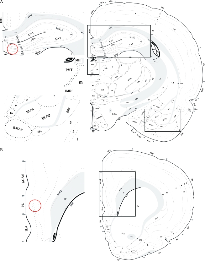
CREB- or pCREB-positive cells in CA1, CA2, CA3, PVT, amygdala (basolateral nucleus amygdala, anterior part [BLAa]; basolateral nucleus amygdala, posterior part [BLAp]; basomedial nucleus amygdala, posterior part [BMAp]; and lateral nucleus amygdala [LA]) (4 slices for each rat, bregma −2.45 to −2.85), ACAd, and ILA (8 slices for each rat, bregma +3.20 to +2.15) (Fig. 1) were blindly quantified using a computerized imaging analysis system (Westenbroek, Den Boer, et al. 2003). The selected areas were digitized by using a Sony CCD camera mounted on a LEICA Leitz DMRB microscope (Leica, Wetzlar, Germany) at ×100 magnification. Regions of interest were outlined with a light pen, measured, and CREB or pCREB positive nuclei were counted using a computer-based image analysis system LEICA (LEICA Imaging System Ltd., Cambridge, England). Each digitized image was individually set at a threshold to subtract the background optical density. Only cell nuclei that exceeded a defined threshold were detected by the image analysis system. The resulting data were reported as number of positive cells/0.1 mm2. All brain regions were quantified bilaterally (Lin et al. 2008). In amygdala, CREB and pCREB positive nuclei were counted in BLAa, BLAp, BMAp, and LA separately, then different subnucleus measures were combined to make an average that is comparable to the results from western blot in a time course.
Figure 1.
Atlas (Swanson 1992) image represents the approximate brain level where proteins levels were analyzed. CREB and pCREB positive nuclei measured by immunohistochemistry were counted in (A) CA1, CA2, CA3, PVT, and amygdala (BLAa, BLAp, BMAp, and LA); (B) ACAd and ILA. BDNF levels in DG (A) and PL (B) (red circle) were measured by ELISA analysis.
BDNF ELISA Analysis
Serial 300-μm coronal sections of the cerebrum were made with a cryostat microtome (−15 °C) and kept frozen on dry ice. Tissue samples were dissected from DG (bregma −2.45 to −2.85) and PL (bregma +3.20 to +2.15) (Fig. 1) by using the “Palkovits Punch” technique (needle diameter 0.94 mm, Stoelting Co., Wood Dale, IL). Two punches per animal (left and right per area, were taken and diluted in 100 μl buffer (50 mM Tris pH 7.0, 500 mM NaCl, 0.2% Triton X-100, 0.1% NaN3, 2 mM ethylenediaminetetraacetic acid, and 1× complete protease inhibitors [Roche, Basel, Switzerland]). The material was sonicated twice for 5 s followed by 30 min centrifugation at 16 000 × g at 4 °C. The supernatant was removed and saved at −20 °C until use. BDNF levels were measured with the BDNF Emax ImmunoAssay System of Promega (G7611).
Statistical Analysis
Data were expressed as means ± standard error of the mean and analyzed with SPSS (SPSS Inc., Chicago, IL) (version 12.0). P < 0.05 was defined as the level of significance. CREB, pCREB, and BDNF levels were analyzed separately in each brain region with an analysis of variance (ANOVA) with treatment group (control or stress) and sex (male or female) as between-subject variables. Bonferroni tests were used to do mean contrasts following ANOVA.
Results
CREB and pCREB Levels in Male and Female CA1, CA2, and CA3 of Hippocampus
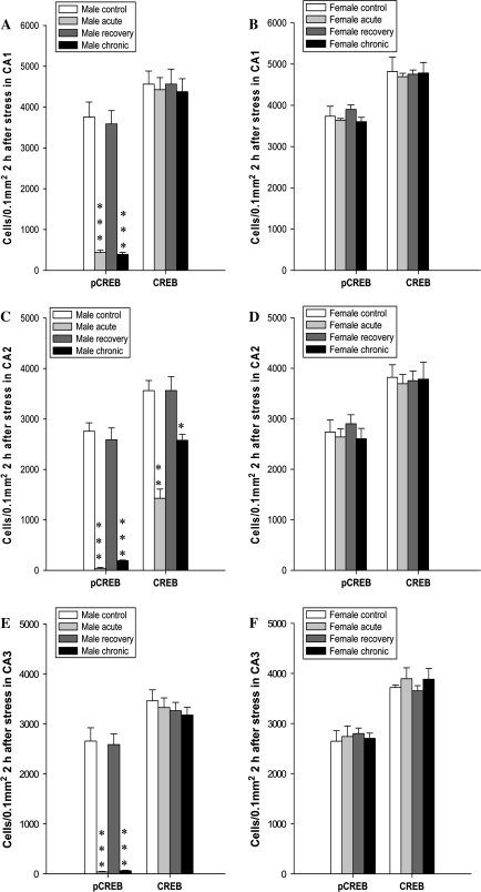
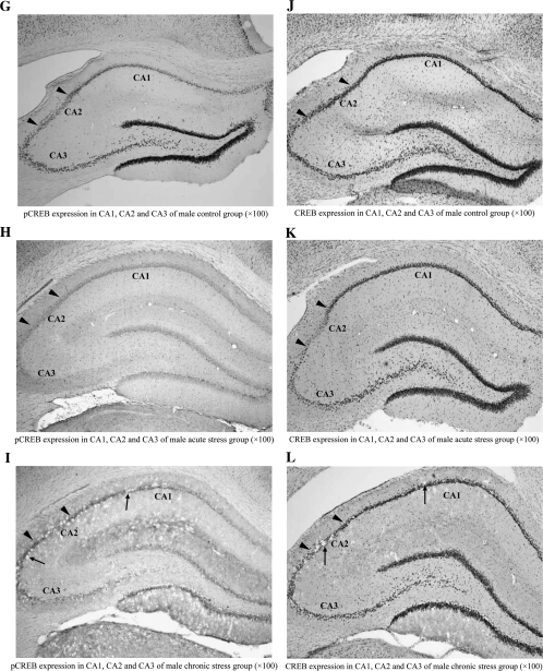
In male rats, acute and chronic stress caused a significant change in the number of pCREB-positive cells in hippocampal areas CA1, CA2, and CA3. The number of pCREB-positive cells was significantly decreased in males from the acute group (F1,8 = 98.473, P < 0.001, Fig. 2A; F1,8 = 112.152, P < 0.001, Fig. 2C; F1,8 = 97.523, P < 0.001, Fig. 2E) and from the chronic group (F1,8 = 106.613, P < 0.001, Fig. 2A; F1,8 = 72.698, P < 0.001, Fig. 2C; F1,8 = 112.745, P < 0.001, Fig. 2E). Patches of morphological abnormalities (Fig. 2I,L; black arrow) were seen in CA1, CA2, and CA3 of male rats from the chronic stress group but not in male rats from the acute stress group (Fig. 2H,K). The size of the patches varied, but in comparison to the surrounding tissue, these patches all appeared very lightly stained and showed no CREB- or pCREB-positive cells. In addition to the effects observed in pCREB levels, CREB levels were reduced by acute stress (F1,8 = 15.562, P < 0.01, Fig. 2C,K) and chronic stress (F1,8 = 4.851, P < 0.05, Fig. 2C,L) in male CA2 but not in male CA1 or CA3. Male rats from the recovery group showed no significant change from the control group in the number of pCREB- or CREB-positive cells on day 43 (Fig. 2A,C,E) and also showed none of the morphologically distinct patches seen in the chronic stress group (data not shown). In female rats, neither acute nor chronic stress had a significant effect on CREB or pCREB levels in CA1, CA2, or CA3 (Fig. 2B,D,F), and neither stress group showed any of the morphologically distinct patches observed in chronically stressed males (data not shown). Male and female control rats did not differ on any measures reported above (Fig. 2A–F).
Figure 2.
Levels of pCREB and CREB in CA1, CA2, and CA3 of hippocampus. (A) Number of pCREB- and CREB-positive cells in male CA1; (B) Number of pCREB- and CREB-positive cells in female CA1; (C) Number of pCREB- and CREB-positive cells in male CA2; (D) Number of pCREB- and CREB-positive cells in female CA2; (E) Number of pCREB- and CREB-positive cells in male CA3; (F) Number of pCREB- and CREB-positive cells in female CA3; (G–I) Representative photomicrographs of pCREB-IR in CA1, CA2, and CA3 of male rat after acute and chronic stress; (J–L) Representative photomicrographs of CREB-IR in CA1, CA2, and CA3 of male rats after acute and chronic stress. Patches were clearly observed (black arrow) in CA1, CA2, and CA3 of male rats exposed to chronic stress. Data were expressed as mean ± standard error of the mean, n = 3. *P ≤ 0.05, **P ≤ 0.01, ***P ≤ 0.001 versus control group.
CREB and pCREB Levels in Male and Female ILA and ACAd
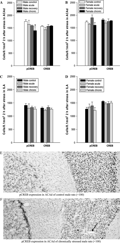
In male rats, stress had a significant effect on the number of cells expressing pCREB in ACAd. Chronic stress significantly decreased the number of cells expressing pCREB in male ACAd compared with control male rats (F1,8 = 2.942, P < 0.05, Fig. 3A). After recovery, there was no significant change in the number of cells expressing pCREB in male ACAd (Fig. 3A). There was no significant change in the number of CREB-positive cells in male ACAd. Numbers of pCREB- and CREB-positive cells were not changed significantly in male ILA (Fig. 3A,C) after chronic stress. Chronic stress also induced morphological abnormalities and irregularities in male ACAd which were reflected by appearance of patches (black arrow) (Fig. 3F). There was no positive staining in these patches, and the background of these patches was bright, which is different from the tissue around. Acute stress exerted no significant effects on CREB and pCREB levels in male ILA and ACAd (Fig. 3A,C). In female ILA and ACAd, no significant change in the number of cells expressing CREB and pCREB was found (Fig. 3B,D) after chronic and acute stress, also no patches were present (data not shown). Male and female control rats did not differ on any measures reported above (Fig. 3A–D).
Figure 3.
Number of cells expressing pCREB and CREB in ILA and ACAd of PFC. (A) Number of pCREB- and CREB-positive cells in male ACAd; (B) Number of pCREB- and CREB-positive cells in female ACAd; (C) Number of pCREB- and CREB-positive cells in male ILA; (D) Number of pCREB- and CREB-positive cells in female ILA; (E) Representative photomicrograph of pCREB-IR in ACAd in control male rats; (F) Representative photomicrograph of pCREB-IR in ACAd in chronically stressed male rats, patches were clearly observed in male ACAd (black arrow) after chronic stress. Data were expressed as mean ± standard error of the mean, n = 3. *P ≤ 0.05 control group versus chronically stressed group.
CREB and pCREB Levels in Male and Female Amygdala
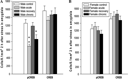
In male rats, acute and chronic stress caused a significant change in the number of pCREB-positive cells in amygdala. The amygdala of male rats showed a significant reduction in the number of pCREB-positive cells following acute (F1,8 = 11.251, P < 0.01, Fig. 4A) and chronic stress (F1,8 = 7.821, P < 0.05, Fig. 4A), but these levels returned to normal in the recovery group (Fig. 4A). However, amygdala from acutely and chronically stressed males showed no significant change in CREB-IR nor did they show any of the morphological abnormalities seen in the hippocampus and ACAd from the same exposure groups (data not shown). Amygdala from acutely and chronically stressed female rats showed neither morphological abnormalities nor any change in pCREB- or CREB-IR. Male and female control rats did not differ on any of the measures reported above (Fig. 4A,B).
Figure 4.
Number of cells expressing pCREB and CREB in amygdala. (A) Number of pCREB- and CREB-positive cells in male amygdala; (B) Number of pCREB- and CREB-positive cells in female amygdala. Data were expressed as mean ± standard error of the mean, n = 3. *P ≤ 0.05, **P ≤ 0.01 versus control group.
CREB and pCREB Levels in Male and Female PVT
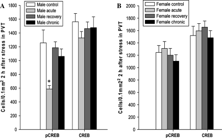
PVT from acutely stressed male rats showed a significant decrease in the number of pCREB-positive cells relative to controls (F1,8 = 6.594, P < 0.05), but the number of CREB-positive cells was not significantly changed (Fig. 5A). Chronic stress, on the other hand, had no significant effect on the number of pCREB- or CREB-positive cells in male PVT (Fig. 5A). PVT from female rats showed no changes in CREB or pCREB levels in either stress group, and neither sex showed any stress-induced morphological changes in the PVT. Male and female control rats did not differ on any of the measures reported above (Fig. 5A,B).
Figure 5.
Number of cells expressing pCREB and CREB in PVT. (A) Number of pCREB- and CREB-positive cells in male PVT; (B) Number of pCREB- and CREB-positive cells in female PVT. Data were expressed as mean ± standard error of the mean, n = 3. *P ≤ 0.05 versus control group.
BDNF Levels in Male and Female PL and DG
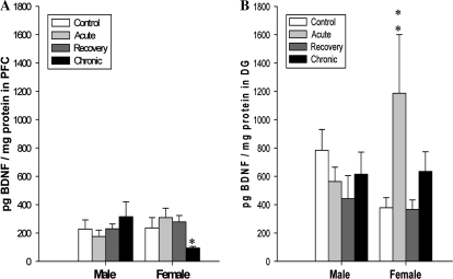
In male rats, neither the DG nor the PL region of PFC showed any significant change in BDNF levels in either acute or chronic stress groups when measured by ELISA (Fig. 6). However, in female rats, the DG showed increased BDNF in the acute stress group only (F1,16 = 24.451, P < 0.01, Fig. 6A), and the PL region of PFC showed decreased BDNF in the chronic stress group only (F1,16 = 5.564, P < 0.05, Fig. 6B). Male control rats showed a higher baseline BDNF level in the DG than did females, but no other sex-related differences in BDNF levels were noted in the control groups.
Figure 6.
Levels of BDNF measured by ELISA in PFC (A) and DG (B) of male and female rats. Data were expressed as mean ± standard error of the mean, n = 5. *P ≤ 0.05, **P ≤ 0.01 versus control group.
Discussion
Sex-specific analysis has much potential to further our understanding of mental health disorders and to improve the treatment of affected patients (Vlassoff and Garcia Moreno 2002). For this reason, we investigated sex differences in response to stress, a risk factor for developing depression. Here we show that the brains of male and female rats respond to stress in different ways. The main results of this study can be summarized with 4 points: First, in male rats, stress reduces pCREB levels in ACAd, CA1, CA2, CA3, PVT, and amygdala and CREB levels in CA2 but has no similar effect in females; Second, stress exerts no effect on BDNF levels in male PFC and DG, but chronic stress decreases BDNF levels in female PFC and acute stress increases BDNF levels in female DG; Third, stress induces morphological alterations in male CA1, CA2, CA3, and ACAd, whereas these changes are not found in females; Fourth, recovery following stress restores these parameters to baseline in both male and female rats.
Hippocampus and PFC are implicated as the key sites of neuropathology in depression (Rajkowska 2000; McEwen 2001; Shansky et al. 2004). Our results reveal morphological alterations (patches) in CA1, CA2, CA3, and ACAd following stress in male rats but not in females, and similar changes are also found in male DG and PL (Lin et al. 2008). The patches observed in male rats may be due to necrosis and/or apoptosis (Sapolsky 2000) and those seen in ACAd may also represent a loss of glial cells (Cotter et al. 2001). Stress not only induces morphological alterations but also decreases levels of pCREB and CREB in hippocampus and ACAd. Similarly, chronic stress suppresses hippocampal neurogenesis in rats, as indicated by decreased numbers of BrdU-labeled and pCREB-positive cells (Luo et al. 2005). In view of the implications of CREB and pCREB in neuronal plasticity, these findings may provide a biological explanation for clinically observed atrophy of neuronal and/or glial cells in depressive patients, particularly in the hippocampus and ACAd (Rantamäki et al. 2006). Our results indicate that the male rat hippocampus is more susceptible to stress-induced changes in morphology and in pCREB and CREB levels than is the female rat hippocampus. Despite these findings, men are still less likely than women to experience depression. In fact, increasing evidence from animal studies suggests that females are relatively resistant to neurobiological effects of acute and chronic stress (Lin et al. 2008). For example, chronic stress over 21 days produces reversible atrophy of apical dendrites of hippocampal pyramidal neurons in males but not in females (Galea et al. 1997; Conrad et al. 1999). Similarly, repeated swim stress over 30 days decreases CA3 and CA4 pyramidal cell numbers in gonadectomized male rats but not in females (Mizoguchi et al. 1992). The modulation of neurogenesis by controllability is evident in males but not in females (Shors et al. 2007). Furthermore, neurogenesis in adult females is reportedly not affected by predator odors, whereas it is significantly decreased in males (Falconer and Galea 2003).
Hippocampus and ACAd are implicated in several higher brain functions, including learning, memory, attention, conflict monitoring, pain, pleasure, and decision making (Genoux et al. 2002; Vogt and Laureys 2005). The reduction in pCREB and CREB levels and the morphological alterations seen in hippocampus and ACAd suggest dysfunction of neurons in these areas, which may contribute to cognitive deficits in mental disorders (Genoux et al. 2002; Sanders et al. 2002). Increasing evidence indicates that there is a strong correlation between depression and cognitive impairments (Song et al. 2006), so dysfunction caused by damage to these areas may significantly contribute to depression. Although chronic stress decreases pCREB levels and induces morphological abnormalities in male hippocampus and ACAd, no such alterations are found in male ILA of PFC. These findings suggest that the effects of stress on the brain are likely to be region specific. However, there is evidence that the ILA has extensive projections to central autonomic nuclei that modulate visceral responses to stress, and ILA has been reported to be activated following stress (Hurley et al. 1991; Nikulina et al. 2004). As such, the evidence presented here should not be considered an argument against the involvement of ILA in depression but rather an elimination of one possible mechanism of it's involvement. We also found an interesting pattern of stress-related changes in the amygdala. Although chronic stress decreases pCREB levels in male amygdala, no concurrent abnormalities in morphology are seen. The mechanisms underlying the discrepancy between amygdala and hippocampus or PFC remain unclear. The amygdala is considered to be a primary site of synaptic plasticity and could be involved in anhedonia, a key symptom of depression (Wallace et al. 2004), and chronic antidepressant administration has been shown to increase CREB and pCREB levels in amygdala (Thome et al. 2000; Wallace et al. 2004). The reduction in pCREB in the present study suggests a role for amygdala dysfunction following chronic stress.
Contrary to the effects of stress on pCREB and CREB levels, stress has no effect on BDNF, a CREB-regulated target gene (Nibuya et al. 1995) in male rats but impacts BDNF levels in female rats. Consistent with these findings, artificial rearing decreases BDNF levels in mPFC in comparison to mother-reared groups in female rats (Burton et al. 2007). These results suggest that BDNF may be regulated by factors other than CREB and pCREB. Changes in BDNF protein levels and CREB activation are not necessarily correlated (Miyata et al. 2001; Aso et al. 2008), and other studies have suggested that nuclear factor of activated T cells may be a more critical regulator of BDNF in the brain (Graef et al. 2003). The transcriptional activity of the CREB/pCREB system is known to be enhanced when the ratio of pCREB to CREB increases (Pardon et al. 2005), but our data failed to show any correlation between such a ratio and the measured outcomes reported here. However, it should be noted that the activation of CREB phosphorylation exhibits a biphasic pattern following stress (Stanciu et al. 2001; Bilang-Bleuel et al. 2002), and our previous study showed that pCREB levels are normal 30 min after stress but significantly decreased after 2 h (Lin et al. 2008). Furthermore, BDNF mRNA shows a biphasic time course following stress (Nanda and Mack 2000; Pizarro et al. 2004), whereas BDNF protein levels show a monophasic response (Nanda and Mack 2000; Pardon et al. 2005). This could arise from the fact that 4 different RNA transcripts, differentially transcribed in the central nervous system, encode for the same BDNF protein (Timmusk et al. 1993) suggesting a possible uncoupling of BDNF transcriptional and translational mechanisms; Further complicating the role of CREB and pCREB in regulation of BDNF responses to stress are findings that CREB-mediated gene expression might occur in the absence of Ser133 phosphorylation (Conkright et al. 2003; Iourgenko et al. 2003). Clearly, more studies are needed to clarify the signal transduction pathways between CREB/pCREB and BDNF. Nevertheless, our results suggest that the changes in BDNF, CREB, and pCREB cannot fully explain the sex differences and morphologic alterations observed in the stress response.
This study did not attempt to address the role of gonadal steroids in the stress response, but such steroids have been implicated as a modulating factor in some of the sex differences (Simerly et al. 1990; Hagihara et al. 1992; Young et al. 2000). For example, clinical evidence indicates that estrogen may contribute to the sex differences (Bebbington et al. 1998; Born et al. 2002; Hayward and Sanborn 2002). Further investigation is needed to determine if sex hormones contribute to the differential regulation of CREB, pCREB, and BDNF in response to stress. It should be noted that in this study 3 weeks of recovery after stress restored all the affected parameters to control levels in both male and female rats, suggesting that the changes in morphology and chemistry seen in hippocampus, PFC, PVT, and amygdala in response to acute and chronic stress are largely reversible. However, other studies have demonstrated that permanent damage may occur in the brain under extreme stress conditions (McEwen 2000). In our experiment, rats reexposed to the adverse environment after 3 weeks of recovery and were expected to show attenuated versions of the responses seen in acutely stressed animals. However, recovery male rats show pCREB and CREB levels and morphology similar to controls. Corticosteroid levels are increased after reexposure in male and female rats, implying that the reexposure to the adverse environment is still stressful (Lin et al. 2008), but the stressful reexposure does not induce changes in pCREB and CREB levels and morphology. One possible explanation is that male rats cannot retrieve the previous adverse event because the neuronal circuitry somehow has adapted, which may help male rats to circumvent negative effects of stress (Shors 1999; Rasmusson et al. 2002). Female rats do not show changes in pCREB and CREB levels and morphology after acute and chronic stress, and thus, it is not surprising that we do not find such changes after reexposure. It remains to be seen whether other signal transduction systems are more critical in stress coping in female rats.
Although sex differences are observed at the biochemical level (CREB, pCREB, and BDNF), no sex differences are seen in behavioral responses to stress as measured with the open-field tests (Lin et al. 2008). This nonsynchronousness may be explained by the evidence that loss of hippocampal BDNF per se is not sufficient to alter locomotor activity, anxiety-like behavior, fear conditioning or depression-related behaviors (Adachi et al. 2008), and conditional or partial deletion of BDNF is not sufficient to produce a depressive phenotype (Duman and Monteggia 2006). Even so, BDNF in DG may be essential in mediating the therapeutic effect of antidepressants. Franklin and Perrot-Sinal (2006) report lower levels of BDNF protein in DG of intact females relative to intact males, which is confirmed in control rats in our study. However, in our experiment, no sex differences in behavior are found in control rats. Interestingly, levels of BDNF in DG of male and female rats are at the same level at the end of our experiment, which may partially explain the lack of sex differences seen in behavior. Although CREB is critical to target gene regulation after chronic drug administration, behavioral responses to antidepressants may occur by CREB-independent mechanisms (Conti et al. 2002). This hypothesis is supported by the evidence that CREBad-homozygous deficient mice exhibit normal activity in a familiar environment (Hebda-Bauer et al. 2004) and indicates that the changes in CREB and BDNF levels are not necessary to induce behavior alteration. Furthermore, the different behavioral effects of CREB levels in different brain regions, such as hippocampus (Chen et al. 2001), nucleus accumbens (Barrot et al. 2002), amygdala (Pandey et al. 2003), or CREB-deficient mice (Valverde et al. 2004), make it possible to counteract the behavior induced by stress-related changes in CREB levels in these brain regions. Accordingly, no sex differences in behavior are found despite the sex differences in CREB levels. Therefore, it is of interest to identify other potential effector genes that could influence behavior (Wallace et al. 2004), and consideration of sex should be emphasized in future studies of the neurobiological mechanisms of depression (Monteggia et al. 2007). Furthermore, our findings showing that chronic stress is not different from acute stress in terms of changes in CREB and pCREB suggest that CREB may be critical for a stress response but not for stress-induced depression which is presumed to occur as a result of chronic but not acute stress as the molecular changes do not correspond to the presumed behavioral changes.
Based on our results, it can be concluded that male and female rats respond to stress in different ways. CREB and pCREB levels and morphology are more sensitive to stress in male rats than in female rats, whereas BDNF is more sensitive to stress in female rats than in male rats. The levels of CREB/pCREB and BDNF are not necessarily correlated. These sex differences may have important implications for development of novel therapeutics for depression.
Funding
Funding for this article came from the Bernouilli grant from the University of Groningen. Funding to pay the Open Access publication charges for this article was provided by University Medical Center Groningen and the University Groningen.
Acknowledgments
We would like to thank Rob Visser and Britta Kuest in the Graduate School Behavioral and Cognitive Neurosciences of the University of Groningen for administrative support. We also would like to thank Dr James Tucker and Carl B. Watt from University of Utah for English improvement. Conflict of Interest: None declared.
References
- Adachi M, Barrot M, Autry AE, Theobald D, Monteggia LM. Selective loss of brain-derived neurotrophic factor in the dentate gyrus attenuates antidepressant efficacy. Biol Psychiatry. 2008;63:642–649. doi: 10.1016/j.biopsych.2007.09.019. [DOI] [PMC free article] [PubMed] [Google Scholar]
- Aso E, Ozaita A, Valdizán EM, Ledent C, Pazos A, Maldonado R, Valverde O. BDNF impairment in the hippocampus is related to enhanced despair behavior in CB1 knockout mice. J Neurochem. 2008;105:565–572. doi: 10.1111/j.1471-4159.2007.05149.x. [DOI] [PubMed] [Google Scholar]
- Bale TL. Stress sensitivity and the development of affective disorders. Horm Behav. 2006;50:529–533. doi: 10.1016/j.yhbeh.2006.06.033. [DOI] [PubMed] [Google Scholar]
- Barrot M, Olivier JD, Perrotti LI, DiLeone RJ, Berton O, Eisch AJ, Impey S, Storm DR, Neve RL, Yin JC, et al. CREB activity in the nucleus accumbens shell controls gating of behavioral responses to emotional stimuli. Proc Natl Acad Sci USA. 2002;99:11435–11440. doi: 10.1073/pnas.172091899. [DOI] [PMC free article] [PubMed] [Google Scholar]
- Bebbington PE, Dunn G, Jenkins R, Lewis G, Brugha T, Farrell M, Meltzer H. The influence of age and sex on the prevalence of depressive conditions: report from the National Survey of Psychiatric Morbidity. Psychol Med. 1998;28:9–19. doi: 10.1017/s0033291797006077. [DOI] [PubMed] [Google Scholar]
- Beck CH, Fibiger HC. Chronic desipramine alters stress-induced behaviors and regional expression of the immediate early gene, c-fos. Pharmacol Biochem Behav. 1995;51:331–338. doi: 10.1016/0091-3057(94)00391-u. [DOI] [PubMed] [Google Scholar]
- Berton O, Nestler EJ. New approaches to antidepressant drug discovery: beyond monoamines. Nat Rev Neurosci. 2006;7:137–151. doi: 10.1038/nrn1846. [DOI] [PubMed] [Google Scholar]
- Bilang-Bleuel A, Rech J, De Carli S, Holsboer F, Reul JM. Forced swimming evokes a biphasic response in CREB phosphorylation in extrahypothalamic limbic and neocortical brain structures in the rat. Eur J Neurosci. 2002;15:1048–1060. doi: 10.1046/j.1460-9568.2002.01934.x. [DOI] [PubMed] [Google Scholar]
- Bissette G, Klimek V, Pan J, Stockmeier C, Ordway G. Elevated concentrations of CRF in the locus coeruleus of depressed subjects. Neuropsychopharmacology. 2003;28:1328–1335. doi: 10.1038/sj.npp.1300191. [DOI] [PubMed] [Google Scholar]
- Blendy J. The role of CREB in depression and antidepressant treatment. Biol Psychiatry. 2006;59:1144–1150. doi: 10.1016/j.biopsych.2005.11.003. [DOI] [PubMed] [Google Scholar]
- Born L, Shea A, Steiner M. The roots of depression in adolescent girls: is menarche the key? Curr Psychiatry Rep. 2002;4:449–460. doi: 10.1007/s11920-002-0073-y. [DOI] [PubMed] [Google Scholar]
- Bubser M, Deutch AY. Stress induces Fos expression in neurons of the thalamic paraventricular nucleus that innervate limbic forebrain sites. Synapse. 1999;32:13–22. doi: 10.1002/(SICI)1098-2396(199904)32:1<13::AID-SYN2>3.0.CO;2-R. [DOI] [PubMed] [Google Scholar]
- Burton CL, Chatterjee D, Chatterjee-Chakraborty M, Lovic V, Grella SL, Steiner M, Fleming AS. Prenatal restraint stress and motherless rearing disrupts expression of plasticity markers and stress-induced corticosterone release in adult female Sprague-Dawley rats. Brain Res. 2007;1158:28–38. doi: 10.1016/j.brainres.2007.05.003. [DOI] [PubMed] [Google Scholar]
- Carlezon WA, Jr, Duman RS, Nestler EJ. The many faces of CREB. Trends Neurosci. 2005;28:436–445. doi: 10.1016/j.tins.2005.06.005. [DOI] [PubMed] [Google Scholar]
- Carter-Snell C, Hegadoren K. Stress disorders and gender: implications for theory and research. Can J Nurs Res. 2003;35:34–55. [PubMed] [Google Scholar]
- Chen ACH, Shirayama Y, Shin KH, Neve RL, Duman RS. Expression of cAMP response element binding protein (CREB) in hippocampus produces an antidepressant effect. Biol Psychiatry. 2001;49:753–762. doi: 10.1016/s0006-3223(00)01114-8. [DOI] [PubMed] [Google Scholar]
- Conkright MD, Canettieri G, Screaton R, Guzman E, Miraglia L, Hogenesch JB, Montminy M. TORCs: transducers of regulated CREB activity. Mol Cell. 2003;12:413–423. doi: 10.1016/j.molcel.2003.08.013. [DOI] [PubMed] [Google Scholar]
- Conrad CR, Magarinos AM, LeDoux JE, McEwen BS. Repeated restraint stress facilitates fear conditioning, independently of causing hippocampal CA3 dendritic atrophy. Behav Neurosci. 1999;113:902–913. doi: 10.1037//0735-7044.113.5.902. [DOI] [PubMed] [Google Scholar]
- Conti AC, Cryan JF, Dalvi A, Lucki I, Blendy JA. cAMP response element-binding protein is essential for the upregulation of brain-derived neurotrophic factor transcription, but not the behavioral or endocrine responses to antidepressant drugs. J Neurosci. 2002;22:3262–3268. doi: 10.1523/JNEUROSCI.22-08-03262.2002. [DOI] [PMC free article] [PubMed] [Google Scholar]
- Cotter D, Mackay D, Landau S, Kerwin R, Everall I. Reduced glial cell density and neuronal size in the anterior cingulate cortex in major depressive disorder. Arch Gen Psychiatry. 2001;58:545–553. doi: 10.1001/archpsyc.58.6.545. [DOI] [PubMed] [Google Scholar]
- Drevets WC. Neuroimaging and neuropathological studies of depression: implications for the cognitive emotional features of mood disorders. Curr Opin Neurobiol. 2001;11:240–249. doi: 10.1016/s0959-4388(00)00203-8. [DOI] [PubMed] [Google Scholar]
- D'Sa C, Duman RS. Antidepressants and neuroplasticity. Bipolar Disord. 2002;4:183–194. doi: 10.1034/j.1399-5618.2002.01203.x. [DOI] [PubMed] [Google Scholar]
- Duman RS, Monteggia LM. A neurotrophic model for stress-related mood disorders. Biol Psychiatry. 2006;59:1116–1127. doi: 10.1016/j.biopsych.2006.02.013. [DOI] [PubMed] [Google Scholar]
- Falconer EM, Galea LAM. Sex differences in cell proliferation, cell death and defensive behavior following acute predator odor stress in adult rats. Brain Res. 2003;975:22–36. doi: 10.1016/s0006-8993(03)02542-3. [DOI] [PubMed] [Google Scholar]
- Franklin TB, Perrot-Sinal TS. Sex and ovarian steroids modulate brain-derived neurotrophic factor (BDNF) protein levels in rat hippocampus under stressful and non-stressful conditions. Psychoneuroendocrinology. 2006;31:38–48. doi: 10.1016/j.psyneuen.2005.05.008. [DOI] [PubMed] [Google Scholar]
- Galea LM, McEwen BS, Tanapat P, Deak T, Spencer RL, Dhabhar FS. Sex differences in dendritic atrophy of CA3 pyramidal neurons in response to chronic restraint stress. Neuroscience. 1997;81:689–697. doi: 10.1016/s0306-4522(97)00233-9. [DOI] [PubMed] [Google Scholar]
- Genoux D, Haditsch U, Knobloch M, Michalon A, Storm D, Mansuy IM. Protein phosphatase 1 is a molecular constraint on learning and memory. Nature. 2002;418:970–975. doi: 10.1038/nature00928. [DOI] [PubMed] [Google Scholar]
- Ghosh A, Ginty DD, Bading H, Greenberg ME. Calcium regulation of gene expression in neuronal cells. J Neurobiol. 1994;25:294–303. doi: 10.1002/neu.480250309. [DOI] [PubMed] [Google Scholar]
- Graef IA, Wang F, Charron F, Chen L, Neilson J, Tessier-Lavigne M, Crabtree GR. Neurotrophins and netrins require calcineurin/NFAT signaling to stimulate outgrowth of embryonic axons. Cell. 2003;113:657–670. doi: 10.1016/s0092-8674(03)00390-8. [DOI] [PubMed] [Google Scholar]
- Hagihara K, Hirata S, Osada T, Hirai M, Kato J. Distribution of cells containing progesterone receptor mRNA in the female rat di- and telencephalon: an in situ hybridization study. Brain Res Mol Brain Res. 1992;14:239–249. doi: 10.1016/0169-328x(92)90179-f. [DOI] [PubMed] [Google Scholar]
- Hayward C, Sanborn K. Puberty and the emergence of gender differences in psychopathology. J Adolescent Health. 2002;30:49–58. doi: 10.1016/s1054-139x(02)00336-1. [DOI] [PubMed] [Google Scholar]
- Hebda-Bauer EK, Watson SJ, Akil H. CREB deficient mice show inhibition and low activity in novel environments without changes in stress reactivity. Eur J Neurosci. 2004;20:503–513. doi: 10.1111/j.1460-9568.2004.03487.x. [DOI] [PubMed] [Google Scholar]
- Hurley KM, Herbert H, Moga MM, Saper CB. Efferent projections of the infralimbic cortex of the rat. J Comp Neurol. 1991;308:249–276. doi: 10.1002/cne.903080210. [DOI] [PubMed] [Google Scholar]
- Iourgenko V, Zhang W, Mickanin C, Daly I, Jiang C, Hexham JM, Orth AP, Miraglia L, Meltzer J, Garza D, et al. Identification of a family of cAMP response element-binding protein coactivators by genome-scale functional analysis in mammalian cells. Proc Natl Acad Sci USA. 2003;100:12147–12152. doi: 10.1073/pnas.1932773100. [DOI] [PMC free article] [PubMed] [Google Scholar]
- Joëls M. Functional actions of corticosteroids in the hippocampus. Eur J Pharmacol. 2008;583:312–321. doi: 10.1016/j.ejphar.2007.11.064. [DOI] [PubMed] [Google Scholar]
- Kessler RC. Epidemiology of women and depression. J Affect Disord. 2003;74:5–13. doi: 10.1016/s0165-0327(02)00426-3. [DOI] [PubMed] [Google Scholar]
- Kessler RC, McGonagle KA, Swartz M, Blazer DG, Nelson CB. Sex and depression in the National Comorbidity Survey. I. Lifetime prevalence, chronicity and recurrence. J Affect Disord. 1993;29:85–96. doi: 10.1016/0165-0327(93)90026-g. [DOI] [PubMed] [Google Scholar]
- Laifenfeld D, Kerry R, Grauer E, Klein E, Ben-Shachar D. Antidepressant and prolonged stress in rats modulate CAM-L1, laminin, and pCREB, implicated in neuronal plasticity. Neurobiol Dis. 2005;20:432–441. doi: 10.1016/j.nbd.2005.03.023. [DOI] [PubMed] [Google Scholar]
- Lin YH, Westenbroek C, Bakker P, Termeer J, Liu AH, Li XJ, Ter Horst GJ. Effects of long-term stress and recovery on the prefrontal cortex and dentate gyrus in male and female rats. Cereb Cortex. 2008;18:2762–2774. doi: 10.1093/cercor/bhn035. [DOI] [PubMed] [Google Scholar]
- Liotti M, Mayberg HS. The role of functional neuroimaging in the neuropsychology of depression. J Clin Exp Neuropsychol. 2001;23:121–136. doi: 10.1076/jcen.23.1.121.1223. [DOI] [PubMed] [Google Scholar]
- Lonze BE, Ginty DD. Function and regulation of CREB family transcription factors in the nervous system. Neuron. 2002;35:605–623. doi: 10.1016/s0896-6273(02)00828-0. [DOI] [PubMed] [Google Scholar]
- Luo C, Xu H, Li XM. Quetiapine reverses the suppression of hippocampal neurogenesis caused by repeated restraint stress. Brain Res. 2005;1063:32–39. doi: 10.1016/j.brainres.2005.09.043. [DOI] [PubMed] [Google Scholar]
- McEwen BS. Effects of adverse experiences for brain structure and function. Biol Psychiatry. 2000;48:721–731. doi: 10.1016/s0006-3223(00)00964-1. [DOI] [PubMed] [Google Scholar]
- McEwen BS. Plasticity of the hippocampus: adaptation to chronic stress and allostatic load. Ann N Y Acad Sci. 2001;933:265–277. doi: 10.1111/j.1749-6632.2001.tb05830.x. [DOI] [PubMed] [Google Scholar]
- Miyata K, Omori N, Uchino H, Yamaguchi T, Isshiki A, Shibasaki F. Involvement of the brain-derived neurotrophic factor/TrkB pathway in neuroprotecive effect of cyclosporin A in forebrain ischemia. Neuroscience. 2001;105:571–578. doi: 10.1016/s0306-4522(01)00225-1. [DOI] [PubMed] [Google Scholar]
- Mizoguchi K, Kunishita T, Chui DH, Tabra T. Stress induces neuronal death in the hippocampus of castrated rats. Neurosci Lett. 1992;138:157–160. doi: 10.1016/0304-3940(92)90495-s. [DOI] [PubMed] [Google Scholar]
- Monteggia LM, Luikart B, Barrot M, Theobold D, Malkovska I, Nef S, Parada LF, Nestler EJ. Brain-derived neurotrophic factor conditional knockouts show gender differences in depression-related behaviors. Biol Psychiatry. 2007;61:187–197. doi: 10.1016/j.biopsych.2006.03.021. [DOI] [PubMed] [Google Scholar]
- Nanda SA, Mack KJ. Seizures and sensory stimulation result in different patterns of brain derived neurotrophic factor protein expression in the barrel cortex and hippocampus. Brain Res Mol Brain Res. 2000;78:1–14. doi: 10.1016/s0169-328x(00)00054-1. [DOI] [PubMed] [Google Scholar]
- Nibuya M, Morinobu S, Duman RS. Regulation of BDNF and trkB mRNA in rat brain by chronic electroconvulsive seizure and antidepressant drug treatments. J Neurosci. 1995;15:7539–7547. doi: 10.1523/JNEUROSCI.15-11-07539.1995. [DOI] [PMC free article] [PubMed] [Google Scholar]
- Nibuya M, Takahashi M, Russell DS, Duman RS. Repeated stress increases catalytic TrkB mRNA in rat hippocampus. Neurosci Lett. 1999;267:81–84. doi: 10.1016/s0304-3940(99)00335-3. [DOI] [PubMed] [Google Scholar]
- Nikulina EM, Covington HE, 3rd, Ganschow L, Hammer RP, Jr, Miczek KA. Long-term behavioral and neuronal cross-sensitization to amphetamine induced by repeated brief social defeat stress: Fos in the ventral tegmental area and amygdala. Neuroscience. 2004;123:857–865. doi: 10.1016/j.neuroscience.2003.10.029. [DOI] [PubMed] [Google Scholar]
- Ostlund H, Keller E, Hurd YL. Estrogen receptor gene expression in relation to neuropsychiatric disorders. Ann N Y Acad Sci. 2003;1007:54–63. doi: 10.1196/annals.1286.006. [DOI] [PubMed] [Google Scholar]
- Pandey SC, Roy A, Zhang H. The decreased phosphorylation of cyclic adenosinemonophosphate (cAMP) response element binding (CREB) protein in the central amygdala acts as a molecular substrate for anxiety related to ethanol withdrawal in rats. Alcohol Clin Exp Res. 2003;27:396–409. doi: 10.1097/01.ALC.0000056616.81971.49. [DOI] [PubMed] [Google Scholar]
- Pardon MC, Roberts RE, Marsden CA, Bianchi M, Latif ML, Duxon MS, Kendall DA. Social threat and novel cage stress-induced sustained extracellular-regulated kinase1/2 (ERK1/2) phosphorylation but differential modulation of brain-derived neurotrophic factor (BDNF) expression in the hippocampus of NMRI mice. Neuroscience. 2005;132:561–574. doi: 10.1016/j.neuroscience.2005.01.033. [DOI] [PubMed] [Google Scholar]
- Pizarro JM, Lumley LA, Medina W, Robison CL, Chang WE, Alagappan A, Bah MJ, Dawood MY, Shah JD, Mark B, et al. Acute social defeat reduces neurotrophin expression in brain cortical and subcortical areas in mice. Brain Res. 2004;1025:10–20. doi: 10.1016/j.brainres.2004.06.085. [DOI] [PubMed] [Google Scholar]
- Popoli M, Gennarelli M, Racagni G. Modulation of synaptic plasticity by stress and antidepressants. Bipolar Disord. 2002;4:166–182. doi: 10.1034/j.1399-5618.2002.01159.x. [DOI] [PubMed] [Google Scholar]
- Rajkowska G. Postmortem studies in mood disorders indicate altered numbers of neurons and glial cells. Biol Psychiatry. 2000;48:766–777. doi: 10.1016/s0006-3223(00)00950-1. [DOI] [PubMed] [Google Scholar]
- Rajkowska G. Depression: what we can learn from postmortem studies. Neuroscientist. 2003;9:273–284. doi: 10.1177/1073858403252773. [DOI] [PubMed] [Google Scholar]
- Rantamäki T, Knuuttila JE, Hokkanen ME, Castrén E. The effects of acute and long-term lithium treatments on trkB neurotrophin receptor activation in the mouse hippocampus and anterior cingulate cortex. Neuropharmacology. 2006;4:421–427. doi: 10.1016/j.neuropharm.2005.10.001. [DOI] [PubMed] [Google Scholar]
- Rasmusson AM, Shi L, Duman R. Downregulation of BDNF mRNA in the hippocampal dentate gyrus after re-exposure to cues previously associated with footshock. Neuropsychopharmacology. 2002;27:133–142. doi: 10.1016/S0893-133X(02)00286-5. [DOI] [PubMed] [Google Scholar]
- Sairanen M, O'leary OF, Knuuttila JE, Castren E. Chronic antidepressant treatment selectively increases expression of plasticity-related proteins in the hippocampus and medial prefrontal cortex of the rat. Neuroscience. 2007;144:368–374. doi: 10.1016/j.neuroscience.2006.08.069. [DOI] [PubMed] [Google Scholar]
- Sanders GS, Gallup GG, Heinsen H, Hof PR, Schmitz C. Cognitive deficits, schizophrenia, and the anterior cingulate cortex. Trends Cogn Sci. 2002;6:190–192. doi: 10.1016/s1364-6613(02)01892-2. [DOI] [PubMed] [Google Scholar]
- Sapolsky RM. Glucocorticoids and hippocampal atrophy in neuropsychiatric disorders. Arch Gen Psychiatry. 2000;57:925–935. doi: 10.1001/archpsyc.57.10.925. [DOI] [PubMed] [Google Scholar]
- Shansky RM, Glavis-Bloom C, Lerman D, McRae P, Benson C, Miller K, Cosand L, Horvath TL, Arnsten AF. Estrogen mediates sex differences in stress-induced prefrontal cortex dysfunction. Mol Psychiatry. 2004;9:531–538. doi: 10.1038/sj.mp.4001435. [DOI] [PubMed] [Google Scholar]
- Sheline YI. Neuroimaging studies of mood disorder effects on the brain. Biol Psychiatry. 2003;54:338–352. doi: 10.1016/s0006-3223(03)00347-0. [DOI] [PubMed] [Google Scholar]
- Shieh PB, Hu SC, Bobb K, Timmusk T, Ghosh A. Identification of a signaling pathway involved in calcium regulation of BDNF expression. Neuron. 1998;20:727–740. doi: 10.1016/s0896-6273(00)81011-9. [DOI] [PubMed] [Google Scholar]
- Shors TJ. Acute stress and re-exposure to the stressful context suppress spontaneous unit activity in the basolateral amygdala via NMDA receptor activation. Neuroreport. 1999;10:2811–2815. doi: 10.1097/00001756-199909090-00021. [DOI] [PubMed] [Google Scholar]
- Shors TJ, Mathew J, Sisti HM, Edgecomb C, Beckoff S, Dalla C. Neurogenesis and helplessness are mediated by controllability in males but not in females. Biol Psychiatry. 2007;62:487–495. doi: 10.1016/j.biopsych.2006.10.033. [DOI] [PMC free article] [PubMed] [Google Scholar]
- Simerly RB, Chang C, Muramatsu M, Swanson LW. Distribution of androgen and estrogen receptor mRNA containing cells in the rat brain: an in situ hybridization study. J Comp Neurol. 1990;294:76–95. doi: 10.1002/cne.902940107. [DOI] [PubMed] [Google Scholar]
- Smith MA, Makino S, Kvetnansky R, Post RM. Stress and glucocorticoids affect the expression of brain-derived neurotrophic factor and neurotrophin-3 mRNAs in the hippocampus. J Neurosci. 1995;15:1767–1777. doi: 10.1523/JNEUROSCI.15-03-01768.1995. [DOI] [PMC free article] [PubMed] [Google Scholar]
- Song L, Che W, Min-Wei W, Murakami Y, Matsumoto K. Impairment of the spatial learning and memory induced by learned helplessness and chronic mild stress. Pharmacol Biochem Behav. 2006;83:186–193. doi: 10.1016/j.pbb.2006.01.004. [DOI] [PubMed] [Google Scholar]
- Stanciu M, Radulovic J, Spiess J. Phosphorylated cAMP response element binding protein in the mouse brain after fear conditioning: relationship to Fos production. Brain Res Mol Brain Res. 2001;94:15–24. doi: 10.1016/s0169-328x(01)00174-7. [DOI] [PubMed] [Google Scholar]
- Sun MK, Alkon DL. Differential gender-related vulnerability to depression induction and converging antidepressant responses in rats. J Pharmacol Exp Ther. 2006;316:926–932. doi: 10.1124/jpet.105.093948. [DOI] [PubMed] [Google Scholar]
- Swanson LW. Brain maps: structure of the rat brain. Amsterdam (the Netherlands): Elsevier; 1992. [Google Scholar]
- Talarovicova A, Krskova L, Kiss A. Some assessments of the amygdala role in suprahypothalamic neuroendocrine regulation: a minireview. Endocr Regul. 2007;41:155–162. [PubMed] [Google Scholar]
- Thome J, Sakai N, Shin K, Steffen C, Zhang YJ, Impey S, Storm D, Duman RS. cAMP response element-mediated gene transcription is upregulated by chronic antidepressant treatment. J Neurosci. 2000;20:4030–4036. doi: 10.1523/JNEUROSCI.20-11-04030.2000. [DOI] [PMC free article] [PubMed] [Google Scholar]
- Timmusk T, Palm K, Metsis M, Reintam T, Paalme V, Saarma M, Persson H. Multiple promoters direct tissue-specific expression of the rat BDNF gene. Neuron. 1993;10:475–489. doi: 10.1016/0896-6273(93)90335-o. [DOI] [PubMed] [Google Scholar]
- Valverde O, Mantamadiotis T, Torrecilla M, Ugedo L, Pineda J, Bleckmann S, Gass P, Kretz O, Mitchell JM, Schütz G, et al. Modulation of anxiety-like behaviour and morphine dependence in CREB-deficient mice. Neuropsychopharmacology. 2004;29:1122–1133. doi: 10.1038/sj.npp.1300416. [DOI] [PubMed] [Google Scholar]
- Vlassoff C, Garcia Moreno C. Placing gender at the centre of health programming: challenges and limitations. Soc Sci Med. 2002;54:1713–1723. doi: 10.1016/s0277-9536(01)00339-2. [DOI] [PubMed] [Google Scholar]
- Vogt BA, Laureys S. Posterior cingulate, precuneal and retrosplenial cortices: cytology and components of the neural network correlates of consciousness. Prog Brain Res. 2005;150:205–217. doi: 10.1016/S0079-6123(05)50015-3. [DOI] [PMC free article] [PubMed] [Google Scholar]
- Wallace TL, Stellitano KE, Neve RL, Duman RS. Effects of cyclic adenosine monophosphate response element binding protein overexpression in the basolateral amygdala on behavioral models of depression and anxiety. Biol Psychiatry. 2004;56:151–160. doi: 10.1016/j.biopsych.2004.04.010. [DOI] [PubMed] [Google Scholar]
- Wang JW, Dranovsky A, Hen R. The when and where of BDNF and the antidepressant response. Biol Psychiatry. 2008;63:640–641. doi: 10.1016/j.biopsych.2008.01.008. [DOI] [PubMed] [Google Scholar]
- Westenbroek C, Den Boer JA, Ter Horst GJ. Gender-specific effects of social housing on chronic stress-induced limbic Fos expression. Neuroscience. 2003;121:189–199. doi: 10.1016/s0306-4522(03)00367-1. [DOI] [PubMed] [Google Scholar]
- Westenbroek C, Ter Horst GJ, Roos MH, Kuipers SD, Trentani A, den Boer JA. Gender-specific effects of social housing in rats after chronic mild stress exposure. Prog Neuropsychopharmacol Biol Psychiatry. 2003;27:21–30. doi: 10.1016/s0278-5846(02)00310-x. [DOI] [PubMed] [Google Scholar]
- Yang CH, Huang CC, Hsu KS. Differential roles of basolateral and central amygdala on the effects of uncontrollable stress on hippocampal synaptic plasticity. Hippocampus. 2008;18:548–563. doi: 10.1002/hipo.20414. [DOI] [PubMed] [Google Scholar]
- Young EA, Midgley R, Carlson NE, Brown MB. Alteration in the hypothalamic-pituitary-ovarian axis in depressed women. Arch Gen Psychiatry. 2000;57:1157–1162. doi: 10.1001/archpsyc.57.12.1157. [DOI] [PubMed] [Google Scholar]


