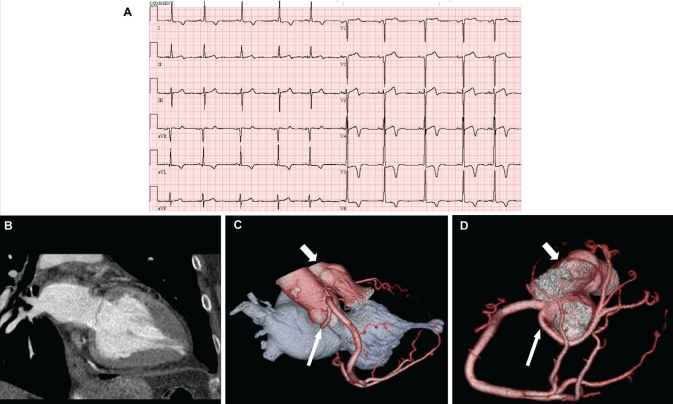
Computed tomographic coronary angiography can noninvasively provide valuable anatomical information that may delineate the mechanism of ischemia (1). The resting electrocardiogram of a 70-year-old woman with a history of hypertension, dyslipidemia and chest pain is shown in Figure 1A. Dipyridamole stress myocardial perfusion imaging revealed distal anterior wall and apical ischemia. Computed tomographic angiography demonstrated the presence of Yamaguchi’s apical variant of hypertrophic cardiomyopathy. The characteristic spade-like configuration of the left ventricular cavity is shown in multiplanar and three-dimensional volume-rendered two-chamber views of the left atrium and left ventricle during diastasis (Figures 1B and 1C). A single coronary artery arising from the right sinus of Valsalva is shown in Figures 1C and 1D. The left main artery (elongated arrow) coursed posterior to the aortic root, supplying a very small left anterior descending artery (right ventricular outflow tract [short arrow]). A large posterior interventricular artery supplied most of the apex. No significant coronary obstructions were detected.
Figure 1.
Both apical hypertrophic cardiomyopathy and anomalous coronary arteries are rare. Yamaguchi’s variant accounts for less than 10% of hypertrophic cardiomyopathy cases in some reports (2) and anomalous coronary arteries occur in less than 1% of live births (3).
REFERENCES
- 1.Berbarie RF, Dockery WD, Johnson KB, Rosenthal RL, Stoler RC, Schussler JM. Use of multislice computed tomographic coronary angiography for the diagnosis of anomalous coronary arteries. Am J Cardiol. 2006;98:402–6. doi: 10.1016/j.amjcard.2006.02.046. [DOI] [PubMed] [Google Scholar]
- 2.Klues HG, Schiffers A, Maron BJ. Phenotypic spectrum and patterns of left ventricular hypertrophy in hypertrophic cardiomyopathy: Morphologic observations and significance as assessed by two-dimensional echocardiography in 600 patients. J Am Coll Cardiol. 1995;26:1699–708. doi: 10.1016/0735-1097(95)00390-8. [DOI] [PubMed] [Google Scholar]
- 3.Kimbiris D, Iskandrian AS, Segal BL, Bemis CE. Anomalous aortic origin of coronary arteries. Circulation. 1978;58:606–15. doi: 10.1161/01.cir.58.4.606. [DOI] [PubMed] [Google Scholar]


