
Fig. 2.
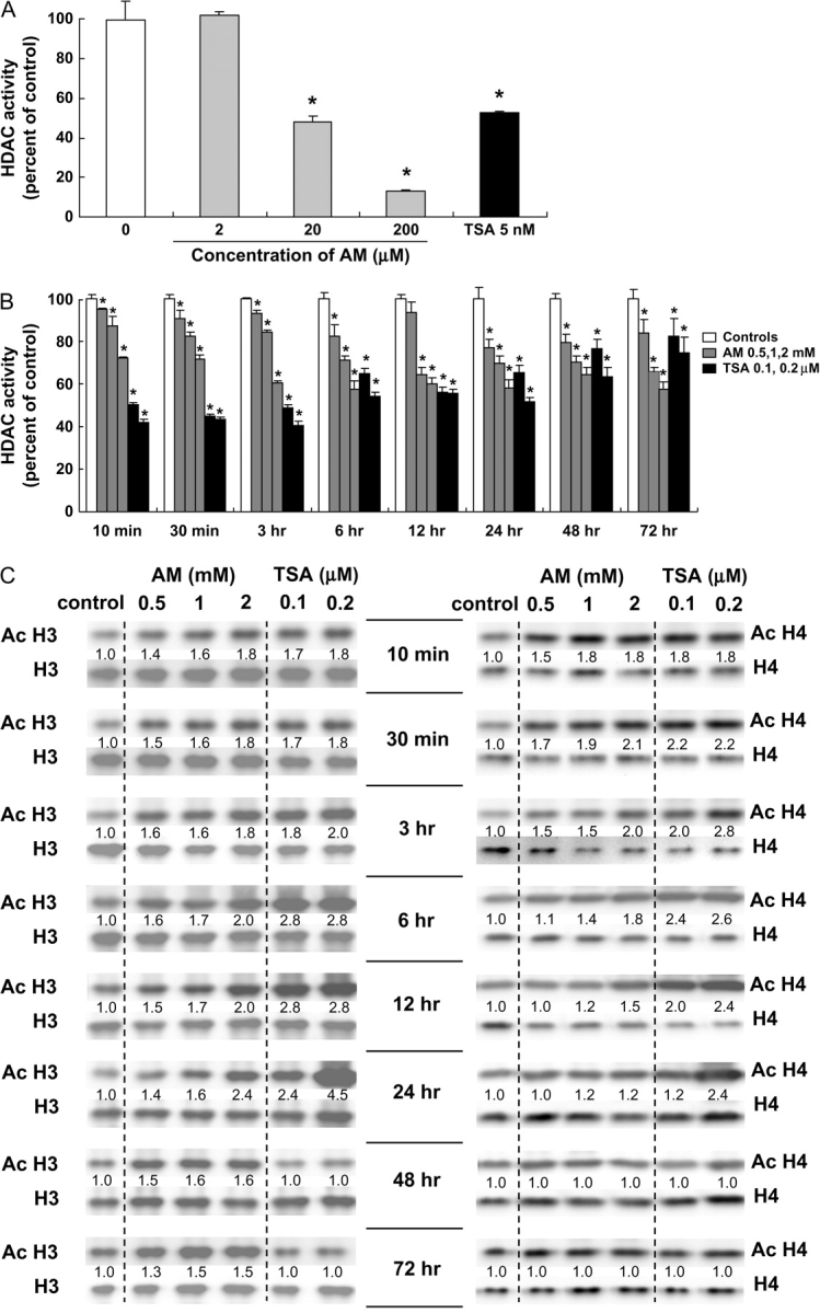
HDAC inhibition and histone acetylation in AM-treated HT29 cells. (A) Whole-cell extracts from human HT29 colon cancer cells were treated directly with the test agents and assayed for HDAC activity (BioMol kit). The IC50 for AM and TSA was 20 μM and 5 nM, respectively. Data = mean ± SD, n = 3, *P < 0.05. (B) HT29 cells were treated with AM (0.5, 1 and 2 mM) or TSA (0.1 and 0.2 μM) for selected times, from 10 min to 72 h, and whole-cell extracts were tested for HDAC activity. Data = mean ± SD, n = 3, *P < 0.05. (C) In the same cell lysates as (B), acetylated histones H3 and H4 were analyzed by immunoblotting. At each time point, acetylated histone expression was normalized to the corresponding non-acetylated histone, and this ratio was assigned an arbitrary value of 1.0 for the vehicle controls. Data are from a single experiment and are representative of findings from experiments repeated at least twice for each time point.