Abstract
Background
The 3-dimensional photonic scan (3DPS) technique has been used during the past decade in the fashion industry and for epidemiologic surveys to estimate human body sizes.
Objective
The objective of the study was to validate the accuracy of a recently developed 3DPS (C9036-02; Hamamatsu Photonics KK, Hamamatsu, Japan) for the measurement of body volume, circumferences, lengths, and percentage body fat with the use of underwater weighing (UWW) and tape measures as criterion methods.
Design
Ninety-two subjects (44 females and 48 males) aged 6–83 y and weighing 23–182 kg (52–400 lbs) participated in the study. The subjects were measured while they wore minimal clothing and a head cap. Similar measurements were performed on a mannequin with and without clothing
Results
All subjects were measured with 3DPS and a tape measure; 63 subjects underwent UWW and residual lung volume measurements. The values obtained with 3DPS were slightly but significantly greater than those obtained with UWW for body volume (81.9 ± 4.0 L compared with 81.5 ± 4.0 L, P < 0.0001) and those obtained with a tape measure for circumferences (P < 0.001), but the values for percentage body fat were not significantly different between 3DPS and UWW (P = 0.648). The values obtained with 3DPS were significantly greater than those obtained by UWW and a tape measure for the clothed mannequin, but the values were not uniformly significantly different for the mannequin without clothing.
Conclusions
The 3DPS measures body volume, circumferences, and length rapidly and accurately. However, to generate an accurate total-body volume measurement with 3DPS to estimate percentage body fat, the subjects must wear close-fitting minimal clothing and be able to stand motionless for 10 s (normal scan mode) while holding their breath, which is done immediately after a maximum expiration.
Keywords: Body composition, 3-dimensional photonic scan, body volume, body dimension, body image
INTRODUCTION
The need for accurate measurements of body shape and dimensions has increased because of accumulating knowledge about their relations with health risks. Body shape is related to growth, age, and fitness in healthy subjects and has been used for centuries as an index for acute or long-term diseases in clinical medicine (1–4). Although measuring body shape, including volumes and dimensions, could provide important information to investigators for research or clinical purposes, no single widely accepted technique can simultaneously measure the multiple variables that determine body shape. Traditional water and air-displacement techniques used to estimate total-body volume require well-trained laboratory staff and extensive cooperation and ability from the participant (5). In addition, total-body volume is the only directly measured value from these methods. Tape measures are usually used for other body sizes or dimensions, such as circumferences and lengths (6). However, the results vary widely depending on the observer’s skill level and measurement protocols (7). Computed tomography and magnetic resonance images generate more accurate total and regional body volumes and dimensions (8, 9), but these procedures are expensive and neither one is risk-free for the subject.
The use of a digitized optical method and computer to generate a 3-dimensional (3-D) photonic image of an object was developed over 4 decades ago (10) and was used as a technology for whole-body surface anthropometric measurements in humans (11). In 1998, Hamamatsu Photonics (Hamamatsu, Japan) used 8 light sources and fast cameras to assemble a previous model of a 3-D photonic image scanner (3DPS), which could obtain a maximum of 102 400 data points from a 12-s scan of a human subject and generate a 3-D image (12). Validation studies for total-body volume from this first generation 3DPS were conducted in children and adults by using underwater weighing (UWW) and air-displacement plethysmography techniques as standards (13, 14). The studies did not include regional body volume or dimension measurements.
The newly developed 3DPS system (Model # C9036-02; Hamamatsu Photonics KK, Hamamatsu, Japan) collects a maximum of 2 048 000 data points over a scan field (200 cm height × 100 cm width × 60 cm depth) in 10 s, which is about 20-fold the number of data points of the previous 3DPS generation. The new system also generates values for regional body volumes and dimensions as well as for total-body volume.
The purpose of our study was to validate the accuracy and reproducibility of the newly developed 3DPS for the measurement of total and regional body volumes and dimensions in human subjects with a wide range of age, weight, and body fatness and in a commercially available mannequin. Measurements made with traditional UWW and a tape measure were used as the criterion methods.
SUBJECTS AND METHODS
The 3-dimensional photonic scanner
The 3DPS used in the present study (model # C9036-02; Hamamatsu Photonics KK) is a noninvasive optical method that uses high-speed digital cameras (charge-coupled device cameras) and triangular mathematics to detect the actual position of eye-safe class-1 laser-light points (664 nm) projected onto the surface of an object and reflected to the cameras. Software then connects the points to generate a 3-D body image as well as values for total and regional body volumes and dimensions, such as body circumferences, lengths, widths, and thicknesses. The 3DPS has 4 photonic image production units mounted on 4 poles in the scanner. Each unit consists of a laser source and a digital camera. The distance between the camera and the point on the surface of the object that reflects the light is calculated with a triangulation algorithm. During the scanning process, the 4 scanning units simultaneously move vertically and produce a maximum of 2560 data points at each of the 800 cross-sections over the 2-m vertical scan field in 10 s with a 2.5 mm pitch interval (200 cm height × 100 cm width × 60 cm depth; total = 2 048 000 data points). In an alternate 5-s scan mode, the scanner uses 5.0 mm pitch intervals at 400 cross-sectional positions to produce 1 024 000 data points. The density of the data points from a 10-s scan of the body surface of a scanned subject is 16 data points/cm2. The size of a cross section on the scanned surface is calculated from all detected data points at the cross section, and the distance between any 2 body surface points is determined by all data points on the line between the 2 points. The measurement accuracy and precision of the scanner is directly related to the number of data points obtained on an object’s surface; the greater the number of data points, the higher the resolution or precision. The software can also calculate volume for any selected region of the body.
Subjects
Ninety-two subjects were recruited for the study: 44 females and 48 males, aged 6–83 y (24 were aged < 18 y), and with body weights ranging from 23 to 182 kg, heights ranging from 113.9 to 195.2 cm, and body mass indexes (BMIs; in kg/m2) ranging from 15.2 to 52.4. Forty-four of the 92 subjects (32 adults and 12 children) were classified as obese. Obesity was classified by using different cutoffs for the adult and pediatric subjects. Adults with a BMI > 30 were in the obese subgroup. Pediatric subjects with a BMI > 80th percentile according to the 2000 Centers for Disease Control and Prevention Growth Curves (15) were in the obese subgroup. The study was approved by the St. Luke’s–Roosevelt Hospital Institutional Review Board. A written consent form was obtained from each adult participant, an assent form and parental consent form were obtained from each participant aged < 18 y.
Mannequin
Because one goal of the study was to validate the scanner for regional as well total-body volume measurements, we performed the measurements on a commercially available fiberglass mannequin (size-6 female Child Man"equin; StoreFixtureExperts, Commack, NY); its arms and legs were detachable and could be measured separately. The height of the mannequin was 119.4 cm. The total-body measurements of the mannequin were performed under 2 conditions: with and without clothing. The 3DPS results were compared with the water displacement and tape measure results. The volume for each arm and leg was only measured without clothing by water displacement and compared with the volumes for the same regions generated by the 3DPS without clothing. The measurements for total-body volume were repeated 14 times, regional measurements were repeated 9 times, and ≤ 3 trials were done on a single workday.
Measurements
Three types of measurements were performed in the present study: anthropometric measurements, 3DPS, and UWW, and all were conducted in this order. The subjects wore close-fitting clothing and a head cap during the measurements.
Anthropometric measurements
Body weight, height, circumferences (chest, waist, hip, thigh, and knee), and length (the distance between the center of the kneecap and midpoint of thigh) were measured by an experienced investigator with the use of a heavy-duty, inelastic, plastic fiber Dritz sewing tape (Prym-Dritz USA, Spartanburg, SC) (16) with the methods described by Lohman (17). The precision of tape measures for body circumferences at our laboratory is ±2% (16). All anthropometric measurements were made directly on the skin, except for chest circumferences for females and hip circumferences for both sexes, which were measured over clothing. The location on the skin for each circumference measurement and the 2 points used to define the length of the leg were marked with a 0.5 cm diameter light-reflective and self-adhesive label so the scanner could produce measurements from identical locations. Similar measurements were performed on the mannequin 3 times/d on 3 d. The 3DPS can calculate the circumference at any location or distance between any 2 locations over the body surface. We selected 5 circumferences and one length for comparison.
3-D photonic image scanning measurements
After the anthropometric measurements were completed, the subject was positioned standing in the center of the scanner (which was clearly marked on the scanner floor) with their legs and arms abducted and no contact between any 2 body surfaces. The subject was told to remain motionless, according to the manufacturer’s instructions. The subjects were scanned 3 times while they breathed normally and then 3 times at the end of a maximum expiration, similar to the procedure in the water tank for the underwater weighing measurement (18). The body volume that was measured at the end of the maximum expiration was corrected for the residual lung volume, as measured by spirometry (19), and then body density was calculated by dividing the body weight measured by air displacement by the corrected body volume. The percentage body fat (%BF) was calculated by using the Siri equation (18). We used the normal scan mode of 10 s per scan. Between scans, the subject was asked to step out of the scanner and was repositioned. Similar scanning procedures were performed 3 times/d on 3 d for the mannequin (but, obviously, without maximum expiration).
Underwater weighing measurements
After completion of the 3DPS measurements, UWW was performed by using standard hydrostatic weighing at the end of a maximum expiration. The residual lung volume was measured by spirometry. Body volume was calculated as the difference in the body weight measured by air-displacement techniques and that measured underwater (corrected for the water temperature in the tank; 18) and then was corrected for the residual lung volume (19). Body density was calculated as the body weight measured by air-displacement techniques divided by the corrected body volume, and then %BF was calculated with the Siri equation (18). The within-person and day-to-day CV of this technique was ±0.3% for BF (20).
The total-body, arm, and leg volumes of the mannequin were measured by water displacement with the use of cylindrical glassware that had an internal volume capacity appropriate for the volume of each of the mannequin body parts. All measures were repeated 3 times/d on 3 different days.
Statistical analysis
Descriptive statistics were calculated for all variables by each method for the entire study group and for the subgroup of 63 subjects who had UWW and residual lung volume measurements. The hypotheses were that the mean circumference and length values measured by tape and by 3DPS at identical locations and the body volumes and %BF measured by UWW and by 3DPS would not be different. These hypotheses were tested by using paired t tests. Reproducibility of the measurements for volumes, circumferences at the 5 sites, and length at one site by 3DPS was determined by calculating the intraclass correlation and the CV. A linear regression analysis was used to model the relations between the 3DPS and the UWW and tape measures for selected body volume and dimension measurements and to test whether age, sex, or obesity had any significant influence on these relations.
All statistical analyses were performed with SAS version 8 (SAS Institute Inc, Cary, NC) and STATA version 8.2 statistical software packages for personal computer (STATA Corp, College Station, TX). The level of significance for all statistical tests of hypotheses was 0.05.
RESULTS
All 92 subjects were measured with 3DPS and a tape measure; 63 subjects were measured by UWW and for residual lung volume as well. Therefore, all required measurements, which included total-body volume and %BF measured by both 3DPS and UWW, were only performed on 63 subjects in the present study. In this 63-subject subgroup, 33 subjects were obese and 30 were not obese. Twenty of the 92 subjects were pediatric subjects; of these, 12 had both UWW and 3DPS measurements and 5 were obese (4 boys and 1 girl). The physical characteristics of the obese and nonobese subjects are shown by sex for the entire adult group and for the subgroup who had all the measurements for the study in Table 1.
TABLE 1.
Physical characteristics of the human subjects1
| All subjects (n = 92)2 | Subgroup (n = 63)3 | |
|---|---|---|
| Normal-weight males4 | ||
| Age (y) | 32.4 ± 16.3 (6–74) | 36.7 ± 16.5 (9–74) |
| Weight (kg) | 67.1 ± 17.8 (23.6–89.2) | 71.2 ± 13.5 (35.2–89.2) |
| Height (cm) | 168.8 ± 16.2 (118.5–191.5) | 171.1 ± 9.4 (139.6–180.1) |
| BMI (kg/m2) | 23.0 ± 3.8 (15.9–29.8) | 24.1 ± 3.3 (18.1–29.8) |
| Normal-weight females5 | ||
| Age (y) | 29.4 ± 19.7 (7–83) | 31.3 ± 24.2 (7–83) |
| Weight (kg) | 56.6 ± 17.9 (24.8–91.4) | 55.0 ± 18.5 (24.8–85.4) |
| Height (cm) | 158.2 ± 16.0 (122.1–195.2) | 153.6 ± 15.0 (122.1–169.4) |
| BMI (kg/m2) | 22.0 ± 4.4 (15.2–29.8) | 22.6 ± 4.9 (16.4–29.8) |
| Obese males6 | ||
| Age (y) | 27.4 ± 18.4 (6–68) | 34.7 ± 18.4 (13–68) |
| Weight (kg) | 92.7 ± 40.1 (22.9–181.9) | 107.2 ± 37.7 (57.6–181.9) |
| Height (cm) | 167.2 ± 18.0 (113.9–188.5) | 174.4 ± 9.1 (158.3–186.4) |
| BMI (kg/m2) | 31.6 ± 9.1 (17.7–52.4) | 34.5 ± 9.1 (22.0–52.4) |
| Obese females7 | ||
| Age (y) | 41.9 ± 14.9 (10–65) | 41.9 ± 15.1 (10–65) |
| Weight (kg) | 100.7 ± 17.2 (47.4–131.1) | 99.8 ± 17.8 (47.4–131.1) |
| Height (cm) | 165.2 ± 7.8 (147.2–180.9) | 166.0 ± 7.4 (147.2–180.9) |
| BMI (kg/m2) | 36.8 ± 5.3 (21.9–50.7) | 35.9 ± 4.4 (21.9–42.3) |
All values are x¯ ± SD; range in parentheses.
n = 25 children in the entire group; 48 subjects were normal weight (13 were children) and 44 subjects were obese (12 children).
Thirty subjects were normal-weight (8 children) and 33 subjects were obese (5 children).
n = 24 for the entire cohort, 16 for the subgroup.
n = 24 for the entire cohort, 14 for the subgroup.
n = 24 for the entire cohort, 15 for the subgroup.
n = 20 for the entire cohort, 18 for the subgroup.
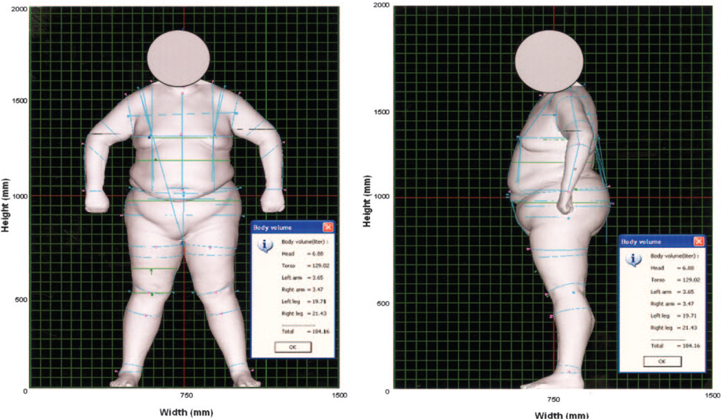
The total and regional body volumes and the frontal and side body dimensions of an adult weighing >400 lb (182 kg) who was measured by the scanner are shown in Figure 1 in proportion to the 2000 × 1500-mm graphic background. The lines indicate the locations for the selected circumferences and length measured with a tape measure and by 3DPS. For subject confidentiality, the scan image above the neck region was immediately deleted from the scan system after all the required dimensional values were generated by the system. The head regional image could not be reproduced.
Figure 1.
Total and regional body volumes and front and side body dimensions of a >182-kg adult measured by 3-dimensional photonic imaging scanner (3DPS) in proportion to the 2000 × 1500-mmgraphic background. The subject was breathing normally at the time of measurement. The dots indicate the selected locations for measurement of circumferences and length by 3DPS and tape measure techniques. The lines were measured by 3DPS.
The reliability of the measurements for volumes, circumferences, and length obtained by the 3DPS in 92 human subjects is shown in Table 2. All have intraclass correlations >0.97. The CV was <0.9 for circumferences and <1.2 for partial thigh length. The CVs were <2.3, <3.2, <2.5, <4.4, <4.5, and <1.9 for head, left and right arms, left and right legs, and torso volumes, respectively, which are significantly lower than the CV of <0.4 for total-body volume.
TABLE 2.
Measurement reliability of the 3-dimensional photonic scanner in 92 human subjects1
| Mean square error | SD | |||||||
|---|---|---|---|---|---|---|---|---|
| Variable | MS (ID) | MSE | Mean | Within-subject | Between-subject | Single reading | IC | CV |
| Circumference (mm) | ||||||||
| Chest | 114 724.51 | 66.79 | 1008.32 | 8.17 | 195.50 | 195.67 | 0.998 | 0.81 |
| Waist | 131 200.81 | 64.39 | 906.98 | 8.02 | 209.07 | 209.23 | 0.999 | 0.88 |
| Hip | 130 537.72 | 28.24 | 1060.83 | 5.31 | 208.57 | 208.64 | 0.999 | 0.50 |
| Thigh | 35 812.55 | 4.32 | 571.11 | 2.08 | 109.25 | 109.27 | 1.000 | 0.36 |
| Knee | 11 040.93 | 4.00 | 402.86 | 2.00 | 60.65 | 60.69 | 0.999 | 0.50 |
| Length (mm) | ||||||||
| Partial thigh | 3886.56 | 6.12 | 207.70 | 2.47 | 35.97 | 36.05 | 0.995 | 1.19 |
| Volume (L) | ||||||||
| Total body | 2952.64 | 0.09 | 80.35 | 0.31 | 31.37 | 31.37 | 1.000 | 0.38 |
| Head | 1.82 | 0.01 | 5.03 | 0.11 | 0.78 | 0.79 | 0.980 | 2.23 |
| Left arm | 1.53 | 0.00 | 2.20 | 0.07 | 0.71 | 0.72 | 0.991 | 3.10 |
| Right arm | 1.64 | 0.00 | 2.28 | 0.06 | 0.74 | 0.74 | 0.994 | 2.45 |
| Left leg | 36.34 | 0.18 | 9.87 | 0.42 | 3.47 | 3.50 | 0.985 | 4.30 |
| Right leg | 37.34 | 0.20 | 10.03 | 0.44 | 3.52 | 3.55 | 0.984 | 4.41 |
| Torso | 1663.19 | 0.86 | 50.95 | 0.93 | 23.54 | 23.56 | 0.998 | 1.83 |
MS (ID), subject mean square; MSE, error mean square; IC, intraclass correlation.
The comparisons between 3DPS and UWW for total-body volumes and %BF and between 3DPS and tape measures for body dimensions in 63 human subjects are shown in Table 3. The body volumes and circumferences measured by 3DPS were significantly larger than those measured by UWW and a tape measure, respectively. No significant difference in %BF was observed between 3DPS and UWW (P = 0.4801). In general, sex, age, and obesity did not influence the above relations.
TABLE 3.
Measurements of body volumes obtained by 3-dimensional photonic scanning (3DPS) and underwater weighing (UWW) and of dimensions measured with 3DPS and tape1
| Variable | 3DPS | UWW or tape | Difference2 | P3 |
|---|---|---|---|---|
| Volume (L) | ||||
| Total body | 81.94 ± 3.97 | 81.48 ± 3.98 | −0.46 ± 0.11 | 0.0001 |
| Total body fat (%) | 28.72 ± 1.69 | 29.45 ± 1.59 | 0.72 ± 1.02 | 0.4801 |
| Circumference (mm) | ||||
| Chest | 1002.0 ± 21.1 | 986.1 ± 20.7 | −15.9 ± 2.2 | 0.0001 |
| Waist | 902.7 ± 22.5 | 891.3 ± 22.3 | −11.4 ± 2.4 | 0.0001 |
| Hip | 1056.3 ± 22.5 | 1037.3 ± 21.8 | −19.0 ± 2.7 | 0.0001 |
| Thigh | 559.5 ± 11.1 | 554.9 ± 10.9 | −4.6 ± 1.3 | 0.0005 |
| Knee | 397.4 ± 6.8 | 382.8 ± 6.2 | −14.5 ± 1.3 | 0.0001 |
| Length (mm) | ||||
| Partial thigh | 206.1 ± 4.0 | 207.8 ± 4.0 | 1.7 ± 0.7 | 0.0146 |
All values are x̄ ± SEM. n = 63, 30 normal-weight subjects (8 children) and 33 obese subjects (5 children).
The difference between UWW or tape and 3DPS measurements.
t test.
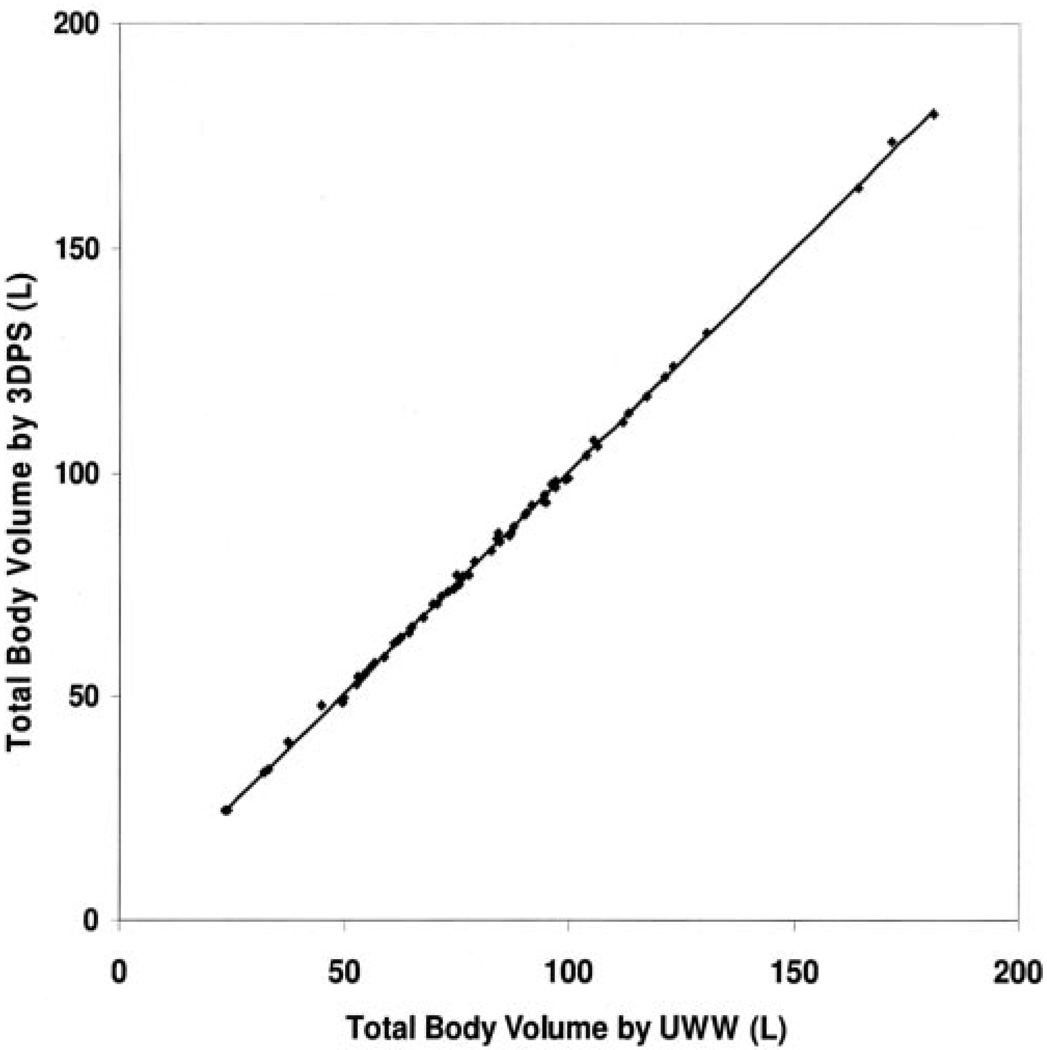
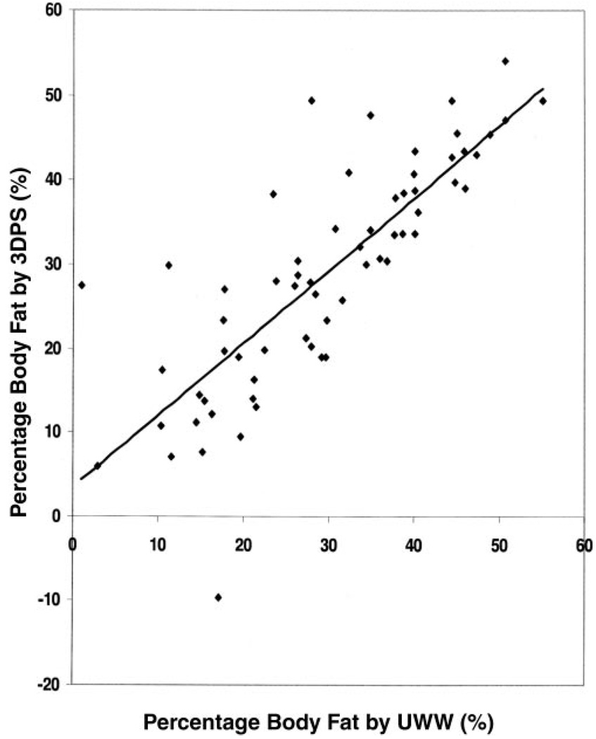
The total-body volumes in the subgroup of 63 subjects who were measured by 3DPS and UWW are shown in Figure 2. The 2 techniques were highly correlated (y =0.9969x+ 0.7103; r2 = 0.999; SEE = 0.892 L). The slope was not significantly different from 1. The constant is significantly larger than zero; however, it was <1% of the mean value. The %BF measured by 3DPS and UWW in the subgroup of 63 subjects who also had residual lung volume measurements is shown in Figure 3 (y = 0.8599x + 3.4012; r2 = 0.6544; SEE = 7.948%). Although there was no significant difference in %BF between 3DPS and UWW, the figure indicates wider variations in %BF than in volume between the 2 techniques. This is a result of estimating %BF with Siri’s equation (18), which uses body density; small differences in body volumes produce differences between the methods in body densities.
Figure 2.
Scatter plot of total-body volume measured by 3-dimensional photonic image scanner (3DPS) and underwater weighing (UWW) in 63 human subjects [30 normal-weight subjects (8 children) and 33 obese subjects (5 children)]. The following equation was obtained by linear regression: y = 0.9969x + 0.7103 (r2 = 0.999, SEE = 0.892 L).
Figure 3.
Scatter plot of percentage body fat estimated by 3-dimensional photonic image scanning (3DPS) and underwater weighing (UWW) in 63 human subjects [30 normal-weight subjects (8 children) and 33 obese subjects (5 children)]. For both methods, body volume was measured at the end of a maximum expiration and was corrected for residual lung volume, which was measured independently by spirometry. The following equation was obtained by linear regression: y = 0.8599x + 3.4012 (r2 = 0.6544, SEE = 7.948%).
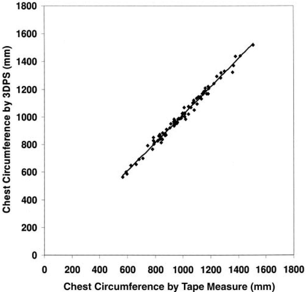
The scatter plot for chest circumferences measured by 3DPS and by a tape measure is shown in Figure 4 (y = 1.0135x + 2.6327; r2 = 0.9889; SEE = 21.064 mm). The slope was not significantly different from 1. Although the constant was >0, it was equivalent to 0.3% of the mean value.
Figure 4.
Scatter plot of chest circumference measured by 3-dimensional photonic image scanning (3DPS) and a tape measure in 89 of 92 human subjects [45 normal-weight subjects (12 children) and 44 obese subjects (12 children)]. The following equation was obtained by linear regression: y = 1.0135x + 2.6327 (r2 = 0.9889, SEE = 21.064 mm).
The data from the mannequin show the effects of clothing on the 3DPS results; the total-body volume measured by 3DPS with clothing was significantly greater than that measured without clothing (24.6 ± 0.11 L compared with 23.87 ± 0.03 L; P = 0.0001); the difference was 0.73 L. The comparisons between 3DPS and water displacement measurements of total and regional body volumes of the mannequin without clothing are shown in Table 4. Although there were significant differences between the techniques, the differences were small: <0.25 L for total-body volume and <0.07 L for regional volumes, and there was no consistent pattern between 3DPS and water displacement. The comparisons between 3DPS and tape measures for body dimensions measured without clothing are shown in Table 5. Similar to the results shown in Table 4, there were significant differences between the 2 techniques, but the differences were small: <5 mm for length and for all circumferences, and there was no consistent pattern between the 3DPS and tape measures.
TABLE 4.
Body volumes of the mannequin measured 9 times (3 times/d) without clothing by the 3-dimensional photonic scanner (3DPS) and water displacement techniques1
| 3DPS | Water displacement | Difference2 | P3 | |
|---|---|---|---|---|
| Volumes (L) | ||||
| Total body | 23.87 ± 0.03 | 23.62 ± 0.03 | −0.25 ± 0.04 | 0.0001 |
| Head (top) | 1.88 ± 0.02 | 1.82 ± 0.01 | −0.06 ± 0.02 | 0.0149 |
| Left arm | 0.51 ± 0.00 | 0.53 ± 0.00 | 0.01 ± 0.00 | 0.0016 |
| Right arm | 0.42 ± 0.00 | 0.40 ± 0.00 | −0.02 ± 0.00 | 0.0001 |
| Left leg | 2.33 ± 0.02 | 2.37 ± 0.01 | 0.04 ± 0.03 | 0.1256 |
| Right leg | 2.08 ± 0.02 | 2.08 ± 0.00 | 0.00 ± 0.02 | 0.9564 |
All values are x¯ ± SEM.
The difference between water displacement and 3DPS measurements.
t test.
TABLE 5.
Body dimensions of the mannequin measured 9 times (3 times/d) without clothing by the 3-dimensional photonic scanning (3DPS) and tape techniques1
| Dimensions | 3DPS | Tape measure | Difference2 | P3 |
|---|---|---|---|---|
| Circumference (mm) | ||||
| Chest4 | 636.4 ± 2.5 | — | ||
| Waist | 549.5 ± 0.5 | 554.3 ± 0.7 | 4.8 ± 0.9 | 0.0001 |
| Hip | 660.4 ± 0.3 | 664.0 ± 1.1 | 3.6 ± 1.1 | 0.0052 |
| Thigh | 376.8 ± 0.5 | 374.2 ± 1.1 | −2.5 ± 1.2 | 0.0538 |
| Knee | 272.3 ± 0.4 | 273.4 ± 0.5 | 1.2 ± 0.7 | 0.0935 |
| Length (mm) | ||||
| Partial thigh | 155.6 ± 0.2 | 156.9 ± 0.4 | 1.2 ± 0.4 | 0.0087 |
All values are x̄ ± SEM.
The difference between tape and 3DPS measurements.
t test.
Because of the body shape at the chest level and the slippery surface of the mannequin, we could not obtain a reliable measurement with tape.
DISCUSSION
The finding that total-body volumes measured by 3DPS with clothing in humans or mannequins were significantly greater than those measured by water or air displacement was expected because water or air can penetrate through most clothing fabric but the data points used by 3DPS to generate body volume were reflected from the surface of the body or the external-surface of the cloth that covers the body surface. The above observations are supported by the results obtained from the mannequin with and without clothing. Two previous studies validated the first generation 3DPS by using UWW and plethysmography techniques and found no significant differences with respect to absolute total-body volume, which is contrary to our results (13, 14). Our subjects varied widely in age, weight, height, and BMI. The intention was to test the measurement limitation of the 3DPS scanner. The results indicated that age, sex, and obesity had no effect on the relation between 3DPS measurements and UWW or tape measurements.
Two significant differences exist between the previous and the current 3DPS scanners. First, the previous model had 8 scan-units (light-sources and cameras), 4 at the front and 4 at the back of the scanned subject. Some of the inner surfaces of the arms and legs might not have been detected accurately (14) because of the asymmetric layout of the scan heads. The current 3DPS has 4 scan heads positioned in a symmetrical manner, so that each scan head can generate an equal quantity of data points. Second, the current 3DPS obtains >20 times the number of data points of the previous model, and the number of data points obtained from a scan has a direct effect on the measurement accuracy. Tapp et al (21) used a different approach by combining a light source technique with electromagnetic tomography to measure human body volume and composition, but its measurement accuracy and reliability have not been tested. It is likely that the most important contribution of the current 3DPS is that it measures body dimensions and total and regional body volumes with high accuracy and precision, as shown by the mannequin results. The body dimensions measured included body circumferences, lengths, widths, and thicknesses. The 3DPS results can be used in studies involving body shape and image. Currently, this is probably the only noninvasive technique with the capacity to accurately and rapidly generate a large number of body composition variables. However, this technique requires that the subject maintain a specified position while standing motionlessness for ≥10 s during the scan. This would limit its use in the very young or the very sick, which are populations that may be of particular interest. In addition, because the scan is performed with the subject positioned differently from the positions used in traditional anthropometric measurements (7, 17), the circumferences measured by the 3DPS for some regions may be different from those measured with traditional anthropometric techniques.
Neither UWW nor tape measures should be considered an ideal criterion for validating the high-tech 3DPS technique for several reasons. The accuracy of UWW for measuring total-body volume relies on several factors: the accuracy of body weight measurement obtained by air and water displacement techniques, the accuracy of the residual lung-volumes measurement, and, especially, the subject’s cooperation and ability to be submerged underwater. Although the UWW and residual lung volume measurements were performed on 63 of the 92 subjects, some of them, especially the younger children, had difficulty in performing the UWW measurements. It is also important to point out that a comparison of %BF measured between the 2 techniques with the use of Siri’s equation (18) could generate questionable conclusions, because small differences in body volumes can produce large differences in %BF between the 2 methods, as shown in Figure 3.
Different technical problems arise when body circumference measurements obtained with a tape measure are compared with those obtained with 3DPS. Measuring circumferences on a human body with a tape measure requires tension on the tape to hold the tape in a horizontal plane for the measurement. Although tension should be minimal with the tape measure, it is not tension-free, unlike measuring with the 3DPS. The 3DPS reconstructs a body circumference from the data points that the scanner obtained at the horizontal plane of the circumference. Therefore, the observed slight differences in circumferences measured by 3DPS and a tape measure may be due to this fundamental difference between the 2 techniques. The results obtained with the mannequin support this point (Table 5). Because the mannequin is made of fiberglass, tension applied to the tape for measurement would not change the dimension of the mannequin. Although there were significant differences between the 3DPS and tape measures, the differences were small and there was no consistent pattern between 3DPS and tape measure.
The mannequin selected for the present study may also not be the ideal tool for validation purposes for 2 reasons. First, although commercially available mannequins range in size from newborn to adult, we selected a size-6 mannequin for the present study based on the ability of our laboratory to accurately measure the water displacement of the total and regional body volumes of the mannequin. The body size of the mannequin is equivalent to a 6-y-old child. Second, because this fiberglass-constructed mannequin has a very smooth and slippery surface, it was extremely difficult to hold the tape measure at some positions to generate reliable circumference measurements. For example, because of the triangular shape of the mannequin’s upper torso, we could not obtain reliable measurements for the chest circumference. Therefore, we could not compare the chest circumference values that were measured by 3DPS and a tape measure. Although there were significant differences in some measurements between the 3DPS and tape measures (Table 5), the differences were small because both techniques have high measurement reproducibility. We were not sure whether the significant differences observed in some measurements were due to measurement error by the 3DPS or by the tape measure.
As described in the Methods, body volumes measured by UWW and 3DPS were measured at the end of a maximum expiration, and the residual lung volume was measured independently by spirometry (19). Although we have no data to document whether a subject could hold a similar residual lung volume in these 2 different environments, a strong relation between the 2 techniques in the corrected total-body volumes was observed (Figure 2), which also indicates that the residual lung volumes in a subject under the 2 different conditions should be similar.
In conclusion, this newly developed, noninvasive, whole-body imaging technique could be an important tool for the biomedical field. It has potential application in the prevention, classification, and monitoring of treatment of diseases that are related to body shape, size, or fatness, such as growth and development, age, weight management, fitness management, and pre- and postsurgical procedures.
Acknowledgments
We thank all the volunteers and the parents of the young volunteers who participated in this study. We thank Mr Hiruma and his colleagues (Hamamatsu Photonics KK, Japan) for their technical and instrumental supports to make the study possible.
Footnotes
Supported by in part by grants PO1 NIDDK-DK42618 and P30-NIDDK DK26687 from the NIH.
JW, DG, JCT, and FXP-S designed the study. All authors participated in conducting the study, data collection and interpretation, and manuscript preparation. None of the authors had personal or financial conflicts of interest.
REFERENCES
- 1.Forbes GB. Human body composition. New York, NY: Springer-Verlag; 1987. [Google Scholar]
- 2.Pouliot MC, Despres JP, Pouliot PJ, et al. Waist circumference and abdominal sagittal diameter: best simple anthropometric indexes of abdominal visceral adipose tissue accumulation and related cardiovascular risk in men and women. Am J Cardiol. 1994;73:460–468. doi: 10.1016/0002-9149(94)90676-9. [DOI] [PubMed] [Google Scholar]
- 3.Kotler DP, Rosenbaum K, Wang J, Pierson RN., Jr Studies of body composition and fat distribution in HIV-infected and control subjects. J Acquir Immune Defic Syndr Hum Retrovirol. 1999;20:228–237. doi: 10.1097/00042560-199903010-00003. [DOI] [PubMed] [Google Scholar]
- 4.Bolton MA, Pruzinky T, Casj TF, Persing JA. Measuring outcomes in plastic surgery: body image and quality of life in medical abnominoplasty patients. Plast Reconstr Surg. 2003;112:619–625. doi: 10.1097/01.PRS.0000070972.57000.08. [DOI] [PubMed] [Google Scholar]
- 5.Buskirk ER. Underwater weighing and body density: a review of procedure. In: Brozek J, Henschel A, editors. Techniques for measuring body composition. Washington, DC: National Academy of Sciences National Research Council; 1961. [Google Scholar]
- 6.Montagu MFA. A handbook of anthropometry. Springfield, Illinois: CC Thomas; 1960. [Google Scholar]
- 7.Wang J, Thornton JC, Kolesnik S, Pierson RN., Jr Anthropometry in body composition An overview. Ann N Y Acad Sci. 2000;904:317–326. doi: 10.1111/j.1749-6632.2000.tb06474.x. [DOI] [PubMed] [Google Scholar]
- 8.Winter J, King W. Basic principles of computerized tomography. In: Greenberg M, Greenberg BM, editors. Essentials of body computed tomography. Philadelphia, PA: WB Saunders Co; 1983. pp. 1–23. [Google Scholar]
- 9.Mitchell AD, Wang PC, Rosebrough RW, Elsasser TH, Schmidt WP. Assessment of body composition of poultry by nuclear magnetic resonance imaging and spectroscopy. Poultry Sci. 1991;70:2494–2500. doi: 10.3382/ps.0702494. [DOI] [PubMed] [Google Scholar]
- 10.Hertzberg HT, Dupertuis CW, Emanual I. Sterophotogrammetry as an anthropometric tool. Photogramm Eng. 1957;23:942–947. [Google Scholar]
- 11.Robinette KM, Daanen H, Paquet E. The Caesar Project: a 3-D surface anthropometry survey. Inst Elec Electronic Eng. 1999:380–385. [Google Scholar]
- 12.Horighchi C. Body line scanner. Internat Arch Photogramm Remote Sensing. 1998;32 part 5. [Google Scholar]
- 13.Dekker L, Douros I, Buxton BF, Treleaven P. Building symbolic information for 3D human body modeling from range data. Second International Conference on 3-D imaging and modeling. Inst Elec Electronic Eng. 1999:388–397. [Google Scholar]
- 14.Wells JCK, Douros I, Fuller NJ, Elia M, Dekker L. Assessment of body volume using three-dimensional photonic scanning. Ann N Y Acad Sci. 2000;904:247–254. doi: 10.1111/j.1749-6632.2000.tb06460.x. [DOI] [PubMed] [Google Scholar]
- 15.Centers for Disease Control and Prevention. [Accessed 8 May 8 2003];CDC Growth Curves. 2000 Internet: http://www.cdc.gov/nchs/about/major/nhanes/growthcharts/clinical_chart.him.
- 16.Wang J, Laferrere B, Thornton JC, Pierson RN, Jr, Pi-Sunyer FX. Regional subcutaneous-fat loss induced by caloric restriction in obese women. Obes Res. 2002;10:885–890. doi: 10.1038/oby.2002.121. [DOI] [PubMed] [Google Scholar]
- 17.Lohman TG, Roche AF, Martorell, editors. Anthropometric standardization reference manual. Champaign, IL: Human Kinetics; 1988. [Google Scholar]
- 18.Siri WE. The gross body composition of the body. Adv Biol Med Phys. 1956;4:239–280. doi: 10.1016/b978-1-4832-3110-5.50011-x. [DOI] [PubMed] [Google Scholar]
- 19.Wilmore JH. The use of actual predicted and constant residual lung volumes in assessment of body composition by underwater weighing. Med Sci Sports Exerc. 1969;1:87–90. [Google Scholar]
- 20.Heymsfield SB, Lichtman S, Baumgartner RN, et al. Body composition of human: comparison of two four-compartment models that differ in expense, technical complexity, and radiation exposure. Am J Clin Nutr. 1990;52:52–58. doi: 10.1093/ajcn/52.1.52. [DOI] [PubMed] [Google Scholar]
- 21.Tapp HS, Goss D, Mackin RO. A combined digital camera-EMT system to measure human body composition. Banff, Canada: Third World Congress on Industrial Process Tomography; 2003. pp. 384–390. [Google Scholar]


