Abstract
Background
Previous studies have shown that the human circadian system is maximally sensitive to short wavelength (blue) light. Whether this sensitivity can be utilized to increase the size of phase shifts using light boxes and protocols designed for practical settings is not known. We assessed whether bright polychromatic lamps enriched in the short wavelength portion of the visible light spectrum could produce larger phase advances than standard bright white lamps.
Methods
Twenty-two healthy young adults received either a bright white or bright blue-enriched 2-hour phase advancing light pulse upon awakening on each of four treatment days. On the first treatment day the light pulse began 8 hours after the dim light melatonin onset (DLMO), on average about 2 hours before baseline wake time. On each subsequent day, light treatment began one hour earlier than the previous day, and the sleep schedule was also advanced.
Results
Phase advances of the DLMO for the blue-enriched (92 ± 78 minutes, n = 12) and white groups (76 ± 45 minutes, n = 10) were not significantly different.
Conclusion
Bright blue-enriched polychromatic light is no more effective than standard bright light therapy for phase advancing circadian rhythms at commonly used therapeutic light levels.
Keywords: Blue Light, Human, Circadian, Phase Shift, Melatonin
Introduction
Scheduled exposure to light and darkness are effective tools for phase shifting the human circadian clock (1, 2). Timed exposure to bright light is recognized by the American Academy of Sleep Medicine as an effective treatment for circadian rhythm sleep disorders, such as shift work disorder (SWD) and delayed sleep phase disorder (DSPD) (3-5). Despite this recommendation, much research on the efficiency of light treatment remains to be done, to establish optimal parameters for the dosage (intensity and duration), timing and wavelength of light treatment in practical schedules that could be used in the home or workplace.
Evidence has recently emerged showing that circadian phase shifts in humans are most sensitive to short wavelength light (6-10). Although rod and cone photoreceptors contribute to non-image forming behaviors such as phase shifting in animal models (11-13), which may also occur in humans (14-16), these responses appear primarily driven by a small population of intrinsically photosensitive retinal ganglion cells (17-19) containing the photopigment melanopsin (20-24). The spectral sensitivity of non image-forming (NIF) responses (e.g., the pupillary light reflex; light-induced melatonin suppression and circadian phase shifting) in humans was not known until 2001, when melatonin suppression was shown to be most sensitive to short wavelength light (25, 26). Consequently, most of the earlier circadian phase shifting studies using polychromatic light did not measure or did not report the amount of energy specifically in the blue portion of the visible light spectrum, but instead reported the illuminance of the light source, which typically ranged from 2,000 to 12,000 lux (27-33). Some specified the type of fluorescent lamp (e.g., “cool white” or “full spectrum”) or provided the correlated color temperature (CCT) [in ° kelvin (K)] of the lamps, which is a metric describing the relative proportion of warm versus cool colors in a light source. Most earlier studies used lamps with a CCT < 7,000 ° K because lamps with a higher CCT (containing more short wavelength energy) were not readily available until recently. In the current study we used fluorescent lamps enriched with blue light, rated by the manufacturer as having a CCT of 17,000 ° K.
The spectral sensitivity of circadian phase shifting in humans has been assessed in carefully controlled studies that have used relatively dim, narrow bandwidth light administered with a specialized light delivery apparatus (6-10). For example, one study pharmacologically dilated subject's pupils, had subjects wear blackout goggles for 90 minutes prior to light exposure, and then administered a 6.5 h light pulse while subject's heads were immobilized in a Ganzfeld dome (6). Controls such as these are important for determining spectral sensitivity, but leave open the question of whether this sensitivity could be utilized in practical protocols to shift circadian rhythms, such as advancing rhythms before flying east to attenuate jet lag, or to treat a patient with DSPD. In addition, it is not known whether this sensitivity could be harnessed to increase the size of the phase shift relative to treatment with standard bright “white” light.
Lamps and light-producing devices emitting exclusively or relatively more short wavelength energy are now commercially available (34). This provides clinicians and patient/consumers with a variety of choices when selecting a device for light treatment, but there remains little evidence from well controlled studies demonstrating the efficacy of those devices for circadian phase shifting.
The goal of the current study was to determine whether bright blue-enriched light could phase advance the circadian clock more than standard bright white light at light levels that are currently being used for therapeutic applications, and using light boxes designed for practical applications.
Methods
Subjects
This was a between-subjects design in which subjects were randomly assigned to receive either white (n = 10) or blue-enriched (n = 12) light. The mean ± SD age (blue-enriched: 27 ± 7; white 28 ± 6), sex (blue-enriched: 7 M, 5 F; white: 6 M, 4 F) and morningness-eveningness (35) (blue-enriched: 55 ± 8; white 55 ± 9) of the groups was similar. Subjects did not report any medical, psychiatric, or sleep disorders as assessed by a telephone interview, an in-person interview, and several screening questionnaires. Subjects were not color blind, according to the Ishihara Color Blindness test. All subjects had body mass indices ≤ 30 kg/m2, were nonsmokers, habitually drank < 300 mg of caffeine/day, and were free from prescription medications. Subjects were also free from recreational drug use as confirmed by a urine toxicology screen at the start of the study. Subjects had not worked a night shift three months prior, nor traveled across more than three time zones one month prior to starting the study. This research was in compliance with the Declaration of Helsinki, and the Rush University Medical Center Institutional Review Board approved the study. All subjects provided written informed consent and were compensated for their participation.
Design
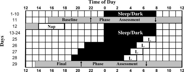
The study began with 10 baseline days in which subjects slept at home on a fixed 8 h schedule similar to their habitual sleep schedule (Fig 1). Subjects were required to remain in bed with the lights out for the specified 8 h. To make the schedule more similar to what most individuals do on their weekends, subjects were required to go to bed one hour later on Friday and Saturday nights, and wake up one hour later on Saturday and Sunday mornings. Weekend naps were permitted within a 3 h time period in the middle of the day at a time centered 12 h from the midpoint of nocturnal week night sleep. Naps at this time are not expected to shift the circadian clock (36). Every morning subjects were required to get at least 10 min of outdoor light between one and 2 h after their baseline weekday wake time. This outdoor light was designed to stabilize circadian phase and also to simulate morning light exposure that many individuals receive on the commute to work. On day 11 subjects came to the lab for a baseline phase assessment, to determine the time of the dim light melatonin onset (DLMO), a marker of the circadian clock. The baseline phase assessment was followed by a compulsory 4 h nap in total darkness in the laboratory, centered 12 hours from the midpoint of the nocturnal week night sleep, to facilitate recovery from the sleep deprivation during the phase assessment. Subjects then returned to the fixed baseline sleep schedule for 13 days at home, while data from the baseline phase assessment was analyzed to determine the baseline DLMO. Previous work in our lab (37) indicated that circadian phase is stable under conditions similar to what these subjects experienced during the baseline portion of the study. Thus, the time of the DLMO during the baseline phase assessment was a good estimate for the time of the DLMO on the first night of sleep in the lab. Apart from the baseline phase assessment and the subsequent nap, we did not restrict the type or duration of light exposure when subjects were outside of the laboratory on days 1-24.
Figure 1.
Representative protocol for a subject sleeping 00:00 - 8:00 at baseline. Grey bars indicate circadian phase assessments. Upward and downward arrows within the grey bars depict the DLMO and DLMOff, respectively. Rectangles with L's represent the times of the 2 h light pulses, which began 8 h after the baseline DLMO on the first treatment day, and began one hour earlier on each subsequent treatment day. In the text, day numbers correspond to the rows in this figure, between 12:00 and 12:00.
On day 25 subjects came to the lab for the first of four consecutive light treatment days. Subjects slept at the lab in individual bedrooms beginning at their scheduled weekday bedtime the first night (day 25). On the first morning in the laboratory, subjects were awakened 8 h after their baseline DLMO, to begin the phase advancing light treatment. Light exposure at this circadian time coincides with the high amplitude portion of the advance region of the light PRC (2, 38), and was expected to produce large phase advances. Because there are individual differences in the position of the DLMO relative to the sleep period, and the light treatment began at the same time relative to the DLMO in all subjects, the advance of wake time on day 25 differed among subjects. All subjects remained in the lab until 6 hours after their waking time on each day of light treatment to control for any outside morning light exposure that could contribute to circadian phase advances. Then subjects were free to leave the lab, but were required to return to the lab 2 hours before their scheduled bedtime on each successive night, and remained in normal room light (< 60 lux) on these evenings before scheduled bedtime.
Wake time and the start of the light pulse were advanced by one hour each successive treatment day to keep up with the expected phase advances of the circadian clock (Fig 1). Bedtime was also advanced on each successive treatment day so that time in bed was 8 h. Bedtime was not advanced on day 25 (the evening before the first morning with light treatment) because it could have then occurred during the `wake maintenance zone' (39), and resulted in subjects not being readily able to fall asleep. Advancing bedtime on day 26 was associated with less risk of this happening because the circadian clock would have advanced from the morning light pulse on day 25, and most subjects were slightly sleep deprived due to an earlier-than-normal awakening on day 25. Following the four treatment days, a final phase assessment was conducted.
Light Treatment
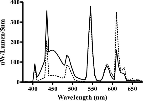
Light treatment consisted of a continuous 2 h light pulse administered upon awakening. The light was produced by a single light box containing fluorescent lamps placed on a desk about 40 cm in front of subjects' eyes. Subjects read or ate breakfast during the light treatment. Light levels were measured from the eye at the angle of gaze at regular intervals. Light boxes in each condition were similar in size and shape (64 cm × 66 cm × 8.5 cm), but differed in spectral composition (Fig 2). The spectral output of the blue-enriched (17,000 °K, Philips Lighting, Eindhoven, The Netherlands) and white (4,100 °K, Enviro-Med, Vancouver, WA) lamps were measured (after passing through the diffuser screen) with an SM240 CCD Spectrometer (Spectral Products, Putnam, CT). At the typical distance and angle of gaze, the white light box delivered slightly more total photons than the blue-enriched light box (4.9 vs. 4.2 × 10¹⁵ photons/cm2/sec) while the blue-enriched light box delivered more than double the number of photons between 400-490nm (1.9 × 10¹⁵ vs. 0.8 × 10¹⁵). For the blue-enriched lamps, the illuminance was ~ 4000 lux and the irradiance was ~ 1640 μW/cm2, while for the white lamps the illuminance was ~ 6000 lux and the irradiance was ~1741 μW/cm2. Subjects remained in normal room light (< 60 lux, 4100 °K fluorescent lamps) at all other times when in the lab during the light treatment days.
Figure 2.
Spectral power distribution of the 4,100 K white lamps (dotted line) and 17,000 K blue-enriched lamps (solid line). The spectral plots overlap at ~550nm.
Phase Assessments
During the phase assessments (Fig 1), subjects remained awake and seated in a recliner chair in dim light (< 5 lux). A baseline phase assessment began 8.5 hours before habitual bedtime. A final phase assessment began 8.5 h before the baseline DLMO. Both phase assessments lasted 22.5 h. Saliva samples were collected every 30 minutes using a salivette (Sarstedt, Newton, NC, USA), as described previously (37, 40, 41). Samples were centrifuged immediately upon collection and frozen. These samples were shipped on dry ice to Pharmasan Labs (Osceola, WI) and radioimmunoassayed for melatonin. The sensitivity of the assay was 0.7 pg/ml; the intra- and inter-assay variability was 12.1% and 13.2%, respectively.
Other Procedures
Daily sleep logs and wrist-worn actigraphy [Actiwatch-L (AWL), Mini-Mitter, Bend, OR] were used to measure total sleep time (TST). A second AWL was attached to a cord and worn around the neck to measure light exposure. The Columbia Jet Lag scale (42) was completed daily before bedtime. A “How Are You Feeling Right Now?” questionnaire (43) was completed four times per day. It contained the Stanford Sleepiness Scale (SSS) (44) and 7 questions assessing physical fatigue, mental fatigue, sadness, anxiety, irritability, and gastrointestinal discomfort on a 10-point ordinal scale. Subjects also completed the SSS during morning light treatment sessions. The first SSS rating was completed within 5 minutes of awakening, and ratings were completed every 30 minutes thereafter for the next 5 hours, yielding a total of 9 ratings per morning.
Data Analysis
To determine the DLMO and dim light melatonin offset (DLMOff), a threshold was calculated for each individual melatonin profile. The threshold was the average of the 5 lowest continuous points during the phase assessment, plus 15% of the average of the five highest continuous points (37, 40, 41). Melatonin profiles were smoothed with a locally weighted least squares (LOWESS) curve using the “fine” level of smoothing (GraphPad Prism, San Diego, CA). The DLMO was defined as the time that this curve exceeded and remained above the threshold. The DLMOff was the time that the smoothed curve dropped and remained below the threshold. The DLMOff of one subject in the blue-enriched group was not included in the analysis because the curve for the melatonin profile of the final phase assessment did not remain below the threshold. The DLMOs for this subject were included in the analysis. Phase shifts were calculated by taking the difference in the time of each phase marker from the baseline to the final phase assessment. Student t-tests were used to compare the phase shift of the DLMO and DLMOff for the blue-enriched and white groups.
Sleep log and actigraphic TST were analyzed with a repeated measure ANOVA. The between-subjects factor “group” had two levels (white and blue-enriched) and the within-subjects factor had 5 levels (the mean TST of baseline days 1-10, and TST on individual days 25-28). Colombia scale and average daily “How Are You Feeling Right Now?” scores (including the SSS question of the “How Are You Feeling Right Now?” questionnaire) were converted into difference from baseline scores and analyzed with a repeated measure ANOVA. The between-subjects factor was group and the within-subjects factor was day (25-28). SSS ratings during morning light sessions were analyzed with a repeated measure ANOVA. The between-subjects factor was group, the first within-subjects factor of day had 4 levels (25-28), and a second within-subjects factor of time-of-day had 9 levels, corresponding to the 9 ratings that were completed each morning of light treatment. To summarize light exposure during the study we calculated three summary statistics of light levels recorded from the AWL worn around the neck. The number of minutes of light exposure greater than 10 lux and greater than 500 lux were determined, as well as the average light exposure (lux/min) for one, two, and three weeks prior to light treatment. Light exposure was also summarized for the first three hours that subjects could be outside the lab on the four treatment days (days 26-29). A significance level of α = .05 was used. Data are presented as mean ± SD.
Results
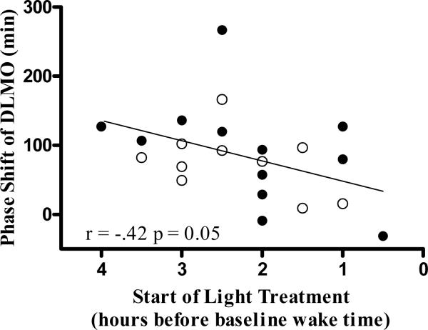
The average advance of wake time on the first treatment day (day 25), relative to each subject's weekday wake time, was 2.2 ± 1.0 h for the blue-enriched group and 2.4 ± 0.8 h for the white group [t(20) = -.46, p = 0.65]. The distribution of the advance of wake time on day 25 is also illustrated in Fig 4.
Figure 4.
Scatterplot showing phase shift versus the start time of light treatment on day 25. Subjects receiving blue-enriched light are represented by filled circles, and subjects receiving white light are represented by open circles. Earlier awakenings this first day of light treatment, and thus all light treatment days, were associated with larger phase advances.
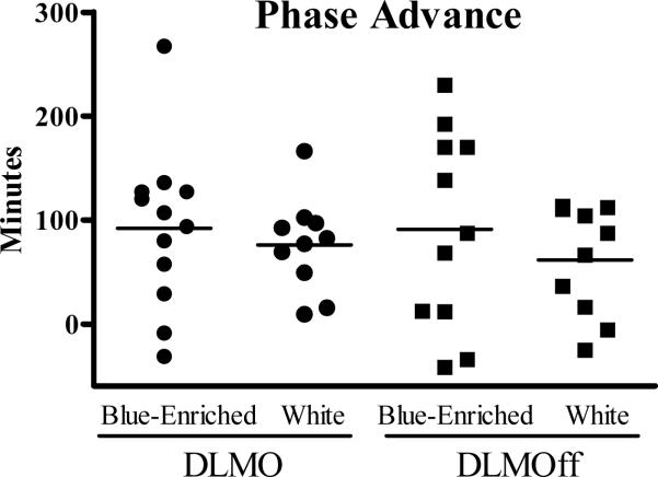
Phase advances of the DLMO and DLMOff for the blue-enriched and white groups were not significantly different (DLMO 92 ± 78 vs. 76 ± 45 min; DLMOff 91 ± 95 vs. 61 ± 53 min, respectively) (Fig 3).
Figure 3.
Phase advance of DLMO (circles) and DLMOff (squares) for individual subjects receiving bright blue-enriched or bright white light treatment. Lines represent the mean.
There were large individual differences in the size of the phase shifts of the DLMO and DLMOff, ranging from advances of over 4 hours to essentially no shift at all. Subjects whose wake time the first day of light treatment (day 25) occurred earlier, relative to their habitual wake time (so that the light treatment could begin 8 h after the baseline DLMO), had larger phase advances (r = -.42, p = 0.05; Fig 4). This was also true for phase advances of the DLMOff (r = -.78, p < 0.001).
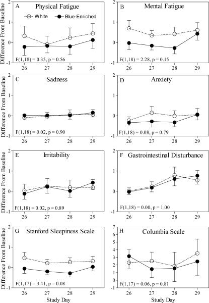
There were no significant group differences for sleep log and actigraphic TST [sleep logs F(1,20) = 0.33, p = 0.57; actigraphy F(1,20) = 0.70, p = 0.41]. Scores on the Colombia Jet Lag scale and the “How Are You Feeling Right Now?” questionnaire during the four treatment days were similar to the each individual's baseline score, and showed no significant group differences (Fig 5). There were no group differences in SSS ratings during morning light treatment sessions [F(1, 15) = 0.13, p = 0.72].
Figure 5.
Subjective well-being ratings for the components of the “How Are You Feeling Right Now?” questionnaire (panels A-G) and the Columbia Jet Lag Scale (panel H) when undergoing phase advancing light treatment. All scores are shown as difference from baseline days 1-10. Panels A-G show the average of the four daily ratings. ANOVA results for the main effect of group are shown for each component. There were no significant main effects of group or group interaction effects.
There were no significant group differences in light exposure history. The average daily number of minutes > 500 lux on days 1-25 was 100.3 ± 12.9 for the white group and 117.8 ± 47.6 for the blue-enriched group [t(20) = -1.13, p = 0.27]. Collapsing the groups, the correlation between phase shift of the DLMO and the average number of minutes > 500 lux on days 1-25 was not significant (r = -.01, p = 0.97). Results using the two other light thresholds (minutes > 10 lux and average lux/minute) and examining different durations of the baseline (e.g., days 18-25) were similar (data not shown). Light exposure outside of the laboratory during the 4 treatment days was not significantly different between the groups. The number of minutes > 500 lux outside of the lab on days 26-29 ranged from 0 to 134 minutes for the blue-enriched group and 6 to 154 minutes for the white group (mean of 45.8 ± 17.1 for the blue-enriched group and 48.8 ± 22.9 for the white group [t(20) = -0.35, p = 0.73]). Collapsing the groups, correlations between phase shift of the DLMO and minutes > 500 lux when outside of the lab on days 26-29 ranged from r = .11 to r = .37, and none were statistically significant.
Discussion
We believe our data are the first comparing the effectiveness of different polychromatic lights for phase shifting human circadian rhythms. We found that the bright blue-enriched polychromatic light box did not produce larger phase advances of the circadian clock than the bright white light box at light levels commonly employed for therapeutic circadian phase shifting.
One other study has tested the impact of different polychromatic lights on human non-image-forming responses. In that study, polychromatic light containing more short-wavelength energy produced greater plasma melatonin suppression than a polychromatic light emitting less short-wavelength energy, but this difference was only significant at one of several low light levels compared (45). The ability of several commercially available devices, primarily emitting light in the blue-green portion of the visible light spectrum, to phase delay the circadian clock has also been compared (34). In this study, all the devices induced phase delays, but no one device produced larger delays than all of the other devices. The largest of the devices compared (a light tower) caused the least amount of eye discomfort and the least difficulty reading or viewing a computer monitor (34). We also used a relatively large light box, which has this advantage, but yet can easily fit on a desk or table. Furthermore, we used a phase advancing rather than a phase delaying protocol because it is more difficult and takes longer to advance human circadian rhythms than to delay them (1).
One possible reason for the lack of a difference in our study was that the light level was saturating (i.e., was so high that altering its spectral composition or increasing the light level further for the given timing and duration of the light pulse would result in no additional phase shift). Action spectra for melatonin suppression using monochromatic light saturate at 1013-1014 photons/cm/sec (25, 26), which is less than the photon densities delivered in this study. However, in those studies pupils were pharmacologically dilated. Phase delays of the melatonin rhythm using “cool white” fluorescent light have been suggested to saturate at ~ 550 lux (46), and the illuminance of our lamps was above this. However, the saturation level reported in that study (46) was in subjects that were kept indoors in < 150 lux for 5 days prior to and maintained in < 10 lux light for the 48 h immediately preceding bright light exposure. Subjects that are exposed to more typical light levels, including bright outside light, in their everyday lives may have a saturation point that is considerably higher. Nonetheless, because very low levels of short wavelength light can produce phase shifts similar in size to very bright polychromatic light (8), it is possible that standard bright “white” polychromatic light treatment already contains a saturating level of short wavelength energy. We thus find no evidence that polychromatic blue-enriched light boxes are any more effective than the standard bright “white” light boxes that are commonly used for circadian phase shifting. Similarly, a recent report suggested that bright blue-enriched light (17,000 °K) conferred no additional benefit for the treatment of seasonal affective disorder, relative to bright white light of a lower color temperature (5,000 °K) (47). However, it is possible that at lower levels [c.f. (45)] or for different clinical applications, blue-enriched light may be more effective than white light for eliciting the desired response.
We timed the light treatment to begin 8 h after the DLMO so that it would start at the same circadian phase in all subjects. This was necessary because the light phase response curve (PRC) shows that the magnitude of the phase shift depends on the time of light exposure (1, 2). Despite this standardization, there was a large range in the magnitude of phase advances. Other carefully controlled laboratory studies (6) have showed a similar large range of phase shifts. In our study we found that part of this variation was due to the shift in bed and wake times so that the light could begin at the same circadian time in all subjects. Subjects with an earlier baseline DLMO, relative to their habitual bedtime, were awakened earlier on the first day of treatment so that the light pulse could begin 8 h after the DLMO. Bedtime on the next day was subsequently advanced more so time in bed returned to 8 h. Thus, a larger advance of the sleep/dark period was associated with a larger advance of the DLMO. The distribution and the average advance of wake time on the first day of light treatment was similar for the two groups, suggesting that this difference did not substantially affect the size of the average phase shift for each group. This observation that the advance of the sleep/dark period significantly affected the advance of the DLMO suggests that in addition to morning light treatment to facilitate phase advances, clinicians should encourage patients and clients to go to bed and awaken earlier.
This study has several limitations. Independent of the lighting condition in this study, the observed phase advances were not as large as expected, and did not advance as fast as the one hour/day advance of the sleep schedule and light pulse. Consequently, especially during the last few days of light treatment, it is conceivable that some subjects received the light pulse on the delay portion of the light phase response curve, which would have diminished the net phase advance. A second limitation of this study is the method for measuring light exposure when outside of the laboratory. We used the AWL device, which measures illuminance (lux), worn around the neck. However, because NIF responses are most sensitive to short wavelengths, quantifying light exposure to these short wavelengths, rather than the photopic system, would be much more relevant. When this study was conducted, we were not aware of any small portable devices with that capacity. Future studies measuring circadian phase shifting would benefit from quantification of the history of light exposure driving the NIF system.
We speculate that the clinical utility of the short-wavelength sensitivity may not be the ability of bright blue-enriched light to augment circadian phase shifts relative to standard bright “white” lamps, but rather may lie in reducing the intensity and/or duration of light pulses without compromising the magnitude of the phase shift. When the relative contribution of other photoreceptors (15, 16) and the apparent photoisomerase capacity of melanopsin (48) in humans are characterized, blue light could be combined or alternated with other wavelengths in patterns to optimize the efficiency of phase shifting responses. This optimized schedule could then be used to decrease the treatment time necessary to achieve the desired change in circadian phase.
Acknowledgments
Supported by National Institutes of Health Grant R01 NR007677. Philips Lighting donated the blue-enriched light boxes. Enviro-Med donated the white light boxes. We thank Thomas Molina, Erin Cullnan, Heather Holly, Meredith Rathert, Jillian Canton, Daniel Alderson, Jonathan Swisher, Clara Lee, Meredith Durkin, Young Cho, Valerie Ellios, and Helen Burgess, Ph. D. for assistance with data collection. We thank Apollo Health for assistance with light measurements.
References
- 1.Eastman CI, Martin SK. How to use light and dark to produce circadian adaptation to night shift work. Ann Med. 1999;31:87–98. doi: 10.3109/07853899908998783. [DOI] [PubMed] [Google Scholar]
- 2.Revell VL, Eastman CI. How to trick mother nature into letting you fly around or stay up all night. J Biol Rhythms. 2005;20:353–65. doi: 10.1177/0748730405277233. [DOI] [PMC free article] [PubMed] [Google Scholar]
- 3.Sack R, Aukley D, Auger R, Carskadon M, Wright K, Vitiello M, et al. Circadian rhythm sleep disorders: Part II, Advanced sleep phase disorder, delayed sleep phase disorder, free-running disorder, and irregular sleep-wake rhythm. Sleep. 2007;30(11):1484–501. doi: 10.1093/sleep/30.11.1484. [DOI] [PMC free article] [PubMed] [Google Scholar]
- 4.Morgenthaler T, Lee-Chiong T, Alessi C, Friedman L, Aurora N, Boehlecke B, et al. Practice parameters for the clinical evaluation and treatment of circadian rhythms sleep disorders. Sleep. 2007;30(11):1445–59. doi: 10.1093/sleep/30.11.1445. [DOI] [PMC free article] [PubMed] [Google Scholar]
- 5.Sack R, Auckley D, Auger R, Carskadon M, Wright K, Vitiello M, et al. Circadian rhythm sleep disorders: Part 1, basic principles, shift work and jet lag disorders. Sleep. 2007;30(11):1460–83. doi: 10.1093/sleep/30.11.1460. [DOI] [PMC free article] [PubMed] [Google Scholar]
- 6.Lockley SW, Brainard GC, Czeisler CA. High sensitivity of the human circadian melatonin rhythm to resetting by short wavelength light. J Clin Endocrinol Metab. 2003;88:4502–5. doi: 10.1210/jc.2003-030570. [DOI] [PubMed] [Google Scholar]
- 7.Revell VL, Arendt J, Terman M, Skene DJ. Short-wavelength sensitivity of the human circadian system to phase-advancing light. J Biol Rhythms. 2005;20:270–2. doi: 10.1177/0748730405275655. [DOI] [PubMed] [Google Scholar]
- 8.Warman VL, Dijk DJ, Warman GR, Arendt J, Skene DJ. Phase advancing human circadian rhythms with short wavelength light. Neurosci Lett. 2003;342:37–40. doi: 10.1016/s0304-3940(03)00223-4. [DOI] [PubMed] [Google Scholar]
- 9.Wright HR, Lack LC. Effect of light wavelength on suppression and phase delay of the melatonin rhythm. Chronobiol Int. 2001;18:801–8. doi: 10.1081/cbi-100107515. [DOI] [PubMed] [Google Scholar]
- 10.Wright HR, Lack LC, Kennaway DJ. Differential effects of light wavelength in phase advancing the melatonin rhythm. J Pineal Res. 2004;36:140–4. doi: 10.1046/j.1600-079x.2003.00108.x. [DOI] [PubMed] [Google Scholar]
- 11.Dkhissi-Benyahya O, Gronfier C, De Vanssay W, Flamant F, Cooper HM. Modeling the role of mid-wavelength cones in circadian responses to light. Neuron. 2007 Mar 1;53(5):677–87. doi: 10.1016/j.neuron.2007.02.005. [DOI] [PMC free article] [PubMed] [Google Scholar]
- 12.Mrosovsky N. Contribution of classic photoreceptors to entrainment. J Comp Physiol A Neuroethol Sens Neural Behav Physiol. 2003 Jan;189(1):69–73. doi: 10.1007/s00359-002-0378-7. [DOI] [PubMed] [Google Scholar]
- 13.Ruby NF, Brennan TJ, Xie X, Cao V, Franken P, Heller HC, et al. Role of melanopsin in circadian responses to light. Science. 2002;298:2211–3. doi: 10.1126/science.1076701. [DOI] [PubMed] [Google Scholar]
- 14.Rea MS, Bullough JD, Figueiro MG. Phototransduction for human melatonin suppression. J Pineal Res. 2002;32:209–13. doi: 10.1034/j.1600-079x.2002.01881.x. [DOI] [PubMed] [Google Scholar]
- 15.Figueiro MG, Bullough JD, Parsons RH, Rea MS. Preliminary evidence for spectral opponency in the suppression of melatonin by light in humans. Neuroreport. 2004;15:313–6. doi: 10.1097/00001756-200402090-00020. [DOI] [PubMed] [Google Scholar]
- 16.Revell VL, Skene DJ. Light-induced melatonin suppression in humans with polychromatic and monochromatic light. Chronobiol Int. 2007;24(6):1125–37. doi: 10.1080/07420520701800652. [DOI] [PubMed] [Google Scholar]
- 17.Dacey DM, Liao HW, Peterson BB, Robinson FR, Smith VC, Pokorny J, et al. Melanopsin-expressing ganglion cells in primate retina signal colour and irradiance and project to the LGN. Nature. 2005;433:749–54. doi: 10.1038/nature03387. [DOI] [PubMed] [Google Scholar]
- 18.Berson DM, Dunn FA, Takao M. Phototransduction by retinal ganglion cells that set the circadian clock. Science. 2002;295:1070–3. doi: 10.1126/science.1067262. [DOI] [PubMed] [Google Scholar]
- 19.Hattar S, Liao HW, Takao M, Berson DM, Yau KW. Melanopsin-containing retinal ganglion cells: architecture, projections, and intrinsic photosensitivity. Science. 2002;295:1065–70. doi: 10.1126/science.1069609. [DOI] [PMC free article] [PubMed] [Google Scholar]
- 20.Qiu X, Kumbalasiri T, Carlson SM, Wong KY, Krishna V, Provencio I, et al. Induction of photosensitivity by heterologous expression of melanopsin. Nature. 2005;433:745–9. doi: 10.1038/nature03345. [DOI] [PubMed] [Google Scholar]
- 21.Panda S, Nayak SK, Camp B, Walker JR, Hogenesch JB, Jegla T. Illumination of the melanopsin signaling pathway. Science. 2005;307:600–4. doi: 10.1126/science.1105121. [DOI] [PubMed] [Google Scholar]
- 22.Fu Y, Zhong H, Wang MH, Luo DG, Liao HW, Maeda H, et al. Intrinsically photosensitive retinal ganglion cells detect light with a vitamin A-based photopigment, melanopsin. Proc Natl Acad Sci U S A. 2005 Jul 19;102(29):10339–44. doi: 10.1073/pnas.0501866102. [DOI] [PMC free article] [PubMed] [Google Scholar]
- 23.Provencio I, Rodriguez IR, Jiang G, Hayes WP, Moreira EF, Rollag MD. A novel human opsin in the inner retina. J Neurosci. 2000;20:600–5. doi: 10.1523/JNEUROSCI.20-02-00600.2000. [DOI] [PMC free article] [PubMed] [Google Scholar]
- 24.Melyan Z, Tarttelin EE, Bellingham J, Lucas RJ, Hankins MW. Addition of human melanopsin renders mammalian cells photoresponsive. Nature. 2004;433:741–5. doi: 10.1038/nature03344. [DOI] [PubMed] [Google Scholar]
- 25.Thapan K, Arendt J, Skene DJ. An action spectrum for melatonin suppression: evidence for a novel non-rod, non-cone photoreceptor system in humans. J Physiol (Lond) 2001;535:261–7. doi: 10.1111/j.1469-7793.2001.t01-1-00261.x. [DOI] [PMC free article] [PubMed] [Google Scholar]
- 26.Brainard GC, Hanifin JP, Greeson JM, Byrne B, Glickman G, Gerner E, et al. Action spectrum for melatonin regulation in humans: evidence for a novel circadian photoreceptor. J Neurosci. 2001;21:6405–12. doi: 10.1523/JNEUROSCI.21-16-06405.2001. [DOI] [PMC free article] [PubMed] [Google Scholar]
- 27.Czeisler CA, Johnson MP, Duffy JF, Brown EN, Ronda JM, Kronauer RE. Exposure to bright light and darkness to treat physiologic maladaptation to night work. N Engl J Med. 1990;322(18):1253–9. doi: 10.1056/NEJM199005033221801. [DOI] [PubMed] [Google Scholar]
- 28.Boivin DB, James FO. Circadian adaptation to night-shift work by judicious light and darkness exposure. J Biol Rhythms. 2002;17:556–67. doi: 10.1177/0748730402238238. [DOI] [PubMed] [Google Scholar]
- 29.Dawson D, Campbell SS. Timed exposure to bright light improves sleep and alertness during simulated night shifts. Sleep. 1991;14:511–6. doi: 10.1093/sleep/14.6.511. [DOI] [PubMed] [Google Scholar]
- 30.Crowley SJ, Lee C, Tseng CY, Fogg LF, Eastman CI. Combinations of bright light, scheduled dark, sunglasses, and melatonin to facilitate circadian entrainment to night shift work. J Biol Rhythms. 2003;18:513–23. doi: 10.1177/0748730403258422. [DOI] [PubMed] [Google Scholar]
- 31.Campbell SS. Effects of times bright-light exposure on shift-work adaptation in middle-aged subjects. Sleep. 1995;18:408–16. doi: 10.1093/sleep/18.6.408. [DOI] [PubMed] [Google Scholar]
- 32.Rosenthal NE, Joseph-Vanderpool JR, Levendosky AA, Johnston SH, Allen R, Kelly KA, et al. Phase-Shifting effects of bright morning light as treatment for delayed sleep phase syndrome. Sleep. 1990;13(4):354–61. [PubMed] [Google Scholar]
- 33.Lewy AJ, Sack RL, Miller LS, Hoban TM. Antidepressant and circadian phaseshifting effects of light. Science. 1987;235:352–4. doi: 10.1126/science.3798117. [DOI] [PubMed] [Google Scholar]
- 34.Paul M, Miller J, Gray G, Buick F, Blazeski S, Arendt J. Circadian phase delay induced by phototherapeutic devices. Aviat Space Environ Med. 2007;78(7):645–52. [PubMed] [Google Scholar]
- 35.Horne JA, Ostberg O. Self-assessment questionnaire to determine morningnesseveningness in human circadian rhythms. Int J Chronobiol. 1976;4:97–110. [PubMed] [Google Scholar]
- 36.Buxton OM, L'Hermite-Baleriaux M, Turek FW, Van Cauter E. Daytime naps in darkness phase shift the human circadian rhythms of melatonin and thyrotropin secretion. Am J Physiol. 2000;278:R373–R82. doi: 10.1152/ajpregu.2000.278.2.R373. [DOI] [PubMed] [Google Scholar]
- 37.Revell VL, Kim H, Tseng CY, Crowley SJ, Eastman CI. Circadian phase determined from melatonin profiles is reproducible after 1 wk in subjects who sleep later on weekends. J Pineal Res. 2005;39:195–200. doi: 10.1111/j.1600-079X.2005.00236.x. [DOI] [PubMed] [Google Scholar]
- 38.Khalsa SBS, Jewett ME, Cajochen C, Czeisler CA. A phase response curve to single bright light pulses in human subjects. J Physiol (Lond) 2003;549.3:945–52. doi: 10.1113/jphysiol.2003.040477. [DOI] [PMC free article] [PubMed] [Google Scholar]
- 39.Lavie P. Ultrashort sleep-waking schedule III. “Gates” and “forbidden zones” for sleep. Electroencephalogr Clin Neurophysiol. 1986;63:414–25. doi: 10.1016/0013-4694(86)90123-9. [DOI] [PubMed] [Google Scholar]
- 40.Revell VL, Burgess HJ, Gazda CJ, Smith MR, Fogg LF, Eastman CI. Advancing human circadian rhythms with afternoon melatonin and morning intermittent bright light. J Clin Endocrinol Metab. 2006;91:54–9. doi: 10.1210/jc.2005-1009. [DOI] [PMC free article] [PubMed] [Google Scholar]
- 41.Lee C, Smith M, Eastman C. A compromise phase position for permanent night shift workers: circadian phase after two night shifts with scheduled sleep and light/dark exposure. Chronobiol Int. 2006;23(4):859–75. doi: 10.1080/07420520600827160. [DOI] [PubMed] [Google Scholar]
- 42.Spitzer RL, Terman M, Williams JB, Terman JS, Malt UF, Singer F, et al. Jet lag: clinical features, validation of a new syndrome-specific scale, and lack of response to melatonin in a randomized, double-blind trial. Am J Psychiatry. 1999;156:1392–6. doi: 10.1176/ajp.156.9.1392. [DOI] [PubMed] [Google Scholar]
- 43.Eastman CI, Gazda CJ, Burgess HJ, Crowley SJ, Fogg LF. Advancing circadian rhythms before eastward flight: A strategy to prevent or reduce jet lag. Sleep. 2005;28:33–44. doi: 10.1093/sleep/28.1.33. [DOI] [PMC free article] [PubMed] [Google Scholar]
- 44.Hoddes E, Zarcone V, Smythe H, Phillips R, Dement WC. Quantification of sleepiness: A new approach. Psychophysiology. 1973;10:431–6. doi: 10.1111/j.1469-8986.1973.tb00801.x. [DOI] [PubMed] [Google Scholar]
- 45.Figueiro MG, Rea MS, Bullough JD. Circadian effectiveness of two polychromatic lights in suppressing human nocturnal melatonin. Neurosci Lett. 2006 Oct 9;406(3):293–7. doi: 10.1016/j.neulet.2006.07.069. [DOI] [PubMed] [Google Scholar]
- 46.Zeitzer JM, Dijk DJ, Kronauer RE, Brown EN, Czeisler CA. Sensitivity of the human circadian pacemaker to nocturnal light: melatonin phase resetting and suppression. J Physiol (Lond) 2000;526.3:695–702. doi: 10.1111/j.1469-7793.2000.00695.x. [DOI] [PMC free article] [PubMed] [Google Scholar]
- 47.Gordijn M, Mannetje D, Meesters Y. The effects of blue enriched light treatment compared to standard light treatment in SAD. Society for Light Treatment and Biological Rhythms Abstracts. 2006;18:6. [Google Scholar]
- 48.Mure LS, Rieux C, Hattar S, Cooper HM. Melanopsin-dependent nonvisual responses: evidence for photopigment bistability in vivo. J Biol Rhythms. 2007 Oct;22(5):411–24. doi: 10.1177/0748730407306043. [DOI] [PMC free article] [PubMed] [Google Scholar]


