Abstract
Recent evidence suggests that fatty acids generated from intracellular triacylglycerol (TAG) hydrolysis may have important roles in intracellular signaling. This study was conducted to determine if fatty acids liberated from TAG hydrolysis regulate peroxisome proliferator-activated receptor α (PPARα). Primary rat hepatocyte cultures were treated with adenoviruses overexpressing adipose differentiation-related protein (ADRP) or adipose triacylglycerol lipase (ATGL) or treated with short interfering RNA (siRNA) targeted against ADRP. Subsequent effects on TAG metabolism and PPARα activity and target gene expression were determined. Overexpressing ADRP attenuated TAG hydrolysis, whereas siRNA-mediated knockdown of ADRP or ATGL overexpression resulted in enhanced TAG hydrolysis. Results from PPARα reporter activity assays demonstrated that decreasing TAG hydrolysis by ADRP overexpression resulted in a 35–60% reduction in reporter activity under basal conditions or in the presence of fatty acids. As expected, PPARα target genes were also decreased in response to ADRP overexpression. However, the PPARα ligand, WY-14643, was able to restore PPARα activity following ADRP overexpression. Despite its effects on PPARα, overexpressing ADRP did not affect PPARγ activity. Enhancing TAG hydrolysis through ADRP knockdown or ATGL overexpression increased PPARα activity. These results indicate that TAG hydrolysis and the consequential release of fatty acids regulate PPARα activity.
Keywords: fatty acids, adipose differentiation-related protein, adipose triglyceride lipase
Lipid droplets are now recognized as important organelles in lipid storage and fatty acid trafficking. Recent studies have characterized a host of proteins involved in lipid droplet biogenesis and turnover, including the perilipin/adipophilin/TIP-47 family of lipid droplet binding proteins and numerous lipases. The expression and activity of these proteins are highly regulated, indicating that turnover of the lipid droplet is also a highly controlled process (1). The regulation of lipid droplet metabolism not only determines the size and morphology of the lipid droplet, but also influences the release of fatty acids from triacylglycerol (TAG) stores. Because intracellular TAG continuously undergoes hydrolysis, this pathway may represent a significant source of intracellular fatty acids.
In addition to their historical roles in energy storage and as membrane constituents, fatty acids are also bioactive molecules that regulate a multitude of physiological processes (2). Fatty acids elicit these biological effects through a host of mechanisms, including the regulation of transcription factors to control gene expression. For instance, fatty acids serve as ligands for peroxisome proliferator-activated receptor α (PPARα) (3), the predominant PPAR isoform in the liver that governs the expression of genes involved in fatty acid oxidation and gluconeogenesis (4). In particular, eicosapentaenoic acid (EPA), an omega-3 fatty acid, elicits the most robust response in PPARα activity (5, 6). The predominant sources of intracellular fatty acids are exogenous uptake, de novo synthesis, chylomicron remnant uptake, and the hydrolysis of TAG and phospholipid. Fatty acids from each of these sources may form separate pools within cells that influence cellular metabolism and signaling activities (7). For example, fatty acids released from phospholipids are preferentially channeled toward eicosanoid production, thereby initiating a multitude of signaling pathways that influence cellular function (8). Furthermore, it has been hypothesized that fatty acids derived from de novo synthesis are important for the activation of PPARα (9). Therefore, it is possible that intracellular fatty acids derived from distinct sources may differentially regulate numerous physiological processes, including gene expression.
Recent data have suggested that manipulations to lipases and lipid droplet proteins, both of which influence TAG turnover, affect gene expression. Gene array analysis of tissues from adipose triacyglycerol lipase (ATGL) null mice revealed that the expression of genes in numerous pathways are altered, including decreased expression of genes involved in fatty acid β-oxidation (10). Similarly, overexpression of lipid droplet proteins decreased the expression of genes related to fatty acid β-oxidation, whereas knockdown of these proteins increased genes involved in fatty acid catabolism (11–16). In this study, we investigated the direct effects of manipulating TAG hydrolysis on PPARα activity. To modulate TAG hydrolysis in primary rat hepatocytes, we either overexpressed or knocked down adipose differentiation-related protein (ADRP) or overexpressed ATGL. Previous studies have shown that overexpression of ADRP results in lipid accumulation, while its knockdown promotes TAG breakdown and fatty acid release (17–20). ATGL is a lipase with high preference for TAG and is highly expressed in adipose tissue, heart, and muscle (21, 22). We found that overexpressing ADRP resulted in decreased TAG hydrolysis and reductions in PPARα reporter activity and target gene expression, whereas hepatocytes with ADRP knocked down or ATGL overexpressed had increased PPARα reporter activity. These data implicate TAG hydrolysis as an important source of fatty acids that serve as signaling molecules to regulate PPARα activity. Because PPARα plays a central role in governing hepatic energy metabolism, findings from this study provide further insight into the underpinnings of the development of fatty liver, insulin resistance, and metabolic diseases in general.
MATERIALS AND METHODS
Materials
Tissue culture plates were from Nunc and media were obtained from Invitrogen. Rat-tail collagen I was obtained from BD Biosciences. Silica gel A plates were from Whatman. EPA was from Cayman Chemical, and [1-14C]oleate was from PerkinElmer Life Sciences. Short interfering RNA (siRNA) duplexes were obtained from Qiagen. All other chemicals were obtained from Sigma-Aldrich.
Primary hepatocyte cell culture, siRNA electroporation, and adenoviral infection
Male Sprague-Dawley Rats (250–300 g) were fed ad libitum prior to primary hepatocyte isolation using the collagenase perfusion method as described previously (23). The protocol for animal use was approved by the University of Minnesota Institutional Animal Care and Use Committee. Hepatocytes were plated on collagen-coated multiwell dishes for 4 h with M199 plating media that contained 23 mM HEPES, 26 mM sodium bicarbonate, 10% FBS, 1% penicillin/streptomycin, 100 nM dexamethasone, 100 nM insulin, and 11 mM glucose. M199 maintenance media contained 23 mM HEPES, 26 mM sodium bicarbonate, 1% penicillin/streptomycin, 5.5 mM glucose, 100 µM carnitine, 10 nM dexamethasone, and 10 nM insulin. For experiments involving ADRP siRNA, hepatocytes were electroporated with either a nontargeting control (Qiagen All-Starts Negative Control) or ADRP siRNA at a concentration of 2.0 µg/106 cells immediately following isolation using a Rat Hepatocyte Nucleofector kit from Amaxa Biosystems. The sense and antisense strands for ADRP siRNA are r(GUCUGUCUGUUCCGAAUAA)dTdT and r(UUAUUCGGAACAGACAGAC)dGdT, respectively. Murine ADRP cDNA (kindly donated by Dr. Ginette Serrero of Harvard Medical School), fused to FLAG cDNA at its N terminus, was cloned into an adenovirus expression system using the AdEasy adenoviral vector system (Stratagene). The methods used to generate the ADRP virus are the same as those used for the ATGL virus production and are previously described (24). For ADRP adenovirus experiments, after a 4 h attachment period, cells were exposed to either 5 multiplicities of infection of adenovirus expressing green fluorescent protein (Ad-GFP) or Ad-ADRP for 2.5 h in M199 maintenance media; the GFP adenovirus was kindly provided by Wade Bresnahan, University of Minnesota, Twin Cities. For ATGL adenovirus experiments, cells were infected with 20 multiplicities of infection of Ad-GFP or Ad-ATGL. Subsequently, all transduced cells were cultured in M199 maintenance media.
Cell radiolabeling, lipid extraction, and analysis
Cells were pulsed with 500 µM [1-14C]oleate bound to fatty acid-free BSA in a 3:1 molar ratio for 1.5 h at 22 h or 43 h postplating for ADRP and ATGL adenovirus or ADRP siRNA experiments, respectively. Following the pulse, some cells were harvested for lipid extractions to measure radiolabel incorporation into lipid classes as described below. The media were removed, cells were washed with PBS, and fresh medium without oleate was added on other cells for a 6 h chase period. Subsequently, cells were collected and lipids were extracted (25). Following extraction, cellular lipids were separated by TLC on silica gel plates in a solvent system of hexane:ethyl ether:acetic acid (80:20:2, v/v). Known standards (Sigma-Aldrich and BioChemika) were used to identify lipids. Radiolabeled lipids were detected by iodine vapor and quantified by scintillation counting.
PPARα and PPARγ reporter gene assays
Hepatocytes were cotransfected with the pSG5-GAL4-hPPARα or pSG5-GAL4-hPPARγ expression plasmids and a TK-MH-UAS-LUC reporter plasmid at 1 μg of each plasmid per million cells. Plasmids were kindly provided by Dr. Philippe Thuillier, Oregon Health and Science University. A renilla luciferase vector (pRL-SV40; Promega) was transfected at a concentration of 20 ng per million cells as an internal control reporter. The plasmids were transfected with Lipofectamine (Invitrogen) upon adenovirus removal or after the 4 h attachment period for the siRNA experiments. Cells were exposed to one of the following fatty acid treatments: BSA, 250 µM oleate, 250 µM EPA, or a combination of 250 µM oleate and 250 µM EPA. EPA was used because it robustly activates PPARα, whereas oleate was used because it is the most abundant fatty acid in tissues. The combination of the two fatty acids represents a more physiological condition in which the liver is exposed to more than a single fatty acid. For overexpression experiments, cells were exposed to the above treatments at 24 h posttransduction unless otherwise noted. For ADRP siRNA experiments, cells were exposed to the fatty acid treatments at 43 h after plating. PPARα or -γ activity was determined in cell lysates by dual luciferase reporter gene assays (Promega) and expressed as relative luciferase units.
Quantitative RT-PCR
For both ADRP overexpression and knockdown experiments, RNA was isolated using Trizol, and reverse transcription and quantitative RT-PCR (qRT-PCR) were performed using the SYBR GreenER Two-step qRT-PCR Universal kit (Invitrogen). Primers for all gene expression experiments are listed in Table 1. Samples were analyzed on an ABI 7300 sequence detection system (Applied Biosystems). Abundance of mRNA was normalized to ribosomal protein L32, and data were analyzed using the delta CT method.
TABLE 1.
Primer sequences for qRT-PCR
| Gene | Forward Primer | Reverse Primer |
|---|---|---|
| ADRP | CTCTCGGCAGGATCAAAGAC | CGTAGCCGACGATTCTCTTC |
| ACOT-1 | GATGGCCTCAAGGATGTTGT | TCCAGTTGTGGTCATCCTGA |
| PEPCK | TGTGCCAGCCAGAGTATATTC | GTGAGAGCCAGCCAACAG |
| UCP-2 | ATGACAGACGACCTCCCTTG | GAAGGCATGAACCCCTTGTA |
| RPL-32 | AAACTGGCGGAAACCCAGAG | GCAGCACTTCCAGCTCCTTG |
ACOT-1, acyl-CoA thioesterase-1; PEPCK, phosphoenolpyruvate carboxykinase; UCP-2, uncoupling protein-2; RPL-32, ribosomal protein L32.
Western blot analysis
Hepatocytes were lysed at 24 h posttransduction, and proteins were separated by gel electrophoresis on a 10% polyacrylamide gel and transferred to an immobilin-P membrane (Millipore). The membrane was incubated with mouse monoclonal anti-FLAG M2-peroxidase (HRP) conjugated antibody at a 1:500 dilution to confirm ADRP overexpression. For ATGL analysis, membranes were incubated with a 1:1,000 dilution of anti-ATGL antibody (24) followed by a 1:5,000 dilution of anti-rabbit HRP-conjugated secondary antibody. Bands were visualized by the ECL Plus chemiluminescent detection reagent (Amersham).
Statistics
Data were analyzed by Student's t-test, and significance was declared at P < 0.05.
RESULTS
ADRP overexpression decreases fatty acid loss from intracellular TAG
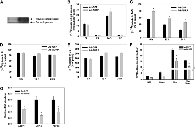
Because previous data show that manipulating ADRP influences TAG content, we chose to overexpress ADRP in order to suppress TAG hydrolysis. Primary hepatocytes were transduced with adenoviruses containing either GFP or ADRP. To confirm ADRP overexpression, we harvested protein from cells 24 h after transduction for Western blot analysis, which showed that ADRP was highly expressed at this time (Fig. 1A). To measure the effects of ADRP overexpression on TAG turnover, we performed pulse-chase experiments with 500 µM [1-14C]oleate. Ad-ADRP overexpression resulted in a 22% increase in [1-14C]oleate incorporation into TAG during the pulse period when compared with cells transduced with Ad-GFP (Fig. 1B). During the chase period in which the exogenous radiolabel was removed, overexpressing ADRP resulted in 46–65% less loss of [14C]TAG compared with cells overexpressing GFP (Fig. 1C). The effects of ADRP on loss of TAG was independent and not influenced by duration of the chase period. Therefore, ADRP overexpression effectively decreased the loss of fatty acids from TAG stores.
Fig. 1.
ADRP overexpression decreases the loss of fatty acids from TAG and inhibits PPARα activity and target gene expression. A: Western blot analysis of ADRP overexpression showing endogenous rat ADRP and overexpressed murine FLAG-tagged ADRP, which ran approximately 2 kDa higher during electrophoresis. B: Incorporation of [1-14C]oleate into cellular lipids. C–E: Chase experiments evaluating turnover of TAG, phospholipids, and cholesterol esters. Statistics for the chase period were analyzed as a percentage of the pulse. F: PPARα reporter gene activity following exposure to fatty acids. G: Expression of PPARα target genes. All data are reported as means ± SE from three to four experiments. *P < 0.05 and **P < 0.01 when compared with Ad-GFP controls.
To characterize the selectivity of ADRP in modulating lipid turnover, we investigated if ADRP overexpression influenced fatty acid incorporation into phospholipids and cholesterol esters and their subsequent turnover. After performing the same pulse-chase experiments, we found that ADRP overexpression did not influence fatty acid incorporation into phospholipids or cholesterol ester (Fig. 1B) or the loss of fatty acids from these lipid species during the chase period (Fig. 1D, E). Moreover, cellular radiolabeled free fatty acids were unaffected during either pulse or chase period. Thus, it appears that ADRP influences only TAG turnover and is a viable method to alter TAG hydrolysis.
Overexpressing ADRP decreases PPARα activity
To determine if blocking TAG hydrolysis influences PPARα activity, we overexpressed ADRP and treated hepatocytes with BSA, 250 µM oleate, 250 µM EPA, or a combination of 250 µM oleate and 250 µM EPA for 6 h and measured PPARα reporter activity. As expected, cells exposed to EPA alone or in combination with oleate demonstrated a 40- and 20-fold increase in PPARα reporter activity, respectively (Fig. 1F). This was a more robust response than observed with oleate alone, which typically only resulted in a 2- to 3-fold induction of PPARα. When ADRP was overexpressed, reporter activity decreased 35–60% in all treatment groups, suggesting that the activation of PPARα requires fatty acids derived from TAG hydrolysis. Moreover, the fold induction in response to fatty acids was similar between Ad-GFP and Ad-ADRP transduced cells. Confirming the effect, overexpressing ADRP decreased basal mRNA abundance of three major targets of PPARα, acyl-CoA thioesterase-1, uncoupling protein-2, and phosphoenolpyruvate carboxykinase from 35% to 70% (Fig. 1G). Therefore, overexpressing ADRP resulted in suppressed TAG hydrolysis, decreased activity of PPARα, and lower expression of PPARα target genes.
ADRP knockdown increases fatty acid loss from intracellular TAG
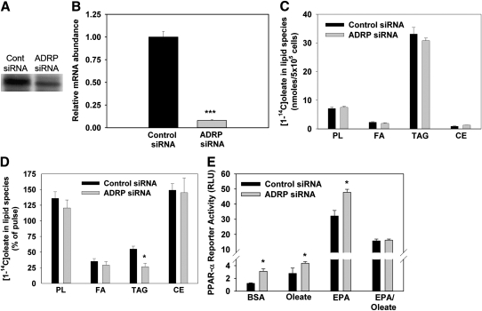
To further manipulate TAG hydrolysis, we electroporated cells with siRNA targeted against ADRP. After 8 h, ADRP mRNA abundance, as measured by qRT-PCR, was decreased 92% compared with cells electroporated with a nontargeting control (Fig. 2B), and protein abundance was also suppressed after 48 h (Fig. 2A). After 43 h, we performed pulse-chase labeling experiments to determine if the decrease in ADRP expression would facilitate fatty acid incorporation or loss from TAG or other lipid species. ADRP knockdown did not significantly alter [1-14C]oleate incorporation into TAG during the pulse (Fig. 2C). However, following the 6 h chase, there was a 75% loss of [14C]TAG in cells with ADRP knocked down, whereas cells exposed to the control siRNA lost only 40% (Fig. 2D). No changes in fatty acid incorporation or loss from other lipid species were observed. Thus, ADRP knockdown enhanced the amount of fatty acid lost from intracellular TAG.
Fig. 2.
ADRP knockdown enhances fatty acid loss from intracellular TAG and increases PPARα activity. A: ADRP protein abundance measured with Western blotting at 48 h after transfection. B: Eight hours after siRNA transfection, cells were lysed and RNA was harvested and analyzed for ADRP mRNA abundance with qRT-PCR. C: Incorporation of [1-14C]oleate into cellular lipids. D: Chase experiments evaluating turnover of lipid species. Statistics for the chase period were analyzed as a percentage of the pulse. Statistics for the chase period were analyzed as a percentage of the pulse. E: PPARα reporter gene activity following exposure to fatty acids. Data are reported as means ± SE, n = 3. *P < 0.05, **P < 0.01, and ***P < 0.001 when compared with siRNA controls.
ADRP knockdown increases PPARα activity
Similar to the overexpression protocol, we exposed hepatocytes to BSA or fatty acids as described above and measured PPARα reporter activity following ADRP knockdown. Hepatocytes with ADRP knocked down demonstrated significantly higher PPARα activity when treated with EPA, oleate, or BSA alone compared with cells treated with control siRNA (Fig. 2E). ADRP siRNA did not influence PPARα activity when cells were exposed to both EPA and oleate, which cannot be readily explained. Taken together, increasing TAG hydrolysis increased PPARα reporter activity in response to exogenous fatty acids and under basal conditions.
Overexpressing ADRP decreases PPARα activity following removal of exogenous fatty acids
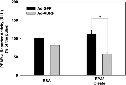
Because of the differences in PPARα activity observed under basal conditions, we wanted to further characterize how TAG hydrolysis influences PPARα activity independent of exogenous fatty acids. To do so, we overexpressed GFP or ADRP and exposed cells to BSA or a mixture of oleate and EPA for 6 h. After this exposure, the exogenous fatty acids were removed for an additional 6 h chase period. By measuring the PPARα reporter activity before and after fatty acid exposure, we could eliminate the effect of the exogenous fatty acids and isolate the role of TAG hydrolysis alone in regulating PPARα activity. For cells exposed to BSA alone, overexpressing ADRP did not influence the change in PPARα reporter activity during the 6 h after the exogenous fatty acids were removed (Fig. 3). Ad-GFP-treated cells that were exposed to fatty acids showed a slight increase in PPARα reporter activity during this time period. However, compared with Ad-GFP transduced cells, overexpressing ADRP resulted in a 50% decrease in reporter activity following removal of exogenous fatty acids. These data show that inhibiting TAG hydrolysis reduces PPARα reporter activity following removal of exogenous ligands. Thus, it appears that fatty acids liberated from TAG hydrolysis may have an important role in maintaining PPARα activity following exposure to exogenous fatty acids.
Fig. 3.
Overexpressing ADRP decreases PPARα reporter activity following removal of exogenous fatty acids. Fourteen hours after transduction, cells were exposed to a combination of 250 μM EPA and 250 μM oleate for 6 h (pulse). The media was removed and replaced with media devoid of fatty acids for an additional 6 h (chase). Reporter gene assays were performed immediately after removal of exogenous fatty acids and 6 h later. Data are presented as PPARα reporter activity after the 6 h chase period expressed as a percentage of the pulse period. Data are reported as means ± SE, n = 3. *P < 0.05 when compared with Ad-GFP controls.
Overexpressing ATGL alters fatty acid incorporation in lipid species and their subsequent turnover
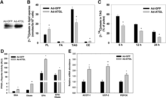
Although ADRP is exclusively localized to TAG droplets and regulates TAG turnover, it is possible that the observed effects on PPARα activity could be due to altering ADRP rather than TAG hydrolysis. Thus, we sought an additional mechanism to modulate TAG hydrolysis. Because ATGL is a lipase with high specificity toward TAG, we overexpressed ATGL and characterized its effects on fatty acid metabolism and TAG turnover. We expected that ATGL overexpression would mimic the effects of ADRP knockdown on TAG hydrolysis. After 24 h of overexpression, we observed a marked increase in ATGL protein abundance (Fig. 4A). Ad-ATGL-treated cells had decreased [1-14C]oleate incorporation in TAG during pulse radiolabeling as expected (Fig. 4B). Overexpressing ATGL also increased oleate incorporation into phospholipids and cholesterol esters during the pulse. Overexpressing ATGL increased loss of radiolabeled fatty acids from TAG (Fig. 4C) but did not influence loss of fatty acids from other lipid species during any time during the chase period (data not shown). Therefore, although ATGL altered metabolism of exogenous fatty acids into other lipids, the effects on hydrolysis were specific to TAG.
Fig. 4.
Overexpressing ATGL enhances the loss of fatty acids from TAG and increases PPARα activity and target gene expression. A: Western blot analysis of ATGL overexpression. B: Incorporation of [1-14C]oleate into cellular lipids. C: Chase experiments evaluating turnover of TAG. Statistics for the chase period were analyzed as a percentage of the pulse. D: PPARα reporter gene activity following exposure to fatty acids. E: Expression of PPARα target genes. All data are reported as means ± SE, n = 3. *P < 0.05 and **P < 0.01 when compared with Ad-GFP controls.
Overexpressing ATGL increases PPARα activity
We next sought to test the effects of overexpressing ATGL on PPARα activity. Hepatocytes were transduced with Ad-GFP and Ad-ATGL for 24 h prior to exposure to fatty acids and reporter gene assays. Compared with cells overexpressing GFP, Ad-ATGL transduced cells treated with BSA alone, oleate, EPA, or a combination of EPA and oleate exhibited 2.2-, 2.7-, 3.1-, and 1.8-fold increases, respectively, in PPARα reporter activity (Fig. 4D). Additionally, overexpressing ATGL increased the mRNA abundance of the PPARα target genes acyl-CoA thioesterase-1, uncoupling protein-2, and phosphoenolpyruvate carboxykinase (Fig. 4E). These experiments further support the ADRP data showing that altering TAG hydrolysis has a direct effect on PPARα activity.
WY-14643 prevents the decrease in PPARα activity following ADRP overexpression
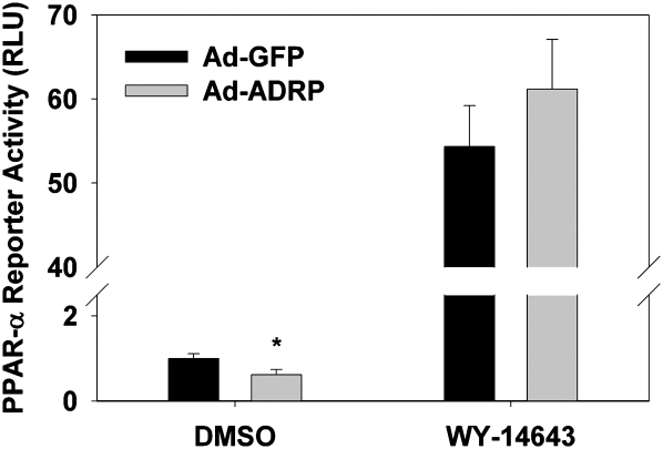
Although manipulating TAG hydrolysis influences the flux of fatty acids, which can act as ligands for PPARα, it is possible that indirect mechanisms could also link TAG hydrolysis to PPARα activity. To determine if a high-affinity PPARα ligand could rescue PPARα activity in cells overexpressing ADRP, we incubated cells with 1 μM WY-14643. Exposure to this PPARα ligand robustly increased PPARα activity. Furthermore, at 6 h, treatment with WY-14643 resulted in similar activities of PPARα in Ad-GFP and Ad-ADRP treated cells (Fig. 5). Therefore, because a potent PPARα ligand was able to normalize PPARα activity, these data suggest that inhibiting TAG hydrolysis decreases PPARα activity by limiting the amount of endogenous fatty acid ligands.
Fig. 5.
The PPARα ligand, WY-14643, normalizes PPARα activity in cells overexpressing ADRP. At 24 h after viral transduction, cells were treated with DMSO or 1 μM WY-14643 for 6 h prior to harvesting for reporter gene assays. Data are reported as means ± SE, n = 3. *P < 0.05 when compared with Ad-GFP controls.
ADRP overexpression does not alter PPARγ activity
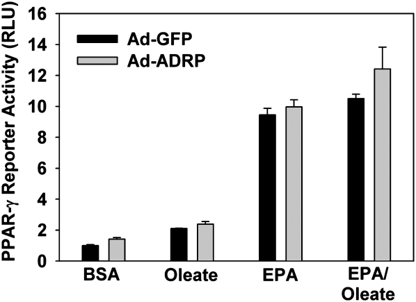
To determine if fatty acids released from TAG hydrolysis can also activate PPARγ, we overexpressed a pSG5-GAL4-hPPARγ expression plasmid together with the TK-MH-UAS-LUC reporter plasmid to specifically determine PPARγ reporter gene activity in response to ADRP overexpression. Exogenous EPA or a combination of EPA and oleate increased PPARγ activity (Fig. 6). However, overexpressing ADRP did not affect PPARγ activity under any condition tested. Since fatty acids from both exogenous and endogenous (i.e., TAG hydrolysis) sources regulated PPARα, but only exogenous fatty acids influenced PPARγ, these data indicate that fatty acids released from TAG hydrolysis form a distinct intracellular pool that differentially regulates hepatic gene expression.
Fig. 6.
ADRP overexpression does not alter PPARγ activity. PPARγ reporter gene activity following exposure to fatty acids. Data are reported as means ± SE, n = 3. There were no significant differences between Ad-GFP and Ad-ADRP transduced cells.
DISCUSSION
Taken as a whole, these data strongly support the conclusion that fatty acids released from TAG hydrolysis activate PPARα as well as the expression of PPARα target genes. The overall findings of the study are that 1) manipulating the intracellular TAG pool by overexpressing ADRP or knocking down ADRP with siRNA modulates the subsequent release of fatty acids; 2) hepatocytes overexpressing ADRP demonstrated a significant reduction in PPARα reporter activity and target gene expression, whereas hepatocytes with ADRP knocked down or ATGL overexpressed had significantly higher PPARα activity; 3) ADRP overexpression caused a more rapid decline in PPARα reporter activity following removal of exogenous fatty acids; 4) exposure to WY-14643 alleviated the suppression of PPARα activity in ADRP overexpressing cells; and 5) ADRP overexpression did not alter PPARγ activity.
In adipocytes, the lipases responsible for hydrolysis of TAG, and glycerolipids in general, have been well characterized. Studies in this area have primarily explored the activities of ATGL, hormone-sensitive lipase, and monoacylglycerol lipase and the signaling cascades responsible for their regulation. Despite this research in adipose tissue, little is known about the enzymes responsible for TAG hydrolysis in the liver or the mechanisms governing this process. Thus, in order to regulate hepatic TAG hydrolysis, we manipulated ADRP or ATGL. ADRP is ubiquitously expressed, and both in vivo and in vitro studies demonstrate that manipulating ADRP specifically influences hepatic TAG content (19, 26, 27). Additionally, ATGL is a lipase that shows specificity to TAG and is expressed in the liver (21, 22). Our metabolic labeling studies are in agreement with these prior studies in that overexpression of ADRP decreased TAG loss, while ADRP knockdown or ATGL overexpression promoted TAG hydrolysis.
Although the exact mechanism through which changes in TAG hydrolysis influences PPARα activity is not known, the most likely explanation is through the supply of ligands. Because the PPARα expression construct used in this study contained only the ligand binding domain (amino acids 167–468) of PPARα, it appears that ligand binding most likely mediates the effects of manipulating TAG hydrolysis on PPARα activity. Previous studies have shown that fatty acids bind and activate PPARα (3, 28). Moreover, exogenous omega-3 polyunsaturated fatty acids elicit the most robust response in PPARα activity both in vitro and in vivo (5, 29, 30).
Although the most direct explanation for the effects observed on PPARα activity is that TAG hydrolysis produces fatty acids that are PPARα ligands, these released fatty acids could have indirect effects on PPARα via lipin-1 or AMP-activated protein kinase (AMPK). Hepatic lipin-1 is activated by fatty acids (31) and physically interacts with the ligand binding domain of PPARα, although the mechanisms through which this interaction enhances PPARα activity are not known (32). Furthermore, altering TAG hydrolysis alters the activation of AMPK (33). In skeletal muscle, it has been demonstrated that AMPK increases fatty acid oxidation by activating PPARα and PGC-1 (34). Although it is not known if AMPK directly phosphorylates the ligand binding domain of PPARα, this could also contribute to the changes in PPARα activity following alterations in TAG hydrolysis. Despite the possible role of lipin-1 or AMPK in modulating PPARα activity, our data showed that exposure of ADRP overexpressing cells to a potent synthetic PPARα ligand, WY-14643, overcame the decrease in PPARα activity. Thus, it appears that the primary mechanism through which TAG hydrolysis regulates PPARα is through the production of fatty acid ligands. Moreover, data from this study suggest that fatty acids derived from TAG hydrolysis constitute a separate and important pool of ligands for PPARα that cannot be substituted by exogenous fatty acids.
The findings that overexpressing ADRP does not influence PPARγ activity, despite activation by exogenous fatty acids, indicates that fatty acids derived from these different sources form distinct pools with unique signaling properties. Although it is inherently difficult to quantify these distinct pools, it is known that fatty acids are differentially channeled depending upon their source. Studies indicate that exogenous fatty acids are preferentially incorporated into TAG, whereas fatty acids synthesized de novo are preferentially channeled to phospholipid and diacylglycerol (35–37). Similarly, fatty acids derived from TAG hydrolysis are more likely to be incorporated into VLDL than exogenous fatty acids or fatty acids synthesized de novo (38). Additionally, manipulating enzymes in fatty acid metabolism, such as acyl-CoA synthetases or stearoyl-CoA desaturase-1, also result in differential trafficking of fatty acids, which influences their signaling properties (7, 39). Our data identify TAG hydrolysis as a critical physiological process that can regulate intracellular fatty acid signaling. Additionally, it has been estimated that the entire pool of TAG in hepatocytes is turned over in <24 h (38). Thus, this rapid turnover supports the concept that fatty acids released from TAG hydrolysis may comprise a significant portion of intracellular fatty acids.
Recent data from other laboratories support our findings that TAG hydrolysis plays an important role in regulating PPARα target genes. Adenoviral overexpression of hormone-sensitive lipase (HSL) and/or ATGL in ob/ob mice results in increased plasma β-hydroxybutyrate levels and increased expression of the PPARα target genes, carnitine palmitoyl transferase-1 and acyl-CoA oxidase, in the liver (40). Additionally, microarray analyses of liver in ATGL or HSL null mice show profound changes in gene expression (10). Genes involved in β-oxidation are downregulated in cardiac muscle of ATGL null mice and brown adipose tissue of both ATGL and HSL null mice. Since ATGL and HSL are highly expressed in the tissues that displayed altered patterns of gene expression, these data suggest that blocking TAG or diacylglycerol hydrolysis diminishes PPARα and -δ controlled pathways of β-oxidation. FSP27 and perilipin null animals also show increased expression of PPAR target genes in numerous tissues and protection from diet-induced insulin resistance (11–15). Similar to ADRP, perilipin is a member of the perilipin/adipophilin/TIP-47 domain family of lipid droplet proteins that influences TAG hydrolysis. FSP27 has been shown to interact with lipid droplets to influence lipolysis and droplet morphology (16, 41). Overexpression of FSP27 decreased TAG hydrolysis and the expression of genes involved in β-oxidation in 3T3-L1 cells, whereas knockdown of FSP27 resulted in increased expression of the same genes (16). These previous results together with the findings of this study show that TAG hydrolysis is a significant factor that controls transcription of catabolic pathways of fatty acid metabolism.
In summary, our data demonstrate that TAG hydrolysis and release of fatty acids regulate PPARα activity and target gene expression in rat hepatocytes. The data suggest that fatty acids liberated from TAG constitute a specialized pool of ligands for PPARα. Because of this novel regulatory role, understanding the regulation of TAG hydrolysis and its alterations in diseased states should provide further insights into the regulation of gene expression and into the etiology of diseases, such as hepatic steatosis and insulin resistance. Moreover, these data implicate that proteins involved in TAG hydrolysis, such as lipases and lipid droplet proteins, have central roles in regulating gene expression and energy metabolism.
Footnotes
Abbreviations:
- Ad-GFP
- adenovirus expressing green fluorescent protein
- ADRP
- adipose differentiation-related protein
- AMPK
- AMP-activated protein kinase
- ATGL
- adipose triacyglycerol lipase
- EPA
- eicosapentaenoic acid
- HSL
- hormone-sensitive lipase
- PPAR
- peroxisome proliferator-activated receptor
- qRT-PCR
- quantitative reverse transcription polymerase chain reaction
- siRNA
- short interfering RNA
- TAG
- triacylglycerol
REFERENCES
- 1.Ducharme N. A., Bickel P. E. 2008. Minireview: lipid droplets in lipogenesis and lipolysis. Endocrinology. 149: 942–949 [DOI] [PubMed] [Google Scholar]
- 2.Jump D. B., Botolin D., Wang Y., Xu J., Christian B., Demeure O. 2005. Fatty acid regulation of hepatic gene transcription. J. Nutr. 135: 2503–2506 [DOI] [PubMed] [Google Scholar]
- 3.Forman B. M., Chen J., Evans R. 1997. Hypolipidemic drugs, polyunsaturated fatty acids, and eicosanoids are ligands for peroxisome proliferator-activated receptors alpha and delta. Proc. Natl. Acad. Sci. USA. 94: 4312–4317 [DOI] [PMC free article] [PubMed] [Google Scholar]
- 4.Beaven S. W., Tontonoz P. 2006. Nuclear receptors in lipid metabolism: targeting the heart of dyslipidemia. Annu. Rev. Med. 57: 313–329 [DOI] [PubMed] [Google Scholar]
- 5.Pawar A., Jump D. B. 2003. Unsaturated fatty acid regulation of peroxisome proliferator-activated receptor alpha activity in rat primary hepatocytes. J. Biol. Chem. 278: 35931–35939 [DOI] [PubMed] [Google Scholar]
- 6.Berge R. K., Madsen L., Vaagenes H., Tronstad K. J., Gottlicher M., Rustan A. C. 1999. In contrast with docosahexaenoic acid, eicosapentaenoic acid and hypolipidaemic derivatives decrease hepatic synthesis and secretion of triacylglycerol by decreased diacylglycerol acyltransferase activity and stimulation of fatty acid oxidation. Biochem. J. 343: 191–197 [PMC free article] [PubMed] [Google Scholar]
- 7.Mashek D. G., Li L. O., Coleman R. A. 2007. Long-chain acyl-CoA synthetases and fatty acid channeling. Future Lipidol. 2: 465–476 [DOI] [PMC free article] [PubMed] [Google Scholar]
- 8.Wymann M. P., Schneiter R. 2008. Lipid signalling in disease. Nat. Rev. Mol. Cell Biol. 9: 162–176 [DOI] [PubMed] [Google Scholar]
- 9.Chakravarthy M. V., Pan Z., Zhu Y., Tordjman K., Schneider J. G., Coleman T., Turk J., Semenkovich C. F. 2005. “New” hepatic fat activates PPARalpha to maintain glucose, lipid, and cholesterol homeostasis. Cell Metab. 1: 309–322 [DOI] [PubMed] [Google Scholar]
- 10.Pinent M., Hackl H., Burkard T. R., Prokesch A., Papak C., Scheideler M., Hämmerle G., Zechner R., Trajanoski Z., Strauss J. G. 2008. Differential transcriptional modulation of biological processes in adipocyte triglyceride lipase and hormone-sensitive lipase-deficient mice. Genomics. 92: 26–32 [DOI] [PubMed] [Google Scholar]
- 11.Puri V., Ranjit S., Konda S., Nicoloro S. M. C., Straubhaar J., Chawla A., Chouinard M., Lin C., Burkart A., Corvera S., et al. 2008. Cidea is associated with lipid droplets and insulin sensitivity in humans. Proc. Natl. Acad. Sci. USA. 105: 7833–7838 [DOI] [PMC free article] [PubMed] [Google Scholar]
- 12.Castro-Chavez F., Yechoor V. K., Saha P. K., Martinez-Botas J., Wooten E. C., Sharma S., O'Connell P., Taegtmeyer H., Chan L. 2003. Coordinated upregulation of oxidative pathways and downregulation of lipid biosynthesis underlie obesity resistance in perilipin knockout mice: a microarray gene expression profile. Diabetes. 52: 2666–2674 [DOI] [PubMed] [Google Scholar]
- 13.Saha P. K., Kojima H., Martinez-Botas J., Sunehag A. L., Chan L. 2004. Metabolic adaptations in the absence of perilipin: increased β-oxidation and decreased hepatic glucose production associated with peripheral insulin resistance but normal glucose tolerance in perilipin-null mice. J. Biol. Chem. 279: 35150–35158 [DOI] [PubMed] [Google Scholar]
- 14.Nishino N., Tamori Y., Tateya S., Kawaguchi T., Shibakusa T., Mizunoya W., Inoue K., Kitazawa R., Kitazawa S., Matsuki Y., et al. 2008. FSP27 contributes to efficient energy storage in murine white adipocytes by promoting the formation of unilocular lipid droplets. J. Clin. Invest. 118: 2808–2821 [DOI] [PMC free article] [PubMed] [Google Scholar]
- 15.Toh S. Y., Gong J., Du G., Li J. Z., Yang S., Ye J., Yao H., Zhang Y., Xue B., Li Q., et al. 2008. Up-regulation of mitochondrial activity and acquirement of brown adipose tissue-like property in the white adipose tissue of fsp27 deficient mice. PLoS ONE. 3: e2890. [DOI] [PMC free article] [PubMed] [Google Scholar]
- 16.Keller P., Petrie J. T., De Rose P., Gerin I., Wright W. S., Chiang S., Nielsen A. R., Fischer C. P., Pedersen B. K., MacDougald O. A. 2008. Fat-specific protein 27 regulates storage of triacylglycerol. J. Biol. Chem. 283: 14355–14365 [DOI] [PMC free article] [PubMed] [Google Scholar]
- 17.Listenberger L. L., Ostermeyer-Fay A. G., Goldberg E. B., Brown W. J., Brown D. A. 2007. Adipocyte differentiation-related protein reduces the lipid droplet association of adipose triglyceride lipase and slows triacylglycerol turnover. J. Lipid Res. 48: 2751–2761 [DOI] [PubMed] [Google Scholar]
- 18.Imamura M., Inoguchi T., Ikuyama S., Taniguchi S., Kobayashi K., Nakashima N., Nawata H. 2002. ADRP stimulates lipid accumulation and lipid droplet formation in murine fibroblasts. Am. J. Physiol. Endocrinol. Metab. 283: E775–E783 [DOI] [PubMed] [Google Scholar]
- 19.Magnusson B., Asp L., Bostrom P., Ruiz M., Stillemark-Billton P., Linden D., Boren J., Olofsson S. O. 2006. Adipocyte differentiation-related protein promotes fatty acid storage in cytosolic triglycerides and inhibits secretion of very low-density lipoproteins. Arterioscler. Thromb. Vasc. Biol. 26: 1566–1571 [DOI] [PubMed] [Google Scholar]
- 20.Larigauderie G., Cuaz-Perolin C., Younes A. B., Furman C., Lasselin C., Copin C., Jaye M., Fruchart J. C., Rouis M. 2006. Adipophilin increases triglyceride storage in human macrophages by stimulation of biosynthesis and inhibition of beta-oxidation. FEBS J. 273: 3498–3510 [DOI] [PubMed] [Google Scholar]
- 21.Zimmermann R., Strauss J. G., Haemmerle G., Schoiswohl G., Birner-Gruenberger R., Riederer M., Lass A., Neuberger G., Eisenhaber F., Hermetter A., et al. 2004. Fat mobilization in adipose tissue is promoted by adipose triglyceride lipase. Science. 306: 1383–1386 [DOI] [PubMed] [Google Scholar]
- 22.Lake A. C., Sun Y., Li J., Kim J. E., Johnson J. W., Li D., Revett T., Shih H. H., Liu W., Paulsen J. E., et al. 2005. Expression, regulation, and triglyceride hydrolase activity of adiponutrin family members. J. Lipid Res. 46: 2477–2487 [DOI] [PubMed] [Google Scholar]
- 23.Kaytor E. N., Shih H., Towle H. C. 1997. Carbohydrate regulation of hepatic gene expression. Evidence against a role for the upstream stimulatory factor. J. Biol. Chem. 272: 7525–7531 [DOI] [PubMed] [Google Scholar]
- 24.Miyoshi H., Perfield J. W., 2nd, Souza S. C., Shen W. J., Zhang H. H., Stancheva Z. S., Kraemer F. B., Obin M. S., Greenberg A. S. 2007. Control of adipose triglyceride lipase action by serine 517 of perilipin A globally regulates protein kinase A-stimulated lipolysis in adipocytes. J. Biol. Chem. 282: 996–1002 [DOI] [PubMed] [Google Scholar]
- 25.Bligh E. G., Dyer W. 1959. A rapid method of total lipid extraction and purification. Can. J. Biochem. Physiol. 37: 911–917 [DOI] [PubMed] [Google Scholar]
- 26.Chang B. H., Li L., Paul A., Taniguchi S., Nannegari V., Heird W. C., Chan L. 2006. Protection against fatty liver but normal adipogenesis in mice lacking adipose differentiation-related protein. Mol. Cell. Biol. 26: 1063–1076 [DOI] [PMC free article] [PubMed] [Google Scholar]
- 27.Varela G. M., Antwi D. A., Dihr R., Yin X., Singhal N. S., Graham M. J., Crooke R. M., Ahima R. S. 2008. Inhibition of ADRP prevents diet-induced insulin resistance. Am. J. Physiol. Gastrointest. Liver Physiol 295: G621–G628 [DOI] [PMC free article] [PubMed] [Google Scholar]
- 28.Xu H. E., Lambert M. H., Montana V. G., Parks D. J., Blanchard S. G., Brown P. J., Sternbach D. D., Lehmann J. M., Wisely G. B., Willson T. M., et al. 1999. Molecular recognition of fatty acids by peroxisome proliferator-activated receptors. Mol. Cell. 3: 397–403 [DOI] [PubMed] [Google Scholar]
- 29.Pawar A., Botolin D., Mangelsdorf D. J., Jump D. B. 2003. The role of liver X receptor-{alpha} in the fatty acid regulation of hepatic gene expression. J. Biol. Chem. 278: 40736–40743 [DOI] [PubMed] [Google Scholar]
- 30.Buettner R., Parhofer K. G., Woenckhaus M., Wrede C. E., Kunz-Schughart L. A., Scholmerich J., Bollheimer L. C. 2006. Defining high-fat-diet rat models: metabolic and molecular effects of different fat types. J. Mol. Endocrinol. 36: 485–501 [DOI] [PubMed] [Google Scholar]
- 31.Martin P. G., Guillou H., Lasserre F., Dejean S., Lan A., Pascussi J. M., Sancristobal M., Legrand P., Besse P., Pineau T. 2007. Novel aspects of PPARalpha-mediated regulation of lipid and xenobiotic metabolism revealed through a nutrigenomic study. Hepatology. 45: 767–777 [DOI] [PubMed] [Google Scholar]
- 32.Finck B. N., Gropler M. C., Chen Z., Leone T. C., Croce M. A., Harris T. E., Lawrence J. C., Jr., Kelly D. P. 2006. Lipin 1 is an inducible amplifier of the hepatic PGC-1alpha/PPARalpha regulatory pathway. Cell Metab. 4: 199–210 [DOI] [PubMed] [Google Scholar]
- 33.Gauthier M. S., Miyoshi H., Souza S. C., Cacicedo J. M., Saha A. K., Greenberg A. S., Ruderman N. B. 2008. AMP-activated protein kinase is activated as a consequence of lipolysis in the adipocyte: potential mechanism and physiological relevance. J. Biol. Chem. 283: 16514–16524 [DOI] [PMC free article] [PubMed] [Google Scholar]
- 34.Lee W. J., Kim M., Park H. S., Kim H. S., Jeon M. J., Oh K. S., Koh E. H., Won J. C., Kim M. S., Oh G. T., et al. 2006. AMPK activation increases fatty acid oxidation in skeletal muscle by activating PPARalpha and PGC-1. Biochem. Biophys. Res. Commun. 340: 291–295 [DOI] [PubMed] [Google Scholar]
- 35.Li L. O., Mashek D. G., An J., Doughman S. D., Newgard C. B., Coleman R. A. 2006. Overexpression of rat long chain acyl-coa synthetase 1 alters fatty acid metabolism in rat primary hepatocytes. J. Biol. Chem. 281: 37246–37255 [DOI] [PubMed] [Google Scholar]
- 36.Mashek D. G., McKenzie M. A., Van Horn C. G., Coleman R. A. 2006. Rat long chain acyl-CoA synthetase 5 increases fatty acid uptake and partitioning to cellular triacylglycerol in McArdle-RH7777 cells. J. Biol. Chem. 281: 945–950 [DOI] [PubMed] [Google Scholar]
- 37.Igal R. A., Wang S., Gonzalez-Baro M., Coleman R. A. 2001. Mitochondrial glycerol phosphate acyltransferase directs the incorporation of exogenous fatty acids into triacylglycerol. J. Biol. Chem. 276: 42205–42212 [DOI] [PubMed] [Google Scholar]
- 38.Wiggins D., Gibbons G. F. 1992. The lipolysis/esterification cycle of hepatic triacylglycerol. Its role in the secretion of very-low-density lipoprotein and its response to hormones and sulphonylureas. Biochem. J. 284: 457–462 [DOI] [PMC free article] [PubMed] [Google Scholar]
- 39.Miyazaki M., Dobrzyn A., Man W. C., Chu K., Sampath H., Kim H. J., Ntambi J. M. 2004. Stearoyl-CoA desaturase 1 gene expression is necessary for fructose-mediated induction of lipogenic gene expression by sterol regulatory element-binding protein-1c-dependent and -independent mechanisms. J. Biol. Chem. 279: 25164–25171 [DOI] [PubMed] [Google Scholar]
- 40.Reid B. N., Ables G. P., Otlivanchik O. A., Schoiswohl G., Zechner R., Blaner W. S., Goldberg I. J., Schwabe R. F., Chua S. C., Jr., Huang L. 2008. Hepatic overexpression of hormone-sensitive lipase and adipose triglyceride lipase promotes fatty acid oxidation, stimulates direct release of free fatty acids, and ameliorates steatosis. J. Biol. Chem. 283: 13087–13099 [DOI] [PMC free article] [PubMed] [Google Scholar]
- 41.Puri V., Konda S., Ranjit S., Aouadi M., Chawla A., Chouinard M., Chakladar A., Czech M. P. 2007. Fat-specific protein 27, a novel lipid droplet protein that enhances triglyceride storage. J. Biol. Chem. 282: 34213–34218 [DOI] [PubMed] [Google Scholar]


