Abstract
Negative regulation of T cell receptor (TCR) signaling is an important mechanism enforcing immunological self-tolerance to prevent inappropriate activation of T cells and thus the development of autoimmune diseases. The lymphoid-restricted serine/threonine kinase DAP-related apoptotic kinase-2 (DRAK2) raises the TCR activation threshold by targeting TCR-induced calcium mobilization in thymocytes and peripheral T cells, and regulates positive thymic selection and peripheral T cell activation. Despite a hypersensitivity of peripheral drak2-deficient T cells, drak2-deficient mice are enigmatically resistant to induced autoimmunity in the model experimental autoimmune encephalomyelitis (EAE). In order to further evaluate the differential role of DRAK2 in central versus peripheral tolerance and to assess its impact on the development of autoimmune diseases, we have generated a transgenic (Tg) mouse strain ectopically expressing DRAK2 via the lck proximal promoter (1017-DRAK2 Tg mice). This transgene led to highest expression levels in double-positive thymocytes that are normally devoid of DRAK2. 1017-DRAK2 Tg mice displayed a reduction of single-positive CD4+ and CD8+ thymocytes in context with diminished negative selection in male HY TCR × 1017-DRAK2 Tg mice as well as peripheral T cell hypersensitivity, enhanced susceptibility to EAE and spontaneous autoimmunity. These findings suggest that alteration in thymocyte signaling thresholds impacts the sensitivity of peripheral T cell pools.
Keywords: autoimmunity, apoptosis, costimulation, DRAK2, EAE/MS, negative regulation of T cell receptor signaling, selection, T cell, thymus, tolerance, lupus
Introduction
As adaptive immunity must maintain a highly diverse and specific T cell repertoire to defeat microbial pathogens and cancer cells while preventing aberrant activation by self-antigens, multiple surveillance mechanisms have evolved to ensure immunological self-tolerance. The mechanisms of central (thymic) T cell tolerance - negative selection of immature thymocytes with high affinity for self-peptide-MHC by apoptosis (1, 2), and development of natural regulatory T cells (Tregs) (3) - cooperate with peripheral tolerance mechanisms including anergy induction, clonal deletion by activation induced cell death (AICD), and suppression by CD4+CD25+ Tregs (4). Since T cell fate at different stages of lymphoid development is determined by the strength, duration and frequency of signals emanating from the T cell antigen receptor (TCR) as well as the costimulatory context, TCR signaling is subject to extensive negative and positive feedback regulation to ensure proper responses (5).
Depending on their expression and activation patterns during T cell development, negative regulators of TCR signaling may impact central and/or peripheral tolerance mechanisms. Examples of such negative regulators that act downstream of the TCR include the E3 ubiquitin ligases c-Cbl, Cbl-b, Itch, and GRAIL; phosphatases Sts-1/2, SHP-1, PEP, PTP-PEST; adaptor proteins Dok-1/-2; the lipid phosphatase PTEN; and the serine/threonine kinase DRAK2 (6). Germ-line deletion of such genes in mice commonly results in T cell hyper-reactivity and, with the exception of DRAK2, increased susceptibility to autoimmune disease. The majority of these negative regulatory proteins support peripheral tolerance by raising the threshold of peripheral T cell activation to enforce the requirement for costimulation, and thereby limit spurious activation by self-antigens, as recently reviewed in (6). In addition, potential effects on the selection of the T cell repertoire in the thymus either via a direct influence on apoptotic signals or by shifting the threshold to rescue T cells with intermediate or stronger affinity for self-peptide/MHC may be superimposed on any impact on peripheral T cell activation. Negative regulators of TCR signaling that influence thymic selection events include c-Cbl (7), SHP-1 (8), PTEN (9) and DRAK2 (10). The importance of the TCR signaling threshold during thymic selection to peripheral T cell function is underscored by the development of spontaneous polyarthritis in SKG mice that bear a point mutation in the gene encoding the tyrosine kinase ZAP-70 (11). Since the SKG mutation diminishes the activity of ZAP-70, it is thought that this autoimmune phenotype results from enhanced positive selection of T cells exhibiting highly avid TCR, allowing for the escape of auto-reactive T cell clones (that should have been deleted) to the periphery.
The serine/threonine kinase DRAK2, also termed STK17b, comprises the fifth member of the death-associated protein (DAP)-like kinase (DAPK) family, and is predominantly expressed in the thymus, lymph nodes and spleen. The kinase is differentially expressed during both T and B cell development, with highest levels of drak2 transcript in the most mature lymphocyte populations, namely single-positive (SP) thymocytes, naïve peripheral T cells and mature B cells (10). In contrast to the reported ability of DRAK2 to promote apoptosis after ectopic expression in cell lines (12, 13), its germ-line deletion in mice does not lead to any discernable reduction in apoptotic sensitivity in the lymphoid system. Instead, drak2-deficient mice revealed a paradoxical and unique role of the kinase in the regulation of T cell activation and tolerance (10). Despite conferring T cell hyper-reactivity to TCR-stimulation in vitro with reduced activation threshold and diminished requirement for CD28-mediated costimulation, drak2-deficiency does not lead to lymphadenopathies or spontaneous autoimmunity in aging mice. Contrarily, loss of drak2 results in an unexpected resistance to autoimmune disease in a model of EAE induced by injection of myelin oligodendrocyte glycoprotein (MOG) peptides.
During thymic T cell development, drak2-deficient mice display slightly increased positive selection of CD4+ T cells and a reduced TCR activation threshold (10, 14), and this impact on central tolerance might contribute to the altered responsiveness of peripheral T cells. Due to their hypersensitivity to antigenic stimulation, mature peripheral drak2-/--T cells are capable of proliferating in the absence of costimulation via CD28, but develop a large apoptotic population (15), indicating that costimulatory signals are necessary to prevent AICD under such conditions. Although the direct substrates and intracellular targets of DRAK2 kinase activity remain to be identified, experiments have shown that DRAK2 exerts its inhibitory effect on TCR signaling by modulating Ca2+ mobilization, as drak2-deficient thymocytes and peripheral T cells display an increase in Ca2+ influx upon TCR crosslinking. DRAK2 activity itself is induced by Ca2+ mobilization, demonstrating that it serves in a negative regulatory loop to temper Ca2+ signaling (16). Similar to the endogenous calcineurin inhibitor calcipressin 1 (Csp1) (17), DRAK2 may thus function in a signaling module to suppress the expression of high-threshold genes such as FasL during initial clonal expansion of T cells, thereby preventing premature death under certain conditions such as repetitive or suboptimal stimulation.
In order to investigate whether DRAK2 impacts thymic selection, the peripheral T cell repertoire and autoimmune susceptibility, we have developed a transgenic (Tg) mouse model in which DRAK2 expression is driven by the lck proximal promoter (1017-DRAK2 Tg mice). In such mice, DRAK2 is ectopically expressed at high levels in DP thymocytes, a subset with normally low levels of this inhibitory kinase. We hypothesized that, similar to SKG mice bearing a mutation in the ZAP-70 gene, a heightened activation threshold imposed by DRAK2 in developing thymocytes might cause enhanced selection of auto-reactive T cells. This hypothesis is based on the premise that with such an enhanced activation threshold, developing thymocytes normally fated to be eliminated by negative selection would instead be rescued and released into the periphery. In contrast to a DRAK2 transgenic mouse strain ubiquitously overexpressing DRAK2 from the β-actin promoter (18), transgenic expression of DRAK2 from the lck proximal promoter leads to T cell lineage- and developmental stage-specific expression. Analysis of T cell subset distribution and function as well as spontaneous and induced autoimmunity in 1017-DRAK2 Tg mice has revealed significant reductions in SP CD4+and CD8+ thymocytes due to altered positive and negative selection, a hyper-reactivity of the resulting peripheral T cell population in context with organ infiltration in aged mice, and an increased susceptibility to EAE. These results demonstrate that DRAK2 enforced negative regulation of TCR signaling during thymocyte development leads to selection of mature T cells with altered activation thresholds for clonal expansion and an increased reactivity towards self-antigens.
Materials and Methods
Generation of 1017-DRAK2 transgenic mice
Full-length murine drak2 cDNA in context with a hemagglutinin (HA)-tag was cloned into the BamHI site of the 1017-lck vector containing the lck proximal promoter and the 3' untranslated region of human Growth hormone (hGH-3'UTR). The NotI fragment of the resulting 1017-drak2 construct was then injected into fertilized CB6F1 mouse oocytes. Screening for transgene incorporation and genotyping was performed using a PCR-based approach with primers specific for drak2 and the hGH-3UTR'. 1017-DRAK2 transgenic mice used for functional experiments have been back-crossed onto the C57BL/6 background for at least 5 generations, and were between 4-8 weeks of age unless otherwise indicated. In all cases, age-matched littermates were used as controls. H-Y TCR transgenic mice (B10.Cg-Tg 71Vbo N12) were purchased from Taconic Farms (Oxnard, CA and Germantown, NY) and bred onto the 1017-DRAK2 transgenic background to assess positive and negative selection. All mice were housed in a pathogen-free environment in accordance with the regulations of the Institutional Animal Care and Use Committee at the University of California, Irvine.
FACS analyses
Antibodies (conjugated with FITC, PE, PerPC or APC) directed against the following surface markers were obtained from eBioscience (San Diego, CA), BD PharMingen (San Diego, CA) or Caltag Laboratories (Burlingame, CA) and used in 1:400 dilution to analyze immune cell subsets in single cell suspensions of thymi, spleens and lymphnodes of DRAK2 transgenic mice: CD3, CD4 (RM4-5), CD8, CD25a (p55/IL-2α) (7D4), CD44, CD62L (L-Selectin) (MEL-14), CD69 (VEA) (H1.2F3), B220/CD45R (RA3-6B2), H-Y TCR (T3.70), and Vβ screening panel. Apoptotic fractions were detected by Annexin-V staining (FITC, PE, APC) (BD Biosciences). To detect DRAK2 by intracellular staining, thymocytes were at first surface stained with anti-CD4-APC and anti-CD8-PE followed by fixation and permeabilization using the Cytofix/Cytoperm kit (BD Biosciences). Subsequently, anti-DRAK2 mAb (Cell Signaling Technologies, Danvers, MA), anti-HA or isotype control Abs were added for 30 min, followed by washing and staining with FITC-conjugated anti-rabbit or anti-mouse Abs. All FACS analyses were performed using a FACSCalibur (Becton Dickinson, San Jose, CA) and CellQuest Software as well as FlowJo Software (TreeStar, Inc., Ashland, OR).
Western Blot
Whole cell extracts (WCE) were prepared from T cell suspensions using WCE-buffer (0,5 % NP-40) as described previously (19). DRAK2 expression was assessed by Western blotting with an anti-DRAK2 mAb or anti-HA Ab to detect either total DRAK2 or transgenic overexpression (all Abs were from Cell signaling Technologies).
Analysis of DRAK2 mRNA expression by Quantitative real-time RT-PCR (QPCR)
Following isolation of total cellular RNA from either thymocytes or splenocytes with TRIzol® solution (Invitrogen Life Technologies Inc., Carlsbad, CA) according to the manufacturer's instructions, cDNA was generated from 1 μg total RNA using the Superscript First-Stand Synthesis System with oligo(dT) primers (Invitrogen). Samples were analyzed in triplicate by QPCR with an iCycler using the iQ™ SYBRGreen Supermix (Biorad, Hercules, CA) and drak2 specific primers for 40 amplification cycles. Expression levels were calculated by normalization of data to β-actin mRNA expression.
Thymocyte apoptosis assay
Thymocytes were plated at a density of 106/well and either left untreated or incubated for 24 hrs with the following inducers of apoptosis: anti-CD3ε Ab (1 μg/ml), ionomycin (1 μM), dexamethasone (1 μM), etoposide (10 μM), anti-Fas/APO-1 mAb (CD95, Jo2) (1 μg/ml) (eBioscience). Following incubation, cell viability was assessed by co-staining with Annexin V and CD4 as well as CD8 and subsequent FACS analysis.
[3H]-thymidine incorporation and IL-2 production in dose-response assays
Peripheral T cells for the proliferation and functional assays were purified from total splenocytes using the Easy-Sep T cell negative selection kit (Stem Cell, Vancouver, Canada) before plating at a density of 1×10⁶ T cells/ml. Typical T cell purity was greater than 95%. Mouse anti-CD3ε Ab (145-2C11) and anti-CD28 Ab (37.51) (both eBioscience) were used for T cell activation as indicated in either plate-bound or soluble form. To assess proliferative capacity, total splenocytes or purified T cells from 1017-DRAK2 transgenics and littermates were plated in triplicate at a density of 105/100 μl in round-bottom 96 well plates and stimulated for 96 hrs with series of various concentrations of soluble or plate-bound or anti-CD3 (2, 0.2 and 0.02 μg/ml) in the presence of absence of anti-CD28. For the last 18 hrs of culture, cells were pulsed with 1 μCi/ml [3H]-thymidine (NEN Research Products, Boston MA), harvested, and [3H] incorporation was then quantified as counts per minute (cpm) using a beta counter. IL-2 levels of supernatants were measured in triplicate by enzyme-linked immunosorbent assay (ELISA) with an anti-mouse IL-2 antibody pair (JES6-1A12 and JES6-5H4) (eBioscience).
Carboxy-fluorescein diacetate, succinimidyl ester (CFSE) Proliferation Assay
Purified T cells (or whole splenocytes) were labeled with 5 μM 5,6-carboxyfluorescein diacetate succinimidyl ester (CFDA-SE) (Molecular Probes, Eugene, OR) and subsequently plated at a density of 1×10⁶/well before stimulation with plate-bound anti-CD3 as described above with or without 1 μg/ml soluble anti-CD28. As CFDA-SE is converted into CFSE by intracellular esterases, CFSE-labeled cells were collected, co-stained with anti-CD4-APC, anti-CD8-PerCP and Annexin V-PE and analyzed by four-color flow cytometric analysis using a FACSCalibur following the indicated culture periods.
Calcium mobilization assays/flux
To assess calcium flux, thymocytes or purified splenic T cells were labeled with the Ca2+ indicator dyes Fura-Red (2 μM) and Fluo-3 (1 μM) in the presence of 0.2% Pluronic (all Molecular Probes, Eugene, OR). Ca2+ mobilization kinetics were analyzed by flow cytometry after preincubation of labeled cells with biotinylated anti-CD3 and anti-CD4 upon crosslinking with streptavidin in different thymocyte subpopulations like DP thymocytes, SP thymocytes, and transitional stages as well as peripheral T cells and compared to maximal calcium release induced by addition of ionomycin.
Experimental autoimmune encephalomyelitis (EAE)
EAE was induced in groups of 8-10 weeks old 1017-DRAK2 transgenics and wild-type littermates as described (10, 20) by immunization at day 0 with 125 μg myelin oligodendrocyte glycoprotein (MOG35-55) peptide (prepared in the laboratory of Prof. Charles Glabe, UCI) emulsified in complete Freund's adjuvant (CFA), containing heat-inactivated H37Ra Mycobacterium tuberculosis (Fisher Scientific, Tustin, CA), in each hind flank combined with intraperitoneal injection of 200 ng Bordetella pertussis toxin (List Biologicals, Campbell, CA) in sterile PBS immediately after immunization and again on day 2. On day 7, a booster immunization with another dose of MOG35-55 in CFA followed by one injection of Pertussis toxin was given. To evaluate disease severity, all mice were monitored each day over a period of at least 30 days for clinical signs of EAE applying the following standardized scoring: 0 = normal, no signs of disease, 0.5 = altered gait and/or hunched appearance; 1 = limp tail, 2 = partial hind limb paralysis, 3 = complete hind limb paralysis, 4 = complete hind limb paralysis and partial forelimb paralysis, 5 = death. On day 21 post immunization, a fraction of mice was sacrificed, and their brain and spinal cord sections were fixed by immersion in 10% normal buffered formalin for 24 hours for paraffin embedding, and subsequently analyzed histologically for signs of demyelination and development of infiltrates by routine techniques (Hematoxylin/Eosin and luxol fast blue staining) as described (20).
Histology and ANA-ELISA
To assess spontaneous autoimmunity, 1017-DRAK2 transgenics and littermates were aged for at least 12 months before removing various major organs to detect cellular infiltrates in paraffin-embedded tissues by Hematoxylin/Eosin (H&E) staining. In addition, auto-antibody titers were determined in the serum of each mouse with an anti-nuclear antibody (ANA) ELISA kit according to the manufacturer's protocol (Alpha Diagnostic, San Antonio, TX).
Results
Generation and immunophenotyping of 1017-DRAK2 Tg mice
In order to further investigate the differential function of the negative regulator of TCR signaling DRAK2 in central versus peripheral tolerance, i.e. in thymic selection, selection of the T cell repertoire and autoimmune disease, 1017-DRAK2 Tg mice ectopically expressing DRAK2 from the lck proximal promoter were generated. These mice were examined for alterations in immune responses as well as the occurrence of spontaneous and induced autoimmunity. The lck proximal promoter exclusively drives gene expression in the T cell lineage with highest activity in the DP thymocyte population (21, 22), and thus serves as a suitable tool to analyze the impact of DRAK2 overexpression on thymic tolerance/selection at a sensitive stage of thymocyte development where DRAK2 is not expressed yet physiologically.
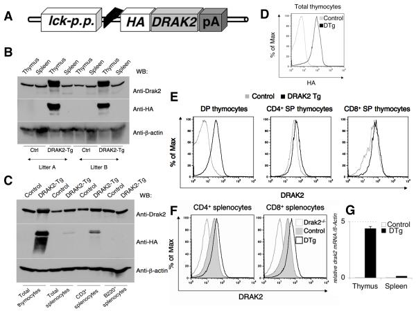
A NotI digested fragment of the 1017-DRAK2 vector construct containing an HA-tagged version of full-length murine drak2 cDNA (Fig. 1a) was injected into fertilized oocytes of CB6F1 mice. Subsequent PCR analysis of tail samples from the resulting progeny identified fifteen positive founders with high-copy integration of the transgene. Among these initial founders, two strains (26.1 and 17.2) with high ectopic expression of HA-DRAK2 in thymocytes were selected for further studies. In contrast to the robust overexpression observed in developing thymocytes, expression of HA-DRAK2 in peripheral splenic T cells was only marginally enhanced compared to endogenous DRAK2 protein levels observed in wild-type (Wt) splenic T cells (Fig. 1b). When fractionated into CD3+ vs. B220+ subsets using magnetic purification, only a small amount of HA-DRAK2 was observed in the former, with no signal observed in the B cell fraction (Fig. 1c); no appreciable increase in total DRAK2 was observed in either splenocyte population. To determine the thymocyte subpopulation(s) bearing transgene expression, we made use of an intracellular staining (ICS) protocol using either anti-HA or anti-DRAK2 mAbs. Using this approach, we observed anti-HA staining in a significant fraction of 1017-DRAK2 Tg thymocytes (Fig. 1d), primarily resulting from HA-DRAK2 expression in DP thymocytes (Supp. Fig. 1). Although western blotting suggested that DRAK2 might be over-expressed in the thymii of 1017-DRAK2 mice, anti-DRAK2 ICS revealed that HA-DRAK2 is instead mis-expressed (Fig. 1e). Overall DRAK2 expression was greatly enhanced only in DP thymocytes, a subset that we have previously observed to be mostly devoid of this immunomodulatory kinase (10, 14). DRAK2 expression was only modestly increased in the CD4+ and CD8+ SP populations, demonstrating that DRAK2 is ectopically expressed during thymocyte development. The modest increase in total DRAK2 in SP thymocyte subsets was also observed in peripheral CD4+ and CD8+ splenocytes. At the messenger RNA level, a significant increase of drak2 mRNA expression over wild-type levels was confirmed by real-time RT-PCR in thymocytes (up to 250 fold), whereas ectopic drak2 mRNA expression in total peripheral splenocytes was only minimal (approximately 4 fold) in these 1017-DRAK2 Tg lines (Fig. 1g). Offspring from these two transgenic founders were backcrossed at least five generations onto the C57BL/6J-background, expanded, and used for detailed studies described below. All 1017-DRAK2 Tg mice were born at Mendelian ratios, viable, fertile and had no gross abnormalities upon visual inspection (not shown).
Figure 1. Generation of 1017-DRAK2 Tg mice and analysis of transgene expression.
A The targeting construct for the transgenic expression of DRAK2 used for microinjection contains the HA-tagged murine drak2 cDNA cloned into the BamHI-site of the 1017 vector under the control of lck proximal promoter and followed by the human growth hormone 3'-UTR. B DRAK2 overexpression was verified by Western blot with anti-HA and anti-DRAK2 Abs in 2 different 1017-DRAK2 transgenic litters: High transgenic overexpression of HA-DRAK2 in thymocytes and minimal overexpression in splenocytes was confirmed in both litters. C Evaluation of transgene enforced DRAK2 expression in splenocyte subsets. Wildtype (control) and 1017-DRAK2 thymocytes and splenocytes were tested for DRAK2 expression using anti-HA and anti-DRAK2 Abs by immunoblotting. Splenocytes were magnetically sorted for T and B cells using anti-CD3 or -B220 magnetic beads. D Intracellular staining of thymocytes reveals high level HA-DRAK2 expression in 1017-DRAK2 Tg (DTg) mice. E Evaluation of thymic subsets for total DRAK2 expression levels. Cells stained as in D were electronically gated on the indicated CD4/CD8 subsets to compare total DRAK2 expression levels vs. wildtype (control) thymocytes. F Evaluation of splenic subsets for total DRAK2 expression levels. Cells stained as in D were electronically gated on the indicated CD4/CD8 subsets to compare total DRAK2 expression levels vs. wildtype (control) splenocytes G Increased drak2 mRNA expression in thymocytes (up to 250 fold) and splenocytes (~4 fold) of 1017-DRAK2 Tg mice compared to control littermates relative to β-actin mRNA expression as assessed by QPCR (samples are in triplicate).
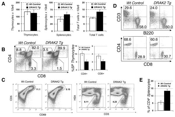
Thymi, spleens and lymphnodes of transgenic and wild-type littermates were harvested at various ages (4-6, and 10 weeks as well as 12 months for aging experiments) and assessed for organ size, weight, total cellularity as well as distributions of various immune and T cell populations. A sub-fraction of 1017-DRAK2 Tg mice displayed splenomegaly early in life (4-6 weeks of age), and most mice developed a splenomegaly with age (see below). Whereas the numbers of total thymocytes were overtly normal in 1017-DRAK2 Tg mice, a moderate, but consistent increase in spleen weight and total cellularity was detectable (Fig. 2a); the number of total splenic T cells after purification was also slightly increased in the transgenic mice. The most striking result was a significant and consistent reduction of the percentages of SP CD4+ and CD8+ T cells and a concomitant increase of the DP population in the thymii of young 1017-DRAK2 mice (Fig. 2b), potentially reflecting impaired positive selection. The decrease in single positive CD4+ and CD8+ T cells was accompanied by reduced numbers of SP CD5Hi/CD69+ and HSALo/CD3Hi thymocytes in DRAK2 transgenic mice, indicating that the intensity of the signal through the TCR was muted during thymocyte selection (Fig. 2c). No obvious differences were observed in double negative thymocyte subsets, as assessed by co-staining of CD4-/CD8- T cells with anti-CD25 and -CD44 (not shown). In the periphery, 1017-DRAK2 Tg mice displayed overtly normal ratios of T and B cells as verified by staining with anti-CD3 and anti-B220 (Fig. 2d). Strikingly, the CD4+ subset expressing CD25 was embellished in all DRAK2 Tg mice examined without concomitant up-regulation of the transient activation marker CD69 (Fig. 2e). In accordance, intracellular staining with an antibody against the transcription factor Foxp3 that serves as a specific marker for the Treg subset, revealed elevated levels of Foxp3, confirming that these CD4+CD25+ T cells possess features of Tregs (not shown). In contrast to a previous report in which DRAK2 expression is mediated by the β-actin promoter (18), the fraction of CD4+ T cells with an activated or memory T cell phenotype (CD44 high/CD62Llow) did not appear to be decreased in 1017-DRAK2 Tg mice. Contrarily, an increase in this T cell population was detectable in most 1017-DRAK2 Tg mice, potentially resulting from activation of peripheral T cells by environmental or self-antigens (see below).
Figure 2. Basic phenotyping of the adaptive immune system of 1017-DRAK2 Tg mice: Altered total cellularities in thymi and spleens and reduction of SP CD4+ and CD8+ thymocytes.
Thymocytes and splenocytes from 10 representative pairs of 4-8 weeks old 1017-DRAK2 transgenics and their wildtype littermates were harvested, counted and subjected to FACS analysis. Diagrams show average data (n > 10), FACS plots show data of representative individual mice. A Slightly decreased total cellularities in thymi and subtle increases in total splenocytes as well as total T cells in young 1017-DRAK2 transgenics. B FACS analysis revealed a significant reduction of CD4+ and CD8+ SP populations and a concomitant increase of DP thymocytes in thymi of 4-8 weeks old 1017-DRAK2 Tg mice. C Grossly normal distribution of T cells (CD3+) versus B cells (B220+) as well as CD4+ versus CD8+ T cells in spleens of 1017-DRAK2 Tg mice. D Decreased CD69+ and CD5+ DP developing thymocytes in 1017-DRAK2 Tg mice as revealed by FACS analysis. E Increased CD25+ splenocytes as a percentage of total CD4+ splenocytes in 1017-DRAK2 Tg mice compared to wildtype (control) mice.
Alterations in positive and negative thymic selection in H-Y 1017-DRAK2 mice
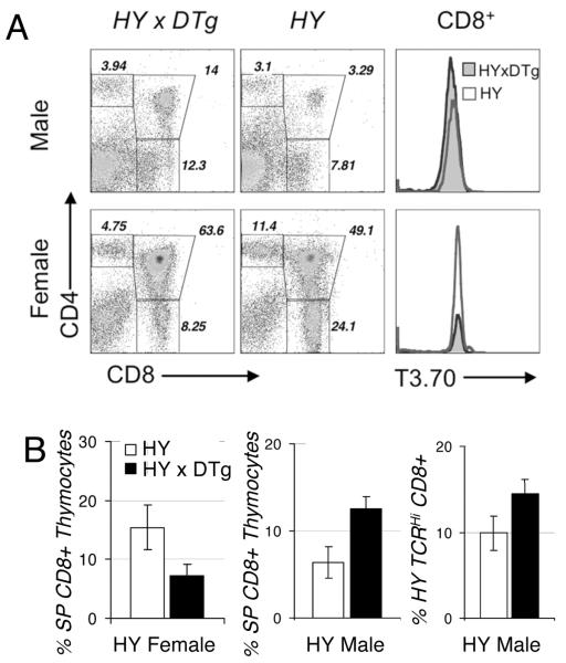
To further explore the potential that ectopic DRAK2 expression limits thymic selection, 1017-DRAK2 Tg mice were crossed onto the H-Y TCR transgenic background. H-Y TCR transgenic (H-Y) mice express a TCR specific for the male H-Y antigen presented in the context of H-2Db molecule. Thymocytes bearing the transgenic TCR are positively selected in females, but deleted by negative selection in male mice (23, 24). After crossing 1017-DRAK2 and H-Y TCR transgenic mice, thymocytes from female and male H-Y TCR x 1017-DRAK2 double Tg mice (H-Y 1017-DRAK2) were analyzed by FACS after staining with anti-CD4, anti-CD8 and clonotypic T3.70 anti-TCR Abs to compare the ratios of DP to SP CD8+ thymocytes as well as the level of transgenic TCR expression (H-Y TCRhigh versus TCRlow). In female H-Y mice, mature CD4-CD8+ T cells predominated over CD4+CD8- T cells in the presence of a selecting H-2Db haplotype in the C57BL/6J, as expected (Fig. 3a). This otherwise augmented proportion of SP CD8+ T cells (average 16%) was significantly reduced in H-Y 1017-DRAK2 female mice (average 8%) reflecting impaired positive selection. Male H-Y mice normally have very small thymi due to deletion of the self-reactive T cells, and thymic sizes and thymocyte numbers were also reduced in H-Y 1017-DRAK2 male mice (not shown). Despite this, we observed an increased proportion of CD4-CD8+ thymocytes bearing the H-Y clonotypic TCR (Fig. 3b). In the periphery, similar proportions of T3.70+ CD4+ and CD8+ T cells were observed (not shown). We conclude that ectopic DRAK2 intensified negative regulation of signals through the TCR in developing thymocytes, leading to partial blockade of thymic positive and negative selection.
Figure 3. Altered positive and negative selection in thymi of 1017-DRAK2 Tg mice in the H-Y TCR transgenic model.
After backcrossing of 1017-DRAK2 Tg mice onto the H-Y TCR transgenic background their thymocytes were harvested at 4-6 weeks of age and stained with anti-CD4, anti-CD8 and clonotypic T3.70 to detect expression of the transgenic TCR. FACS plots show representative data of individual mice, diagrams show averages and standard deviations (n > 10). A In female mice, a lack of the increase in CD8+ SP thymocytes indicates a defect in positive selection in 1017-DRAK2 transgenics. B In male H-Y DRAK2 dTg mice, the fraction of CD8 expressing as well as of H-Y TCRhigh CD8+ thymocytes was increased compared to H-Y littermates, reflecting alterations in negative selection.
Normal apoptotic sensitivity but altered TCR signaling in 1017-DRAK2 Tg mice
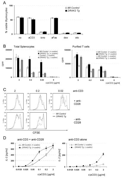
As ectopic expression of DRAK2 in cell lines has previously been shown to correlate with increased apoptosis (12, 13), 1017-DRAK2 thymocytes were tested for their apoptotic sensitivity following treatment under a variety of conditions. No increased spontaneous apoptosis or altered sensitivity to apoptogenic stimuli could be detected by Annexin V-staining, and cell recovery was grossly similar to control thymocytes (Fig. 4a). To investigate whether the changes in thymic selection detected in 1017-DRAK2 Tg mice translate into altered function of mature peripheral T cells, we subsequently examined T cell activation to optimal and suboptimal stimuli in dose-response assays assessing proliferation, apoptotic sensitivity and IL-2 production. Our data indicated that unpurified and purified 1017-DRAK2 T cells hyper-proliferated in vitro in response to suboptimal concentrations of anti-CD3 (+/- anti CD28), as measured by [3H]-thymidine incorporation (Fig. 4b). To distinguish between increased cell division rates and reduced apoptosis, proliferation and apoptosis of CD4+ and CD8+ T cells were quantified simultaneously in the same cultures using carboxy-fluorescein diacetate, succinimidyl ester (CFDA-SE)-labeling and annexin-V staining after 72 hrs stimulation in similar dose-response assays with plate-bound anti-CD3 stimulation as described above (Fig. 4c). Proliferation in response to suboptimal doses of anti-CD3 was enhanced, but was not a consequence of diminished apoptosis of proliferating 1017-DRAK2 T cells since the proportion of annexin-V positive cells was similar or slightly increased compared to controls (Supp. Fig. 2). Consistent with a diminished activation threshold for proliferation, purified 1017-DRAK2 T cells also produced elevated levels of IL-2 in response to suboptimal stimulation as measured by ELISA after 24h of culture (Fig. 4d).
Figure 4. Functional analysis of 1017-DRAK2 Tg T cells revealed a hyper-responsiveness of peripheral T cells in response to suboptimal stimulation, but no significant changes in the apoptotic sensitivity of thymocytes and peripheral T cells.
To assess proliferation and IL-2 production, total splenocytes or purified T cells from either DRAK2 Tg or wildtype mice were seeded in triplicate for each condition at a density of 106/ml and stimulated with indicated amounts of plate-bound or soluble anti-CD3 in the presence or absence of anti-CD28 (1 μg/ml) for the times indicated. Data represent averages of at least 3 individual experiments. A Apoptosis is not increased in 1017-DRAK2 Tg thymocytes as assessed in a thymocyte apoptosis assay after in vitro incubation under various conditions for 24 hrs as indicated. The following concentrations were used: untreated (no), anti-CD3ε Ab (2C11) [1 μg/ml], ionomycin [1 μM], anti-Fas mAb [1 μg/ml], dexamethasone (dex) [1 μM], etoposide (eto) [10 μM]. B 3H-Thymidine uptake. Hyperproliferation of DRAK2 Tg total splenocytes and purified T cells in response to suboptimal stimulation with plate-bound anti-CD3 in the presence or absence of anti-CD3 in vitro (dose-response-assay) (n > 9). Proliferating cells were pulsed with 3H-Thymidine during the last 18 hrs of a 96 hrs incubation period, and Tritium-Incorporation was measured as cpm using a β-Szintilation counter. C CFSE dilution assay to simultaneously monitor proliferation and apoptosis. Purified T cells from either Wt littermates (solid grey lines) or DRAK2 transgenics (dashed black lines) were labeled with CFDA-SE as described, stimulated with indicated concentrations of plate-bound anti-CD3 for 72 hrs and then stained with anti-CD4, anti-CD8 and Annexin V for FACS analysis. Increased proliferation of DRAK2 Tg T cells as indicated by higher fractions of cells with more diluted CFSE was not due to a reduction of apoptosis. D Increased IL-2 production of peripheral DRAK2 Tg T cells in response to suboptimal stimulation as measured by ELISA in culture supernatants after 24 hrs of stimulation.
Reduced calcium mobilization in developing 1017-DRAK2 DP thymocytes, but not in mature SP thymocytes
Since previous studies have demonstrated that endogenous DRAK2 negatively regulates TCR-induced Ca2+ mobilization in transitional and SP thymocytes (14), we sought to evaluate the impact of ectopic DRAK2 expression on this signaling cascade. Thymocytes labeled with fluorescently tagged anti-CD4 and anti-CD8 as well as the Ca2+ indicator dyes Fluo-3 and Fura-Red were analyzed by flow cytometry upon TCR/CD4 cross-linking with streptavidin after incubation with biotinylated anti-CD3 and anti-CD4 (Fig. 5a). In thymocytes, the overall diminished Ca2+ mobilization in response to anti-CD3/anti-CD4 crosslinking reflected a strong reduction in the DP thymocyte population that expresses the highest amounts of the DRAK2 transgene (see above). In contrast, mature SP CD4+ thymocytes displayed grossly similar Ca2+ mobilization kinetics as controls. These results indicate a correlation of DRAK2 transgene expression level and TCR-induced calcium signaling threshold. Maximal Ca2+ release induced by ionomycin was comparable between 1017-DRAK2 and wildtype cells in all populations analyzed. In contrast, ectopic DRAK2 expression in the thymus did not affect other signaling pathways induced by CD3/CD4 crosslinking; no differences in Erk, Jnk or tyrosine phosphorylation were observed in 1017-DRAK2 Tg thymocytes following stimulation with increasing concentrations of anti-CD3 [plus constant anti-CD4] (Fig. 5b). In line with the relatively normal Ca2+ mobilization of 1017-DRAK2 CD4+ SP thymocytes, peripheral CD4+ and CD8+ T cell Ca2+ mobilization following anti-CD3 crosslinking was indistinguishable between wildtype and transgenic T cells (Fig. 5c). Remarkably, we observed consistently enhanced basal Erk and tyrosine phosphorylation in 1017-DRAK2 transgenic peripheral T cells (Fig. 5d). As well, treatment with a suboptimal dose (0.3 μg/ml) of anti-CD3 induced strong Erk and tyrosine phosphorylation in 1017-DRAK2 T cells, whereas this treatment failed to do so in wildtype T cells. We note that despite these signaling differences, no appreciable differences in the expression of CD3 were observed in 1017-DRAK2 thymocytes (Supp. Fig. 3a). Also, a significant fraction of peripheral T cells had slightly but consistently reduced CD3 or TCβ chain expression (Supp. Fig. 3b), demonstrating that the enhanced signaling of these cells is not due to embellished expression of CD3 or TCR molecules on the surface of 1017-DRAK2 T cells. Our data thus indicate that ectopic DRAK2 in the thymus negatively influences calcium-signaling pathways downstream of the TCR. This artificially high activation threshold, as revealed under strong TCR crosslinking conditions (Supp. Fig. 4), leads to the development or selection of T cells with enhanced signaling properties, likely a result of altered selection in the thymus since DRAK2 expression is relatively normal in transgenic peripheral T cells.
Figure 5. Reduced Calcium flux in DP thymocytes from 1017-DRAK2 Tg mice stimulated with anti-CD3 and anti-CD4, but not in SP thymocytes.
A To detect alteration in Calcium signaling upon TCR stimulation by flow cytometry, thymocytes labeled with Fluo-3 and Fura red and stained with CD4 and CD8 were incubated with biotinylated antibodies to CD3 and CD4 before cross-linking with streptavidin as described. Black lines indicate Calcium flux in DRAK2 transgenic thymocytes, grey lines represent Wt controls. For FACS analysis, thymocytes were first sampled for ~60 s to establish baseline Ca2+-levels before stimulation with streptavidin (arrow “SA”). After ~480 s of stimulation, maximal Calcium release was induced by addition of ionomycin (arrow “iono”). Data represent the kinetics of calcium mobilization in response to stimulation in total thymocytes, DP thymocytes and CD4+ thymocytes plotted as ratio of FL-1 (Fluo-3) to FL-3 (Fura red). B Unaltered signalling in 1017-DRAK2 Tg thymocytes in response to TCR crosslinking. Total thymocytes were incubated with the indicated amounts of biotinylated anti-CD3 plus biotinylated anti-CD4, followed by crosslinking for 2 min. with streptavidin. Western blotting was performed on lysates using anti-phospho- and total-Erk1/2, anti-phospho- and total-Jnk1/2, anti-phospho-Tyrosine, anti-DRAK2 and anti-α-tubulin as a loading control. C Detection of calcium signalling as performed in A. on total splenocytes, except that incubation was with anti-CD3 alone before crosslinking with streptavidin as described. D Altered signalling in 1017-DRAK2 Tg splenocytes in response to TCR crosslinking. Total splenocytes were incubated with the indicated amounts of biotinylated anti-CD3 +/- biotinylated anti-CD28, followed by crosslinking for 2 min. with streptavidin. Western blotting was performed as in B.
Splenomegaly and enhanced susceptibility to spontaneous and induced autoimmunity in 1017-DRAK2 Tg mice
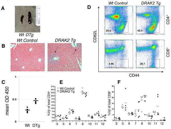
To assess the manifestation of spontaneous autoimmunity, groups of 1017-DRAK2 Tg mice and littermates were aged for 12 months, and examined for the presence of cellular infiltrates in their major organs by routine histology. In addition, amounts of anti-nuclear auto-antibodies (ANA) were measured in sera from these aged mice. Splenomegaly was detectable in a sub-fraction of young 1017-DRAK2 Tg mice, and five out of six aged transgenic mice displayed overt splenomegaly, with an altered histological architecture and hyperplasia of the red and white pulp. In contrast, only one wild-type mouse developed increased spleen size; a representative example of this degree of splenomegaly is shown (Fig. 6a). Mononuclear infiltrates were detected in livers and pancreata of all 1017-DRAK2 Tg mice, while no signs of infiltration were apparent in wild-type littermates (Fig. 6b). 5 out of 6 1017-DRAK2 transgenic mice also displayed slightly elevated levels of anti-nuclear auto-antibodies as determined by ELISA (Fig. 6c). These results suggest an increased prevalence of spontaneous autoimmunity in aged 1017-DRAK2 Tg mice, likely caused by activation of auto-reactive T cells that may have escaped thymic negative selection, and/or possess diminished activation thresholds. Consistent with either scenario, we observed a dramatic increase in the proportion of CD44Hi/CD62LLoCD4+ and CD8+ T cells in 1017-DRAK2 Tg mice (Fig. 6d), suggesting an enhanced proportion of autoreactive effector or memory T cells. However, the ratio of CD3+ vs. B220+ cells in aged 1017-DRAK2 Tg mice with splenomegaly was similar to wildtype littermates lacking splenomegaly, suggesting that cytokine release may have lead to increases in multiple splenocyte subsets (not shown). Since altered selection in the thymus may lead to gross changes in the peripheral TCR repertoire, including the release of autoreactive clones (11), we analyzed TCR Vβ usage to grossly assess the repertoire in wildtype vs. 1017-DRAK2 mice. We observed no major differences in Vβ usage in the thymii or spleens of young (4-6 weeks) mice (Supp. Fig.5). In older mice (8-16 weeks), no significant differences in the Vβ usage of splenic CD4+ subsets from 1017-DRAK2 mice were observed (Fig. 6e). While the Vβ repertoire expressed on CD8+ 1017-DRAK2 T cells was mostly indistinguishable from wildtypes, we note that there was greater variability in the usage of Vβ5, 7 and 11, with these Vβs being highly prevalent in some 1017-DRAK2 mice (Fig. 6f). Although we are currently uncertain about the significance of these results, we believe that these results suggest that the variable differences in Vβ usage in 1017-DRAK2 CD8+ T cells are likely a consequence of expansion of autoreactive clones in these mice as they age.
Figure 6. Increased spontaneous autoimmunity in aged 1017-DRAK2 transgenic mice.
A Significant splenomegaly in aged 1017-DRAK2 transgenics (representative spleens). B Infiltrates in livers of 12 months 1017-DRAK2 Tg mice detected by H&E staining (representative data). C Sera of aged DRAK2 Tg mice were analysed for the presence of anti-nuclear antibodies (ANA) by routine ELISA. The mean OD450 was slightly increased compared to wild-type levels in 5 out of 6 DRAK2 transgenics. D Significantly increased CD44hi/CD62Llo memory cells in 1017-DRAK2 transgenic splenocytes (representative spleens from 12 wk. old littermates). E and F Altered Vβ repertoire in CD8+ SP splenocytes (F) but not CD4+ SP splenocytes (E) in 8-12 wk. old 1017-DRAK2 Tg mice compared to littermate controls. Total splenocytes were stained with CD4, CD8 and the indicated Vβ chains and analyzed by flow cytometry.
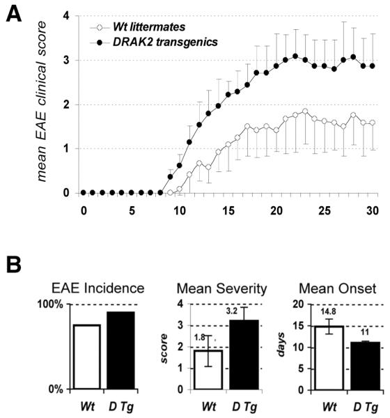
The susceptibility of 1017-DRAK2 Tg mice to induced autoimmune disease was tested in the multiple sclerosis model experimental autoimmune encephalomyelitis (EAE), one of the best-characterized murine models of organ-specific autoimmunity (25). Mice were immunized with a peptide containing residues 35-55 from myelin oligodendrocyte glycoprotein (MOG) emulsified in complete Freund's adjuvant, and were evaluated for clinical manifestation of EAE as described previously (10, 20). The mean clinical severity of the EAE was exacerbated in 1017-DRAK2 Tg mice compared to wild-type littermates, as reflected by the average clinical scores (Fig. 7a). In addition, 1017-DRAK2 Tg mice displayed an earlier onset of disease (average of the day when each mouse first scored one) and a greater number of transgenics exhibited profound signs of disease, scoring two or higher (incidence) (Fig. 7b). Infiltrate density in the central nervous system correlated with clinical scores in 1017-DRAK2 transgenic mice as well as wildtype controls at day 21 post-immunization (not shown). In light of the phenotypic changes detected in the thymus of 1017-DRAK2 transgenics, it is possible that altered thymic selection resulted in a shift in the generated mature T cell pool. Such T cells would possess an increased affinity and/or or avidity to foreign and self-antigens (and/or with altered signaling properties and gene expression profiles), leading to an enhanced sensitivity to autoimmunity.
Figure 7. Increased susceptibility of 1017-DRAK2 Tg mice to induced autoimmune disease in the EAE model.
EAE was induced in DRAK2 Tg mice by immunization with MOG peptide emulsified in CFA as described and compared to responses of wildtype littermates. Severity of EAE was assessed daily for a period of 30 days by a standard scoring system (0-4): 0, no signs of disease; 0.5, altered gait and/or hunched appearance; 1, limp tail; 2, partial hind limb paralysis; 3, complete hind limb paralysis; 4, complete hind limb paralysis and partial forelimb paralysis. The experiment was performed twice, and data were pooled together. A Mean EAE clinical scores were significantly increased in DRAK2 Tg mice. Data represent average clinical scores for 9 DRAK2 Tg mice and 8 Wt littermates. B DRAK2 Tg mice displayed a significantly increased incidence of EAE in two independent experimental sets, as well as significantly exacerbated mean severity (p-value = 0.04) and earlier mean onset (p-value = 0.014) compared to Wt littermates. Statistically-significant results indicated with *.
Discussion
1017-DRAK2 Tg mice serve as a model to examine the effect of overexpression of a negative regulator of immunoreceptor signaling in developing T cells, specifically at the DP thymocyte stage that is critical for positive and negative selection. The major findings of the present survey of these newly generated 1017-DRAK2 Tg mice are: a) reduced generation of SP CD4+ and CD8+ thymocytes that correlated with changes in positive and negative selection in the H-Y TCR transgenic model; b) hyper-responsiveness of peripheral T cells to suboptimal anti-CD3 stimulation reflected by hyper-proliferation and increased IL-2 production, as well as alterations in the composition of the peripheral T cell pool; c) enhanced susceptibility to spontaneous and induced autoimmunity. We conclude that strong suppression of TCR signals during thymic selection caused by ectopic DRAK2 expression alters the responsiveness of peripheral T cells. Although this likely may lead to an altered TCR repertoire, our results also demonstrate that T cells that are capable of overcoming this artificially high selection threshold in the thymus possess substantially enhanced signaling properties in the periphery. The resulting enhanced autoimmune susceptibility is likely a consequence of this diminished negative selection, as well as the reduced activation threshold of 1017-DRAK2 peripheral T cells. As expression of the DRAK2 transgene was only marginally increased in the periphery compared to its high levels in DP thymocytes, the changes in peripheral T cell reactivity observed can likely be attributed to altered central tolerance in 1017-DRAK2 Tg mice. This conclusion is supported by the observation of an enlarged population of T3.70+ CD8+ T cells observed in H-Y 1017-DRAK2 male mice. In addition, mature 1017-DRAK2 T cells possess considerably diminished levels of HA-DRAK2 compared to thymocytes. Thus, it is tenable that only those thymocytes possessing intrinsically enhanced signaling overcome this artificially high activation threshold. By virtue of diminished HA-DRAK2 expression in peripheral T cells, where the lck proximal promoter is known to be quenched, this high activation barrier is removed - a scenario that could contribute to the peripheral hyper-reactivity. Consistently, positive thymic selection has been associated with the development of an increased activation threshold to the selecting self-antigen within the thymus to ensure unresponsiveness of the mature T cell in the periphery (26). A critical role for DRAK2 in thymic positive selection has previously been suggested, since drak2-deficient thymi display an increased fraction of CD4+ SP thymocytes with concomitant up-regulation of CD5 and CD69, indicating enhanced signal intensity downstream of the TCR (10, 14). These changes in central tolerance may contribute to the T cell hypersensitivity, increased fraction of memory-like circulating T cells and the autoimmune resistance in drak2-deficient mice. Our data from 1017-DRAK2 Tg mice confirm that DRAK2 impacts thymic selection, and that its enforced expression partially blocks positive and negative selection, likely resulting in the selection of auto-reactive T cells. DRAK2 expression is tightly regulated in a stage-specific fashion during thymopoiesis (10). Further, it is a primary response gene in DP thymocytes following antigen receptor and co-receptor crosslinking (14). The results here suggest that this tight regulation of DRAK2 expression is necessary for proper positive selection and central tolerance. The manifestation of T cell mediated autoimmunity is currently thought to be influenced by a combination of TCR avidity to auto-antigens, negative regulation of signaling, concentration of self-peptide-MHC complexes presented on APCs, and the costimulatory/inflammatory context (27). Further, the distinct signaling thresholds necessary to induce positive selection (survival), negative selection (apoptosis) or peripheral activation/clonal expansion also impact immunological tolerance (26). During thymic T cell development, only DP thymocytes possessing a TCR with low affinity/low avidity for self peptide-MHC presented by thymic epithelial cells in the cortex (cTECs) receive a sufficient survival signal to be rescued from death by neglect, and subsequently undergo positive selection (1, 2). Subsequent negative selection via apoptosis ensures the intrathymic clonal deletion of immature self-reactive thymocytes with high affinity/low avidity for peripheral tissue-restricted self-antigens ectopically expressed in medullary thymic stomal APCs (mTECs) via the transcription factor AIRE (autoimmune regulator) (28-30). Clonal deletion (negative selection) in response to strong signals is mediated by direct activation of the pro-apoptotic BH3-only protein Bim as well as transcriptional upregulation of Bim and other pro-apoptotic genes (31, 32). Since the altered selection pattern detected on the H-Y TCR transgenic background did not correlate with enhanced apoptotic sensitivity in 1017-DRAK2 Tg mice, DRAK2 does not appear to directly influence the apoptotic pathways involved in thymic development, and likely transduces non-apoptotic signals during thymocyte differentiation.
We propose that excessive negative regulation of TCR signaling by ectopically expressed DRAK2 shifts the threshold for thymic positive and negative selection. Consequently, its hyper-expression may block positive selection, potentially resulting in death by neglect of a population with low avidity for MHC plus self-peptides. On the other hand, it may also rescue thymocytes with a higher affinity/avidity for self-antigen that would otherwise undergo negative selection. Therefore, the resulting peripheral T cell pool in 1017-DRAK2 Tg mice may possess an altered TCR repertoire skewed toward self-reactivity and equipped with a diminished activation threshold. In accord with this model, negative regulation of TCR signaling has been implicated in “tuning” T cell responses to allow high-avidity/low-affinity, self-reactive T cells to avoid negative selection in the thymus (32). Consequently, negative regulation provides non-deleting mechanisms to control the avidity with which T cells recognize self-antigens (“tuning”) to maximize the peripheral T cell repertoire, allowing for survival of T cells that can respond to self, but only at concentrations that are normally not reached in vivo (33). This hypothesis is also supported in models such as SKG mice (11), as well as in the observation that CD28 costimulation can enhance thymic negative selection for certain Vβ subsets (34-36). Further, inhibition of miR-181a expression in immature T cells has recently been demonstrated to reduce intrinsic antigen sensitivity and impair both positive and negative selection via up-regulation of multiple phosphatases (37). Taken together, these findings suggest that activation threshold must be carefully tuned to achieve appropriate responsiveness of peripheral T cell pools.
TCR-induced signaling pathways that mediate positive and negative selection by regulating the balance of pro-/anti-apoptotic factors and apoptotic sensitivity in thymocytes include Ca2+-influx, calcineurin-NFAT-, MAPK- and PKC- pathways. Notably, modulation of signal strength and duration likely determines cell fate, with high-intensity signals required promote execution of cell death and low-intensity stimuli driving positive selection (33). A function of DRAK2 in controlling the threshold for calcium mobilization in the thymus and peripheral T cells has previously been established, since positively selected drak2-deficient thymocytes as well as mature peripheral display enhanced Ca2+ mobilization following suboptimal TCR crosslinking (10, 14). In accord with the notion that DRAK2 sets the threshold for Ca2+ mobilization in developing thymocytes, we observed reduced Ca2+ mobilization in 1017-DRAK2 DP positive thymocytes (but normal MAPK activation and tyrosine phosphorylation), cells that express the highest levels of the transgene, but not in SP thymocytes and peripheral T cells that have modest levels of transgene expression. Recent data indicate that calcium signals are necessary for the induction of positive as well as negative selection: strong TCR-signals lead to Ca2+-dependent transcriptional induction of pro-apoptotic Bim via PKC activation (38). In contrast, weaker signals activate the Ca2+-NFAT pathway to mediate positive selection of thymocytes via MAPK/Erk sensitization (39). Consistently, T cell-specific deletion of NFATc3 or calcineurin in mice causes defects in positive selection (40). The defect in Ca2+ mobilization detected in 1017-DRAK2 DP thymocytes may thus explain the effects of this transgene on positive and negative selection. However, a more complete understanding of the consequences of ectopic DRAK2 expression during thymic selection awaits full description of its kinase substrates in thymocytes.
In transgenic mice expressing DRAK2 via the β-actin promoter (18), a careful analysis of thymic development has not been described, nor have expression patterns of the transgene in thymocyte and other immune subsets been examined. Instead, that study has focused entirely on peripheral T cells. Due to ubiquitous overexpression of DRAK2 in that strain, changes in multiple immune and accessory cells could potentially impact T cell responses and contribute to the phenotype observed. Similar to our results with 1017-DRAK2 transgenics, this previous report described hyper-responsiveness of peripheral T cells, with hyper-proliferation and augmented production of IL-2 (and IL-4) in response to anti-CD3 stimulation. In contrast to our findings described here, Mao et al. also described increases in peripheral T cell apoptosis mediated by IL-2 and a defect in memory cell development. Although 1017-DRAK2 peripheral T cells produce elevated amounts of IL-2, no major increases in apoptosis were detectable in response to anti-CD3 stimulation. Memory T cell responses in 1017-DRAK2 Tg mice are currently under investigation, but we detected an increase, rather than a reduction, in the fraction of the activated or memory type T cell subset (CD44highCD62Llow) in transgenic mice up to 14 weeks of age. That DRAK2 is critical for memory T cell function has also been established by previous analysis of anti-viral responses to murine hepatitis virus (MHC) in drak2-deficient mice (41).
The shift in TCR signaling threshold and peripheral repertoire likely accounts for the enhanced susceptibility of 1017-DRAK2 Tg mice to spontaneous and induced autoimmunity. As discussed above, elevated responses of 1017-DRAK2 peripheral T cells to suboptimal anti-CD3 may represent an indirect effect due to an altered threshold acquired in the course of positive selection with high levels of this negative regulator of TCR signaling. To overcome the higher peripheral activation threshold, costimulation via CD28 normally serves as an amplifier to quantitatively and qualitatively support the TCR signal to induce a proliferative response while preventing anergy and AICD (42); such costimulation is dispensable for the activation of drak2-deficient T cells (10). T cells in 1017-DRAK2 Tg mice that have the potential to recognize self-antigen might be activated more readily, resulting in enhanced spontaneous and induced autoimmune susceptibility. The spontaneous autoimmune hallmarks detected in DRAK2 transgenics have characteristics of a lupus-like disease, being marked by splenomegaly, liver infiltration and increased ANA (43).
The 1017-DRAK2 transgenic mouse model has confirmed that expression of negative regulators of TCR signaling during thymic development may significantly impact central tolerance and the peripheral T cell repertoire by shifting the thresholds for positive and negative selection. Such a shift in TCR repertoire to higher avidity may allow for survival of T cells with intermediate or high affinity/avidity for self-peptide/MHC that may provoke autoimmunity. Taken together, these results suggest that therapeutic efforts to interfere with T cell signaling must be taken with caution to prevent the export of highly auto-reactive clones from the thymus.
Supplementary Material
Acknowledgements
The authors wish to thank the Transgenic Mouse Core Facility at UCI for the professional support with the oocyte injection and generation of DRAK2 transgenic mice, especially Tom Fielder for competent help with all questions, as well as Animal Husbandry Staff and Veterinarians. Many thanks as well to Jennifer Hernandez and the laboratory of Charles Glabe of the University of California, Irvine for providing the MOG35-55 peptide, Si-Yi Chen for additional reagents and support, and to past and present members of the Walsh lab for helpful discussion. MG, RHN and CMW designed the experiments; MG and RH conducted the experiments; MG and CMW wrote the manuscript; CMW edited the manuscript. The authors do not have any conflicting financial interests.
This work was supported by awards from the Arthritis National Research Foundation (ANRF-39494 to MG) and NIH grant R01-AI63419 (to CMW).
Abbreviations
- AICD
activation-induced cell death
- DP, SP
double positive, single positive thymocyte
- DRAK2
DAP-related apoptotic kinase-2
- EAE
experimental autoimmune encephalomyelitis
- TCR
T cell receptor
- Tg, dTg
transgenic, double transgenic
References
- 1.Hogquist KA, Baldwin TA, Jameson SC. Central tolerance: learning self-control in the thymus. Nat Rev Immunol. 2005;5:772–782. doi: 10.1038/nri1707. [DOI] [PubMed] [Google Scholar]
- 2.Marrack P, Kappler J. T cell tolerance. Semin Immunol. 1990;2:45–49. [PubMed] [Google Scholar]
- 3.Sakaguchi S, Yamaguchi T, Nomura T, Ono M. Regulatory T cells and immune tolerance. Cell. 2008;133:775–787. doi: 10.1016/j.cell.2008.05.009. [DOI] [PubMed] [Google Scholar]
- 4.Melero I, Arina A, Chen L. The many sounds of T lymphocyte silence. Immunol Res. 2005;33:135–147. doi: 10.1385/IR:33:2:135. [DOI] [PubMed] [Google Scholar]
- 5.Singer AL, Koretzky GA. Control of T cell function by positive and negative regulators. Science. 2002;296:1639–1640. doi: 10.1126/science.1071551. [DOI] [PubMed] [Google Scholar]
- 6.Gatzka M, Walsh CM. Negative Regulation of TCR Signaling in Immunological Tolerance: Taming Good and Evil. Curr Immunol Rev. 2008 In press. [Google Scholar]
- 7.Naramura M, Kole HK, Hu RJ, Gu H. Altered thymic positive selection and intracellular signals in Cbl-deficient mice. Proc Natl Acad Sci U S A. 1998;95:15547–15552. doi: 10.1073/pnas.95.26.15547. [DOI] [PMC free article] [PubMed] [Google Scholar]
- 8.Carter JD, Neel BG, Lorenz U. The tyrosine phosphatase SHP-1 influences thymocyte selection by setting TCR signaling thresholds. Int Immunol. 1999;11:1999–2014. doi: 10.1093/intimm/11.12.1999. [DOI] [PubMed] [Google Scholar]
- 9.Suzuki A, Yamaguchi MT, Ohteki T, Sasaki T, Kaisho T, Kimura Y, Yoshida R, Wakeham A, Higuchi T, Fukumoto M, Tsubata T, Ohashi PS, Koyasu S, Penninger JM, Nakano T, Mak TW. T cell-specific loss of Pten leads to defects in central and peripheral tolerance. Immunity. 2001;14:523–534. doi: 10.1016/s1074-7613(01)00134-0. [DOI] [PubMed] [Google Scholar]
- 10.McGargill MA, Wen BG, Walsh CM, Hedrick SM. A deficiency in Drak2 results in a T cell hypersensitivity and an unexpected resistance to autoimmunity. Immunity. 2004;21:781–791. doi: 10.1016/j.immuni.2004.10.008. [DOI] [PMC free article] [PubMed] [Google Scholar]
- 11.Sakaguchi N, Takahashi T, Hata H, Nomura T, Tagami T, Yamazaki S, Sakihama T, Matsutani T, Negishi I, Nakatsuru S, Sakaguchi S. Altered thymic T-cell selection due to a mutation of the ZAP-70 gene causes autoimmune arthritis in mice. Nature. 2003;426:454–460. doi: 10.1038/nature02119. [DOI] [PubMed] [Google Scholar]
- 12.Sanjo H, Kawai T, Akira S. DRAKs, novel serine/threonine kinases related to death-associated protein kinase that trigger apoptosis. J Biol Chem. 1998;273:29066–29071. doi: 10.1074/jbc.273.44.29066. [DOI] [PubMed] [Google Scholar]
- 13.Matsumoto M, Miyake Y, Nagita M, Inoue H, Shitakubo D, Takemoto K, Ohtsuka C, Murakami H, Nakamura N, Kanazawa H. A serine/threonine kinase which causes apoptosis-like cell death interacts with a calcineurin B-like protein capable of binding Na(+)/H(+) exchanger. J Biochem (Tokyo) 2001;130:217–225. doi: 10.1093/oxfordjournals.jbchem.a002975. [DOI] [PubMed] [Google Scholar]
- 14.Friedrich ML, Wen BG, Bain G, Kee BL, Katayama C, Murre C, Hedrick SM, Walsh CM. DRAK2, a lymphoid-enriched DAP kinase, regulates the TCR activation threshold during thymocyte selection. Int Immunol. 2005;17:1379–1390. doi: 10.1093/intimm/dxh315. [DOI] [PubMed] [Google Scholar]
- 15.Ramos SJ, Hernandez JB, Gatzka M, Walsh CM. Enhanced T cell apoptosis within Drak2-deficient mice promotes resistance to autoimmunity. J. Immunol. 2008 doi: 10.4049/jimmunol.181.11.7606. In press. [DOI] [PMC free article] [PubMed] [Google Scholar]
- 16.Friedrich ML, Cui M, Hernandez JB, Weist BM, Andersen HM, Zhang X, Huang L, Walsh CM. Modulation of DRAK2 autophosphorylation by antigen receptor signaling in primary lymphocytes. J Biol Chem. 2007;282:4573–4584. doi: 10.1074/jbc.M606675200. [DOI] [PubMed] [Google Scholar]
- 17.Ryeom S, Greenwald RJ, Sharpe AH, McKeon F. The threshold pattern of calcineurin-dependent gene expression is altered by loss of the endogenous inhibitor calcipressin. Nat Immunol. 2003;4:874–881. doi: 10.1038/ni966. [DOI] [PubMed] [Google Scholar]
- 18.Mao J, Qiao X, Luo H, Wu J. Transgenic drak2 overexpression in mice leads to increased T cell apoptosis and compromised memory T cell development. J Biol Chem. 2006;281:12587–12595. doi: 10.1074/jbc.M600497200. [DOI] [PubMed] [Google Scholar]
- 19.Gatzka M, Piekorz R, Moriggl R, Rawlings J, Ihle JN. A role for STAT5A/B in protection of peripheral T-lymphocytes from postactivation apoptosis: insights from gene expression profiling. Cytokine. 2006;34:143–154. doi: 10.1016/j.cyto.2006.04.003. [DOI] [PubMed] [Google Scholar]
- 20.Ramos SJ, Hardison JL, L.N. S, T.E. L, C.M. W. Anti-viral effector T cell responses and trafficking are not dependent upon DRAK2 signaling following viral infection of the central nervous system. Autoimmunity. 2007;40:54–65. doi: 10.1080/08916930600996700. [DOI] [PubMed] [Google Scholar]
- 21.Chaffin K, Beals C, Wilkie T, Forbush K, Simon M, Perlmutter R. Dissection of thymocyte signaling pathways by in vivo expression of pertussis toxin ADP-ribosyltransferase. EMBO J. 1990;9(12):3821–3829. doi: 10.1002/j.1460-2075.1990.tb07600.x. [DOI] [PMC free article] [PubMed] [Google Scholar]
- 22.Shimizu C, Kawamoto H, Yamashita M, Kimura M, Kondou E, Kaneko Y, Okada S, Tokuhisa T, Yokoyama M, Taniguchi M, Katsura Y, Nakayama T. Progression of T cell lineage restriction in the earliest subpopulation of murine adult thymus visualized by the expression of lck proximal promoter activity. Int Immunol. 2001;13:105–117. doi: 10.1093/intimm/13.1.105. [DOI] [PubMed] [Google Scholar]
- 23.Kisielow P, Bluthmann H, Staerz U, Steinmetz M, von Boehmer H. Tolerance in T-cell-receptor transgenic mice involves deletion of nonmature CD4+8+ thymocytes. Nature. 1988;333:742–746. doi: 10.1038/333742a0. [DOI] [PubMed] [Google Scholar]
- 24.Kisielow P, Teh H, Bluthmann H, von Boehmer H. Positive selection of antigen-specific T cells in thymus by restricting MHC molecules. Nature. 1988;335:730–733. doi: 10.1038/335730a0. [DOI] [PubMed] [Google Scholar]
- 25.Baxter AG. The origin and application of experimental autoimmune encephalomyelitis. Nat Rev Immunol. 2007;7:904–912. doi: 10.1038/nri2190. [DOI] [PubMed] [Google Scholar]
- 26.Kretz-Rommel A, Rubin RL. Disruption of positive selection of thymocytes causes autoimmunity. Nat Med. 2000;6:298–305. doi: 10.1038/73152. [DOI] [PubMed] [Google Scholar]
- 27.Gronski MA, Boulter JM, Moskophidis D, Nguyen LT, Holmberg K, Elford AR, Deenick EK, Kim HO, Penninger JM, Odermatt B, Gallimore A, Gascoigne NR, Ohashi PS. TCR affinity and negative regulation limit autoimmunity. Nat Med. 2004;10:1234–1239. doi: 10.1038/nm1114. [DOI] [PubMed] [Google Scholar]
- 28.Anderson MS, Venanzi ES, Klein L, Chen Z, Berzins SP, Turley SJ, von Boehmer H, Bronson R, Dierich A, Benoist C, Mathis D. Projection of an immunological self shadow within the thymus by the aire protein. Science. 2002;298:1395–1401. doi: 10.1126/science.1075958. [DOI] [PubMed] [Google Scholar]
- 29.Liston A, Lesage S, Wilson J, Peltonen L, Goodnow CC. Aire regulates negative selection of organ-specific T cells. Nat Immunol. 2003;4:350–354. doi: 10.1038/ni906. [DOI] [PubMed] [Google Scholar]
- 30.Anderson MS, Venanzi ES, Chen Z, Berzins SP, Benoist C, Mathis D. The cellular mechanism of Aire control of T cell tolerance. Immunity. 2005;23:227–239. doi: 10.1016/j.immuni.2005.07.005. [DOI] [PubMed] [Google Scholar]
- 31.Gatzka M, Walsh CM. Apoptotic signal transduction and T cell tolerance. Autoimmunity. 2007;40:442–452. doi: 10.1080/08916930701464962. [DOI] [PubMed] [Google Scholar]
- 32.Opferman JT. Apoptosis in the development of the immune system. Cell Death Differ. 2008;15:234–242. doi: 10.1038/sj.cdd.4402182. [DOI] [PubMed] [Google Scholar]
- 33.Anderton SM, Wraith DC. Selection and fine-tuning of the autoimmune T-cell repertoire. Nat Rev Immunol. 2002;2:487–498. doi: 10.1038/nri842. [DOI] [PubMed] [Google Scholar]
- 34.Punt J, Osborne B, Takahama Y, Sharrow S, Singer A. Negative selection of CD4+CD8+ thymocytes by T cell receptor-induced apoptosis requires a costimulatory signal that can be provided by CD28. J Exp Med. 1994;179(2):709–713. doi: 10.1084/jem.179.2.709. [DOI] [PMC free article] [PubMed] [Google Scholar]
- 35.Punt JA, Havran W, Abe R, Sarin A, Singer A. T cell receptor (TCR)-induced death of immature CD4+CD8+ thymocytes by two distinct mechanisms differing in their requirement for CD28 costimulation: implications for negative selection in the thymus. J Exp Med. 1997;186:1911–1922. doi: 10.1084/jem.186.11.1911. [DOI] [PMC free article] [PubMed] [Google Scholar]
- 36.Buhlmann JE, Elkin SK, Sharpe AH. A role for the B7-1/B7-2:CD28/CTLA-4 pathway during negative selection. J Immunol. 2003;170:5421–5428. doi: 10.4049/jimmunol.170.11.5421. [DOI] [PubMed] [Google Scholar]
- 37.Li QJ, Chau J, Ebert PJ, Sylvester G, Min H, Liu G, Braich R, Manoharan M, Soutschek J, Skare P, Klein LO, Davis MM, Chen CZ. miR-181a is an intrinsic modulator of T cell sensitivity and selection. Cell. 2007;129:147–161. doi: 10.1016/j.cell.2007.03.008. [DOI] [PubMed] [Google Scholar]
- 38.Cante-Barrett K, Gallo EM, Winslow MM, Crabtree GR. Thymocyte negative selection is mediated by protein kinase C- and Ca2+-dependent transcriptional induction of bim [corrected] J Immunol. 2006;176:2299–2306. doi: 10.4049/jimmunol.176.4.2299. [DOI] [PubMed] [Google Scholar]
- 39.Gallo EM, Winslow MM, Cante-Barrett K, Radermacher AN, Ho L, McGinnis L, Iritani B, Neilson JR, Crabtree GR. Calcineurin sets the bandwidth for discrimination of signals during thymocyte development. Nature. 2007;450:731–735. doi: 10.1038/nature06305. [DOI] [PMC free article] [PubMed] [Google Scholar]
- 40.Cante-Barrett K, Winslow MM, Crabtree GR. Selective role of NFATc3 in positive selection of thymocytes. J Immunol. 2007;179:103–110. doi: 10.4049/jimmunol.179.1.103. [DOI] [PubMed] [Google Scholar]
- 41.Schaumburg CS, Gatzka M, Walsh CM, Lane TE. DRAK2 regulates memory T cell responses following murine coronavirus infection. Autoimmunity. 2007;40:483–488. doi: 10.1080/08916930701651139. [DOI] [PubMed] [Google Scholar]
- 42.Michel F, Attal-Bonnefoy G, Mangino G, Mise-Omata S, Acuto O. CD28 as a molecular amplifier extending TCR ligation and signaling capabilities. Immunity. 2001;15:935–945. doi: 10.1016/s1074-7613(01)00244-8. [DOI] [PubMed] [Google Scholar]
- 43.Fairhurst AM, Wandstrat AE, Wakeland EK. Systemic lupus erythematosus: multiple immunological phenotypes in a complex genetic disease. Adv Immunol. 2006;92:1–69. doi: 10.1016/S0065-2776(06)92001-X. [DOI] [PubMed] [Google Scholar]
Associated Data
This section collects any data citations, data availability statements, or supplementary materials included in this article.


