Abstract
Neutral lipids fulfill multiple specialized roles in cellular function. These roles include energy storage and utilization, the synthesis of complex lipids in cellular membranes, lipid second messengers for cellular signaling, and the modulation of membrane molecular dynamics. We have developed a novel mass spectrometric technology, now termed shotgun lipidomics, that can identify the types and amounts of thousands of lipids directly from extracts of biological samples. Shotgun lipidomics is well suited for the identification and measurement of the types and amounts of neutral lipid classes and individual molecular species through the use of multidimensional mass spectrometry. This review summarizes the basic principles underlying the use of shotgun lipidomics for the direct measurement of neutral lipids from extracts of biological tissues or fluids. Through exploiting the high information content inherent in shotgun lipidomics, this technology promises to greatly facilitate advances in our understanding of alterations in neutral lipid metabolism in health and disease.
Keywords: electrospray ionization mass spectrometry, intrasource separation, multidimensional mass spectrometry, triacylglycerol
the major pathologies of the 21st century, including heart disease, stroke, and hypertension, reflect, in large part, the increased prevalence of calorically dense diets and sedentary lifestyles in industrialized societies. The deleterious effects of obesity and diabetes lead to adaptive changes in cellular bioenergetics and signaling that are substantially different from those that were present during evolutionary adaptation. The resultant alterations in neutral lipid metabolism lead to multiple deleterious changes in cellular function that are prominent features of many lipid-related diseases in industrialized societies. Moreover, alterations in cellular lipid metabolism result in multiple changes in both metabolic and signaling networks that coordinately regulate cellular energy balance, signaling cascades, and insulin sensitivity and predispose to inflammatory changes in the vasculature. These interrelated factors have been recognized as a constellation of pathologies contributing to a group of lipid-mediated diseases that are now collectively termed the “metabolic syndrome” (17, 18, 25).
Obesity, diabetes, and other aspects of the metabolic syndrome are spreading in epidemic proportions in industrialized nations (25). One emerging aspect of lipid-related diseases has been the recognition of neutral lipid accumulation in ectopic locations (e.g., heart, muscle, pancreas) (24–26). It has become increasingly appreciated that the excessive accumulation of neutral lipid deposits in critical organs is a progenitor of pathological functional changes in many of these organs and a common finding in obesity lipid-related diseases. Altered lipid metabolism and accumulation of ectopic lipid deposits have been highly correlated with end organ damage, including cardiomyopathy and heart failure, insulin resistance, β-cell decompensation leading to overt diabetes, and the generation of a proinflammatory state in the vasculature and other tissues. However, the causal role of these ectopic lipid deposits in cellular pathology has been the subject of intense debate. On the one hand, the accumulation of triacylglycerols in ectopic locations has been proposed to result in “lipotoxicity,” where intermediates of triacylglycerol hydrolysis (i.e., diacylglycerols) serve signaling functions through activation of protein kinases. On the other hand, these accumulations of ectopic lipids have been proposed to be the products of a salutary metabolic sink that removes potentially toxic lipid metabolites. It should be recognized that these two features are not mutually exclusive, as cellular compartmentation can potentially accommodate both mechanisms. The dual roles of neutral lipids in energy metabolism and cellular signaling as facilitators of metabolic transitions are now well recognized.
Energy contained in the carbon-carbon bond of dietary substrates is either utilized directly through oxidative pathways or stored in triacylglycerols for use during energy depletion. It is the maintenance of the critical balance of energy reserves through integrated metabolic transitions that constitute a major challenge to human health and treatment of lipid-related disease states in the 21st century. To critically assess the role of alterations in neutral lipid metabolism and the role of the accumulation of neutral lipids in ectopic locations, it is first necessary to identify the types of lipid molecular species that accumulate and next correlate their presence and abundance to pathological changes in cellular function. Finally, it is important to causally prove that the observed chemical perturbations are mechanistically responsible for the pathological functional changes and do not merely represent serendipitous changes that coexist or are adaptive responses to the pathological states of interest. Whether primary mechanistic mediators of pathological alterations or serendipitous markers of alterations in cellular energy metabolism and signaling, alterations in neutral lipids are likely to serve as important biomarkers of altered cellular function, facilitating early diagnosis of disease and potentially providing a valuable measure of treatment efficacy.
Neutral lipids are characterized on the basis of their class (e.g., triacylglycerols, diacylglycerols, etc.), subclass (covalent linkage at the sn-1 position), and individual molecular species (the type of aliphatic chain constituents that are present and their regiospecificity). Previously, it was difficult to obtain accurate information on the types and amounts of the many hundreds of discrete neutral lipid chemical species in biological tissues. Typically, one would rely on only limited information (e.g., the amount of total triacylglycerols, the fatty acid composition present in total triacylglycerols, etc.) since identification and quantitation of individual molecular species and regioisomers were in many cases either technically demanding or impossible using previous technologies. For example, information on individual triacylglycerol molecular species composition and amount was available only through multistep procedures that may contain propagated errors, leaving difficulties on potential mechanistic interpretations. Through recent advances in mass spectrometry [MS; e.g., electrospray ionization (ESI) and multidimensional MS], it has now become possible to determine detailed information on the type and amount of neutral molecular species from minute amounts of biological samples (2, 8, 9, 12). The emergence of this technology provides new perspectives on the types of mechanistic information that can be obtained through use of shotgun lipidomics to identify alterations in cellular neutral lipid metabolism. It should be emphasized that any technology possesses certain limitations. Instruments used to perform multidimensional MS-based shotgun lipidomics analysis are relatively expensive compared with the classically used gas chromotography (GC) or GC-MS instruments. Enantiomers of diacylglycerols and triacylglycerols are not resolved in the current stage of this technology, which is an important factor in investigation of protein kinase C activation. It should also be pointed out that, although the recognition of this technology becomes well known, practical utilization currently is still limited to the laboratory, but intense efforts are being made to translate this technology into the clinical arena.
First, this article will review the underlying principles of shotgun lipidomics. Second, it will focus on the role of multidimensional MS in neutral lipid analyses. Third, it will provide two examples in which shotgun lipidomics analyses of biological tissues have been used to interrogate altered lipid metabolism in pathophysiological processes. No attempt will be made to provide historical coverage of the decades of previously developed methods used in nonpolar lipid analysis (4), but rather, attention will be focused on the opportunities that have emerged for new insights into neutral lipid metabolism through the use of shotgun lipidomics.
The Chemical Principles Underlying Shotgun Lipidomics
Lipidomics begins with the identification and quantitation of different lipid classes and individual molecular species to determine the biochemical flux through cellular metabolic networks (7, 15). Shotgun lipidomics uses the unique physical properties of lipids and the underlying diversity of their different lipid chemical structures to identify and quantitate discrete lipid molecular species directly from extracts of biological tissue or fluids (9, 10). In early work, we recognized that lipid species are linear combinations of aliphatic chains and headgroups attached to a glycerol or a sphingoid backbone, each of which represents a building block of the lipid molecular species under consideration (10). For example, the three acyl moieties in triacylglycerols linked to the hydroxyl groups of glycerol can be recognized as three individual building blocks that are present in biological samples in varying amounts. If each building block is identified in each individual triacylglycerol molecule and their regiochemistry defined, then each individual molecular species from a biological sample can be determined (10). The use of the building block principle has particular relevance in tandem MS, where ionized triacylglycerols can be fragmented by collision-induced dissociation. An emergent development in the analysis of triacylglycerols is the use of shotgun lipidomics with multidimensional MS for the detailed analysis of triacylglycerol molecular species.
Shotgun lipidomics can be viewed as an integrated technology that uses four component processes that each use a unique chemical and physical property of lipids. The first component of shotgun lipidomics is a series of multiplexed extractions. For routine lipid analysis, Bligh and Dyer extractions are routinely used. Second, a process termed intrasource separation, which allows the effective resolution of lipids in the ion source on the basis of their ionization propensities (13), is used. Intrasource separation allows the effective resolution of different lipid classes in the ion source depending upon the ionization conditions employed and the electrical propensities of the individual lipids examined. Thus, intrasource separation of different lipid classes can be viewed as a rapid and inexpensive surrogate for chromatography. Lipid extraction against aqueous solvents removes many unwanted contaminants and ions (through partitioning into the aqueous phase during extraction), allowing the direct interrogation of samples by MS. In the case of triacylglycerols and other neutral lipids where no endogenous charge is present, charge separation can be induced by the addition of oxytropic ions (e.g., lithium ions) into the extract. The resultant association of the lithium ion with the carbonyl facilitates charge separation, allowing the effective ionization of triacylglycerols, although they contain no endogenous charge. Thus, electrospray ionization of neutral lipids and the resultant analysis of lithiated molecular species provide information on the types and amounts of triacylglycerols within each sample of interest. However, we specifically point out that, since the aliphatic chain length and the degree of unsaturation can influence the ionization efficiency of neutral lipids, it is important to employ algorithms that correct for differences in ionization efficiency in triacylglycerol molecular species that contain different chain lengths and degrees of unsaturation (8). The third step in shotgun lipidomics of triacylglycerols is the use of tandem MS with neutral loss scanning that allows the construction of a multidimensional spectrum that has units of mass for each axis. For example, the mass of the lithiated triacylglycerol molecular species is the x-axis, and the mass of each scanned fatty acid is used as the y-axis for each naturally occurring fatty acid of interest (Fig. 1). The intensity of the resultant peak (ion counts) reflects the amount of that fatty acid present in each of the identified lithiated triacylglycerol molecular species. Finally, data are analyzed through a series of array processing programs that allow the assignment of molecular species and their relative abundance. This is achieved through a series of operator-interactive algorithms that correct for the natural abundance of 13C (9). Through these four steps, the facile identification and quantitation of hundreds of individual lipid molecular species can be made through ratiometric comparisons with internal standards (typically, tri 17:1 glycerol is used as standard for quantitation of triacylglycerol molecular species since it is commercially available and the endogenous content of tri 17:1 glycerol is negligible). Detailed descriptions of the instrument settings and experimental protocols have been previously published in detail (8, 12).
Fig. 1.
Positive ion electrospray ionization mass spectrum and neutral loss (NL) mass spectra of lipid extracts from rat myocardium. Lipid samples from rat myocardium (∼20 mg of wet tissue) were extracted by a modified method of Bligh-Dyer in the presence of 50 mM LiCl in the aqueous phase. Aliquots of the extracts in 1:1 chloroform-methanol were infused directly into the electrospray ionization (ESI) source using a Harvard syringe pump at a flow rate of 4 μl/min. A: positive ion ESI mass spectrum of lipid extracts. Positive ion ESI tandem mass spectra of triacylglycerol (TAG) in lipid extracts with NL of palmitoleic acid (16:1; B), palmitic acid (16:0; C), oleic acid (18:1; D), linoleic acid (18:2; E), and arachidonic acid (20:4; F) were acquired through simultaneous scanning both 1st and 3rd quadrupoles at a fixed different mass values (NL). All NL mass spectra were displayed after normalization to the base peak in the individual spectrum. *The internal standard peak (i.e., lithiated T17:1 TAG) for TAG quantification. m/z, Mass-to-charge ratio.
Cholesterol and Cholesteryl Esters
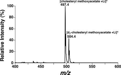
Cholesterol is a prominent constituent of biological membranes that is typically highly localized to the plasma membrane of cells. Alterations in cholesterol content in the plasma membrane have profound effects on membrane molecular dynamics and therefore the activity of transmembrane proteins. Accordingly, identification of alterations in cholesterol content is of fundamental importance to understand the roles of altered cholesterol content in different physiological and pathophysiological contexts. Due to its well-known importance as a risk factor in atherosclerosis, many methods have been developed for the measurement of cholesterol in biological samples. Traditional methods have relied heavily on either enzymatic assays or derivatization of cholesterol and GC-MS. Recently, we have developed a shotgun lipidomics approach that is simple and substantially increases the accuracy and sensitivity of cholesterol measurements in cells (3). Our strategy employed the derivatization of cholesterol with methoxyacetic acid in the presence of d7-cholesterol as an internal standard. After extraction by the Bligh and Dyer method (or other appropriate procedures), a small portion of the lipid extract is treated with methoxyacetic acid. Next, back extraction against a small amount of aqueous LiOH (50 pmol/μl) facilitates charge separation through polarization of the carbonyl moiety present in the derivatized cholesterol (3). The solution is appropriately diluted (depending upon the content of cholesterol in each sample) and directly infused into the ESI mass spectrometer using a Harvard syringe pump at a flow rate of 4 μl/min. Through the use of multidimensional MS, precursor ions that result in a fragment of mass-to-charge ratio (m/z) 97.1 (corresponding to a fragment ion of the derivatized cholesterol after collision-induced dissociation) are acquired in the positive ion mode using mass spectrometric parameters previously described in detail (3). Quantitative analysis can be achieved by direct comparison of the peak intensity of the derivatized endogenous cholesterol to that of the deuterated cholesterol (Fig. 2). The content of total esterified cholesterol in the lipid extracts of samples can be obtained by subtraction of the content of free cholesterol from that of total cholesterol in the sample, as described previously (3). The fatty acyl profile of cholesterol ester molecular species can be readily obtained through precursor ion scanning of m/z 369 (corresponding to positively charged dehydrocholesterol), as described previously (5, 21). It should be pointed out that other alternative derivatization methods can also be employed for the measurement of cholesterol and cholesterol esters (14, 20).
Fig. 2.
Quantitation of cholesterol in mouse cerebellar lipidomes by ESI-mass spectrometry (MS)-MS analyses after derivatization. Mouse cerebellum lipid extracts were prepared, and each of the diluted lipid extract solutions was individually modified with methoxyacetic acid. The reaction-workup solution of each lipid extract was analyzed in the positive ion mode in the presence of LiOH. Precursor ion scanning of m/z 97.1 was acquired in the positive ion mode and used for quantitation of cholesterol in the lipid extract.
Diacylglycerols
Historically, quantitation of diacylglycerol molecular species has been an area of intense effort since diacylglycerols are extremely low-abundance constituents of cellular membranes and their quantitation has been technically difficult. Identification of the roles of diacylglycerols in different physiological and pathological contexts is particularly challenging since diacylglycerols serve dual roles in cellular bioenergetics and signaling. Diacylglycerols activate the classic protein kinase C and participate in proximal signaling steps during many pathophysiological perturbations. Recently, Callender et al. (1) developed a direct infusion approach for diacylglycerol quantitation directly from extracts of biological samples. After extraction of cellular lipids, direct infusion of lipid extracts into a TSQ quantum triple quadrupole mass spectrometer resulted in the appearance of multiple peaks corresponding to discrete diacylglycerol molecular species. Quantitation was accomplished through ratiometric comparisons with a linear regression algorithm similar to that previously employed to correct for the nonlinearities in ionization of other neutral lipid species (8). This is necessary since neutral lipids, in contrast to polar lipids, undergo substantive differences in ionization efficiency with modest changes in aliphatic chain length and unsaturation due to their low intrinsic dipole. In addition, Li et al. (16) have developed an approach to quantitate diacylglycerol molecular species in lipid extracts by ESI-MS after chemical separation and one-step derivatization. We specifically point out that increases in diacylglycerol content are not necessarily accompanied by activation of protein kinase C. It is likely that this critical signaling step is compartmentalized in a spatiotemporal specific manner in different cell types. Thus, although molecular species comparisons with different ligands can provide mechanistic clues to cellular neutral lipid signaling, direct correlations to protein kinase C activation should be interpreted with caution. For example, alterations in cellular diacylglycerol molecular species content often provide mechanistic information that can be used for metabolic network analysis.
Insight into Neutral Lipid Metabolism Using Shotgun Lipidomics
Example 1: shotgun lipidomics identifies alterations in neutral lipid metabolism in diabetic cardiomyopathy.
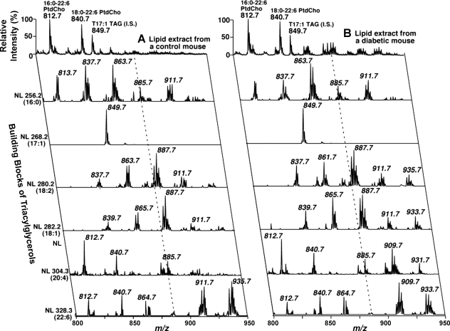
Diabetic cardiomyopathy is a metabolic myopathy that is precipitated by alterations in the structure and function of cellular membranes that lead to bioenergetic inefficiency, mitochondrial dysfunction, and alterations in excitation-contraction coupling (19, 22, 27). Although the historical perception of this disease focused on the role of accelerated atherosclerosis and elevated glucose levels (leading to advanced glycation products), we hypothesized that many of the hallmarks of the disease could be due to lipid-mediated alterations in membrane function, leading to impaired diastolic filling and inefficient myocardial bioenergetic function. In 1995, we hypothesized that cardiac cellular membranes are altered in diabetic myocardium due to the maladaptive and inappropriate activation of myocardial phospholipases. Of course, the first step in proving this hypothesis was the demonstration that alterations in membranes of diabetic myocardium did in fact occur. In early studies using shotgun lipidomics, we demonstrated that substantial alterations in the molecular species composition of triacylglycerols and increases in the plasmalogen content of rat myocardium were present in rats rendered diabetic by streptozotocin treatment (6). These alterations were entirely reversible by administration of insulin. These results identified substantial alterations in both polar and nonpolar lipid metabolism in diabetic rat myocardium. Since alterations in membrane composition result in changes in membrane molecular dynamics, we hypothesized that membrane-mediated alterations in the activity of critical transmembrane enzymes were responsible, at least in part, for the altered calcium homeostasis and diastolic dysfunction present in diabetic myocardium. To gain further insight into the molecular mechanisms underlying lipid alterations in diabetic myocardium, we used multidimensional MS to identify changes in triacylglycerol molecular species composition in murine myocardium rendered diabetic by streptozotocin treatment (11). As can be seen in the molecular ion tracing of multidimensional mass spectra (top trace in control and diabetic samples), substantial increases in triacylglycerols were present in diabetic myocardium (Fig. 3). These results suggested that an energy imbalance in the diabetic heart was present since the diabetic heart accumulated triacylglycerols and apparently was not able to oxidize the amount of fatty acids transported into the myocyte efficiently. During this time, Unger and Orci (24–26) recognized the accumulation of ectopic deposits of triacylglycerols in myocardium and pancreas, which were temporally correlated with physiological dysfunction of these organs. However, it should be specifically pointed out that it is not known whether these ectopic deposits are mechanistically linked to altered diastolic dysfunction or reflect a compensatory mechanism that is engaged to compensate for compromised mitochondrial dysfunction in diabetic myocardium. To gain further insight into the mechanisms underlying altered triacylglycerol mass, we used multidimensional MS with neutral loss scanning of ionized triacylglycerol molecular species (neutral loss of individual fatty acids shown on the y-axis) of triacylglycerols (Fig. 3). The results demonstrated dramatic differences in the individual molecular species of triacylglycerols present in diabetic myocardium. Of particular relevance was the massive increase in arachidonic acid in the triacylglycerol pool (compare row corresponding to neutral loss 20:4 in control vs. diabetic) demonstrating altered eicosanoid metabolism in diabetic myocardium (Fig. 3). The presence of altered arachidonic acid content in the triacylglycerol pool suggests pathological alterations in cellular eicosanoid signaling in diabetic myocardium. Collectively, shotgun lipidomics examination of triacylglycerols in diabetic myocardium identified 1) increases in total triacylglycerol content, 2) profound changes in the molecular species content of triacylglycerols in diabetic myocardium, and 3) altered myocardial eicosanoid metabolism and signaling in the diabetic heart. The biological significance of these findings in the pathogenesis of myocardial dysfunction in diabetic myocardium is actively being examined in many laboratories.
Fig. 3.
Comparison of TAG molecular species in control and diabetic murine myocardium by 2-dimensional electrospray ionization MS. First-dimension spectra were obtained (top trace) in the positive ion mode using intrasource separation. Next, NL scanning of all naturally occurring aliphatic chains (i.e., the building blocks of TAG molecular species) of myocardial chloroform extracts of control (left) and diabetic mice (right) was utilized to identify the molecular species assignments, deconvolute isobaric molecular species, and quantify TAG individual molecular species by comparisons with a selected internal standard. The results were remarkable for the increased content of total TAGs (demonstrated by the increased intensities of the molecular ions at m/z 842–950) and a dramatic alteration in the molecular species distribution of 20:4-containing TAG molecular species (NL scan at 304.3) in diabetic myocardium. Collectively, these results demonstrate alter energy homeostasis (increased TAG content) and altered signaling (alterations in eicosanoid metabolism) in the diabetic state. All mass spectral traces were displayed after normalization to the base peak in the individual spectrum.
Example 2: sequentially ordered fatty acid α-oxidation in preadipocytes undergoing differentiation.
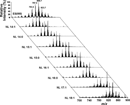
Adipocytes transport fatty acids across their cellular membranes and trap the transported fatty acid through thioesterification to CoA. Typically, the thioesterified fatty acid is used in either de novo synthetic pathways for lipid synthesis or for remodeling through deacylation/reacylation cycling. Alternatively, glucose can be initially metabolized to acetate, which then can be used for the de novo synthesis of fatty acids through sequential chain elongation reactions. Through this mechanism, the resultant pool of triacylglycerols serves as a metabolic reservoir for cellular and organismal energy during times of caloric deprivation. The hormone-induced differentiation of NIH-3T3 preadipocytes to adipocytes has served as a model of adipocyte differentiation for decades, since the resultant adipocytes cannot be distinguished from existing adipocytes after being grafted into athymic mice. Typically, since de novo fatty acid synthesis results in even-chain fatty acids and β-oxidation results in removal of two carbon atoms in each cycle, only even-chain-length fatty acids are typically present in cells. Since plants contain some branched-chain fatty acids, α-oxidation of branched-chain fatty acids is required to render the fatty acid susceptible to β-oxidation distal to the branched chain. Previously, it had been considered a dogma that only branched-chain fatty acids are α-oxidized and that straight-chain fatty acids do not serve as substrates for fatty acid α-oxidation. To determine the temporal sequence of alterations in the lipidome during hormone-induced differentiation of adipocytes, we used shotgun lipidomics with multidimensional MS. The advantage of this technique is that a systems biology approach to examine alterations in adipocyte lipid metabolism can be used without any preconceived notions of changes that occur or pathways that are employed. Shotgun lipidomics examination of differentiating 3T3-L1 cells identified the unanticipated accumulation of large amounts of odd-chain-length fatty acids in many different lipid classes, including triacylglycerols (23). For example, examination of triacylglycerols from differentiated adipocytes by multidimensional MS demonstrated a series of peaks that differed by 14 atomic mass units in the full MS scan of triacylglycerols (x-axis for the top spectrum in Fig. 4). Tandem MS through neutral loss scanning identified large amounts of odd-chain-length fatty acids (e.g., 15:0, 15:1, 17:0, and 17:1) that accumulate during the differentiation of 3T3-L1 cells (y-axis for the spectrum in Fig. 4). Independent analysis by multiple techniques confirmed the presence of dramatic increases in α-oxidation in differentiating adipocytes. This pathway is of interest from multiple perspectives. First, it serves as a potential metabolic pathway to liberate the Gibbs free energy present in carbon-carbon bonds as heat since α-oxidation does not result in the production of high-energy compounds, such as NADH, that are synthesized during β-oxidation. Second, the increase in α-oxidation with resultant increases in odd-chain-length fatty acids and their metabolic intermediates can potentially serve as regulators of cellular metabolic responses. Third, the presence of odd-chain-length fatty acids can potentially serve as a biomarker signaling the degree of peroxisomal proliferation and lipid overload present in adipocytes. Previously, α-oxidation was thought to be used only to facilitate the removal of branched-chain fatty acids. Through the use of shotgun lipidomics, which embraces a systems biology approach, new insights into neutral lipid metabolism in differentiating adipocytes have been realized. The importance of these alterations in adipocyte biology is an attractive area of future investigation.
Fig. 4.
Positive ion electrospray MS and tandem mass spectra of TAGs from hormone-induced differentiation of 3T3-L1 cells (adipocytes). Lipids from day 8 3T3-L1 cells were extracted by a modified Bligh-Dyer technique utilizing 50 mM LiCl in the aqueous layer and were diluted in 1:1 methanol-chloroform with addition of LiOH (50 nmol/mg protein) just prior to direct infusion into the ESI source using a syringe pump at a flow rate of 2 μl/min. Positive ion ESI tandem mass spectra by NL scanning of 226.2 (14:1), 228.2 (14:0), 240.2 (15:1), 242.2 (15:0), 254.2 (16:1), 256.2 (16:0), 268.2 (17:1), and 282.2 (18:1) atomic mass units were acquired through simultaneous scanning of the 1st and 3rd quadrupoles. All NL scanning mass spectra were displayed after normalization to the most intense peak in the individual spectrum. The results are remarkable for the presence of odd-chain-length fatty acids in TAGs after differentiation of preadipocytes to adipocytes by insulin, dexamethasone, and isobutylmethylxanthine.
Conclusion
Through exploiting the high information content inherent in shotgun lipidomics, this technology has greatly facilitated advances in our understanding of neutral lipid metabolism in several disease states. The power of shotgun lipidomics is anticipated to increase as continued refinements in technology result in increases in sensitivity, mass accuracy, and integrated array analyses of the metabolic networks involved in neutral lipid metabolism. Furthermore, it seems likely that the increased use of stable isotope methods in both intact cells and organs will continue to provide rapid improvements in the understanding of the alterations in neutral lipid metabolic networks that participate in cellular bioenergetics, membrane synthesis, and cellular signaling in health and disease.
GRANTS
This work was supported by National Heart, Lung, and Blood Institute Grants P01-HL-57278 and HL-41250.
REFERENCES
- 1.Callender HL, Forrester JS, Ivanova P, Preininger A, Milne S, Brown HA. Quantification of diacylglycerol species from cellular extracts by electrospray ionization mass spectrometry using a linear regression algorithm. Anal Chem 79: 263–272, 2007. [DOI] [PubMed] [Google Scholar]
- 2.Cheng H, Guan S, Han X. Abundance of triacylglycerols in ganglia and their depletion in diabetic mice: implications for the role of altered triacylglycerols in diabetic neuropathy. J Neurochem 97: 1288–1300, 2006. [DOI] [PMC free article] [PubMed] [Google Scholar]
- 3.Cheng H, Jiang X, Han X. Alterations in lipid homeostasis of mouse dorsal root ganglia induced by apolipoprotein E deficiency: a shotgun lipidomics study. J Neurochem 101: 57–76, 2007. [DOI] [PMC free article] [PubMed] [Google Scholar]
- 4.Christie WW Lipid Analysis: Isolation, Separation, Identification and Structural Analysis Of Lipids. Bridgwater, England: The Oily Press, 2003.
- 5.Duffin K, Obukowicz M, Raz A, Shieh JJ. Electrospray/tandem mass spectrometry for quantitative analysis of lipid remodeling in essential fatty acid deficient mice. Anal Biochem 279: 179–188, 2000. [DOI] [PubMed] [Google Scholar]
- 6.Han X, Abendschein DR, Kelley JG, Gross RW. Diabetes-induced changes in specific lipid molecular species in rat myocardium. Biochem J 352: 79–89, 2000. [PMC free article] [PubMed] [Google Scholar]
- 7.Han X, Gross RW. Global analyses of cellular lipidomes directly from crude extracts of biological samples by ESI mass spectrometry: a bridge to lipidomics. J Lipid Res 44: 1071–1079, 2003. [DOI] [PubMed] [Google Scholar]
- 8.Han X, Gross RW. Quantitative analysis and molecular species fingerprinting of triacylglyceride molecular species directly from lipid extracts of biological samples by electrospray ionization tandem mass spectrometry. Anal Biochem 295: 88–100, 2001. [DOI] [PubMed] [Google Scholar]
- 9.Han X, Gross RW. Shotgun lipidomics: electrospray ionization mass spectrometric analysis and quantitation of the cellular lipidomes directly from crude extracts of biological samples. Mass Spectrom Rev 24: 367–412, 2005. [DOI] [PubMed] [Google Scholar]
- 10.Han X, Gross RW. Shotgun lipidomics: multidimensional MS analysis of cellular lipidomes. Expert Rev Proteomics 2: 253–264, 2005. [DOI] [PubMed] [Google Scholar]
- 11.Han X, Yang J, Cheng H, Yang K, Abendschein DR, Gross RW. Shotgun lipidomics identifies cardiolipin depletion in diabetic myocardium linking altered substrate utilization with mitochondrial dysfunction. Biochemistry 44: 16684–16694, 2005. [DOI] [PubMed] [Google Scholar]
- 12.Han X, Yang J, Cheng H, Ye H, Gross RW. Towards fingerprinting cellular lipidomes directly from biological samples by two-dimensional electrospray ionization mass spectrometry. Anal Biochem 330: 317–331, 2004. [DOI] [PubMed] [Google Scholar]
- 13.Han X, Yang K, Yang J, Fikes KN, Cheng H, Gross RW. Factors influencing the electrospray intrasource separation and selective ionization of glycerophospholipids. J Am Soc Mass Spectrom 17: 264–274, 2006. [DOI] [PubMed] [Google Scholar]
- 14.Jiang X, Ory DS, Han X. Characterization of oxysterols by electrospray ionization tandem mass spectrometry after one-step derivatization with dimethylglycine. Rapid Commun Mass Spectrom 21: 141–152, 2007. [DOI] [PMC free article] [PubMed] [Google Scholar]
- 15.Lagarde M, Géloën A, Record M, Vance D, Spener F. Lipidomics is emerging. Biochim Biophys Acta 1634: 61, 2003. [DOI] [PubMed] [Google Scholar]
- 16.Li YL, Su X, Stahl PD, Gross ML. Quantification of diacylglycerol molecular species in biological samples by electrospray ionization mass spectrometry after one-step derivatization. Anal Chem 79: 1569–1574, 2007. [DOI] [PMC free article] [PubMed] [Google Scholar]
- 17.Miranda PJ, DeFronzo RA, Califf RM, Guyton JR. Metabolic syndrome: definition, pathophysiology, and mechanisms. Am Heart J 149: 33–45, 2005. [DOI] [PubMed] [Google Scholar]
- 18.Moller DE, Kaufman KD. Metabolic syndrome: a clinical and molecular perspective. Annu Rev Med 56: 45–62, 2005. [DOI] [PubMed] [Google Scholar]
- 19.Poornima IG, Parikh P, Shannon RP. Diabetic cardiomyopathy: the search for a unifying hypothesis. Circ Res 98: 596–605, 2006. [DOI] [PubMed] [Google Scholar]
- 20.Sandhoff R, Brugger B, Jeckel D, Lehmann WD, Wieland FT. Determination of cholesterol at the low picomole level by nano-electrospray ionization tandem mass spectrometry. J Lipid Res 40: 126–132, 1999. [PubMed] [Google Scholar]
- 21.Schwudke D, Liebisch G, Herzog R, Schmitz G, Shevchenko A. Shotgun lipidomics by tandem mass spectrometry under data-dependent acquisition control. Methods Enzymol 433: 175–191, 2007. [DOI] [PubMed] [Google Scholar]
- 22.Su X, Han X, Mancuso DJ, Abendschein DR, Gross RW. Accumulation of long-chain acylcarnitine and 3-hydroxy acylcarnitine molecular species in diabetic myocardium: identification of alterations in mitochondrial fatty acid processing in diabetic myocardium by shotgun lipidomics. Biochemistry 44: 5234–5245, 2005. [DOI] [PubMed] [Google Scholar]
- 23.Su X, Han X, Yang J, Mancuso DJ, Chen J, Bickel PE, Gross RW. Sequential ordered fatty acid alpha oxidation and Delta9 desaturation are major determinants of lipid storage and utilization in differentiating adipocytes. Biochemistry 43: 5033–5044, 2004. [DOI] [PubMed] [Google Scholar]
- 24.Unger RH Diabetic hyperglycemia: link to impaired glucose transport in pancreatic beta cells. Science 251: 1200–1205, 1991. [DOI] [PubMed] [Google Scholar]
- 25.Unger RH Lipotoxic diseases. Annu Rev Med 53: 319–336, 2002. [DOI] [PubMed] [Google Scholar]
- 26.Unger RH, Orci L. Lipotoxic diseases of nonadipose tissues in obesity. Int J Obes 24: S28–S32, 2000. [DOI] [PubMed] [Google Scholar]
- 27.Wang J, Song Y, Wang Q, Kralik PM, Epstein PN. Causes and characteristics of diabetic cardiomyopathy. Rev Diabet Stud 3: 108–117, 2006. [DOI] [PMC free article] [PubMed] [Google Scholar]


