Medulloblastoma is a common tumour of the posterior fossa, representing 20%–25% of all pediatric neoplasms.1,2 The tumour often occurs in the cerebellar vermis and at the apex of the fourth ventricle.2,3 It commonly presents with rapidly progressive clinical manifestations of raised intracranial pressure and cerebellar dysfunction.1,3 There are only a few reported cases of cerebellopontine (CP)-angle medulloblastoma in the literature, with most being intra-axial and occurring in the pediatric population. We present a rare case of primary extra-axial CP-angle medulloblastoma occurring in an adult.
Case report
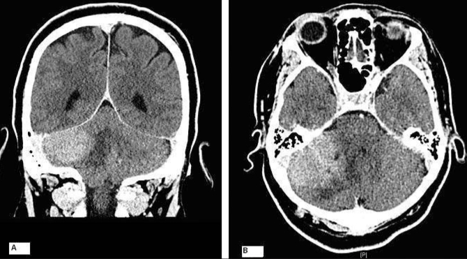
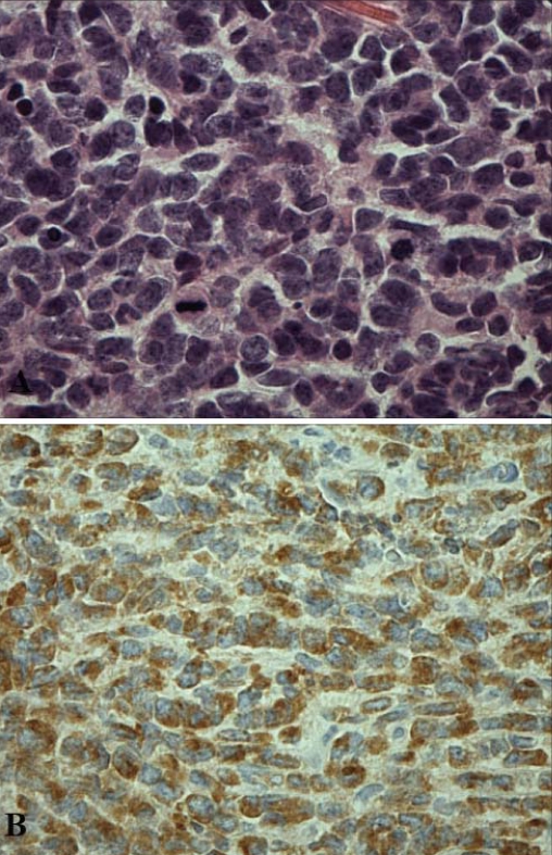
A 47-year old-man presented with a recent history of headaches, nausea and vomiting. There was no associated neurologic deficit. A computed tomography (CT) scan depicted a large well-demarcated, extra-axial lesion adjacent to the right tentorium in the posterior fossa (Fig. 1). Given its shape, location and homogenous pattern of contrast uptake, the lesion was most consistent with a benign lesion such as a CP-angle meningioma. We performed a right suboccipital, infratentorial craniotomy for tumour resection. The tumour was visible extra-axially, extended medially into the CP angle and was attached to the tentorium superiorly. Histopathology results showed small, highly cellular neoplastic cells with round hyperchromatic nuclei, scanty cytoplasm and indistinct cellular borders visible with hematoxylin and eosin staining. We identified no Homer–Wright rosettes. Immunohistochemical staining demonstrated diffuse positivity for synaptophysin and neuron-specific enolase (Fig. 2). These features were consistent with medulloblastoma, although the tumour was completely extra-axial and involved no cerebellar tissue. Adjuvant therapy included postoperative radiation.
Fig. 1.
(A) Coronal computed tomography (CT) scan with contrast showing an extra-axial enhancing mass lesion involving the right cerebellopontine angle overlying the cerebellar hemisphere and with mild mass effect. (B) Axial CT scan with contrast demonstrating a well-circumscribed right infratentorial homogenously enhancing well-defined lesion adjacent to the dura.
Fig. 2.
(A) Highly cellular neoplastic cells with round hyperchromatic nuclei, scanty cytoplasm and indistinct cellular borders (hematoxylin and eosin stain, original magnification ×400). (B) Immunohistochemical stains demonstrated diffuse positivity for synaptophysin (original magnification ×200).
Discussion
Medulloblastoma is a predominantly pediatric tumour commonly occurring in the cerebellar vermis. The lack of association with any cerebellar tissue and the extra-axial location of the tumour made our patient’s case quite rare. Apart from the presentation of the tumour in an adult, other factors that contributed to our high index of suspicion for meningioma included the homogenous contrast uptake, which demonstrated a well-demarcated lesion. Common CP-angle lesions usually include acoustic neuromas, followed by meningiomas, primary cholesteatomas and epidermoid tumours.4,5 Collectively, these account for up to 98% of tumours in this region.4,5
There have been only 8 reported cases of extra-axial medulloblastoma in the adult literature.4,6,7 However, they are likely under-reported owing to publication bias. This tumour is nearly twice as common in men. Patient age at diagnosis ranges from 21 to 52 years, with tumours most often occurring among patients in their late 20s and early 30s.4,6,7 Extra-axially, the tumours have been localized in 2 areas: the tentorial region and the CP angle.4,6,7 Most commonly, they manifest as heterogeneously enhancing lesions upon contrast administration.4,6,7 However, in at least 2 patients, including ours, the tumours have demonstrated a homogenous enhancement pattern, which can lead to misdiagnosis.7
Conclusion
Medulloblastoma is predominantly a childhood tumour that almost always presents intra-axially. However, it is important to consider the rare possibility of medulloblastoma presenting extra-axially. Its occurrence in the CP angle can confuse the nature of the lesion and affect the course of treatment.
Footnotes
Competing interests: None declared.
References
- 1.Albright L, editor. Medulloblastoma. New York (NY): Thieme Medical Publishers, Inc; 1999. [Google Scholar]
- 2.Russell D, Rubinstein L, editors. Pathology of tumours of the nervous system. Baltimore (MD): Williams & Wilkins; 1994. [Google Scholar]
- 3.Greenberg MS. Handbook of neurosurgery. 5th ed. New York (NY): Thieme Medical Publishers, Inc; 2001. [Google Scholar]
- 4.Barkovich AJ. Pediatric neuroimaging. 3rd ed. Philadelphia (PA): Lippincott Williams & Wilkins; 2000. [Google Scholar]
- 5.Moffat DA, Saunders JE, McElveen JT, et al. Unusual cerebellopontine angle tumours. J Laryngol Otol. 1993;107:1087–98. doi: 10.1017/s0022215100125393. [DOI] [PubMed] [Google Scholar]
- 6.Becker RL, Becker AD, Sobel DF. Adult medulloblastoma: review of 13 cases with emphasis on MRI. Neuroradiology. 1995;37:104–8. doi: 10.1007/BF00588622. [DOI] [PubMed] [Google Scholar]
- 7.Gil-Salu JL, Rodriguez-Pena F, Lopez-Escobar M, et al. Medulloblastoma presenting as an extra-axial tumour in the cerebellopontine angle. Neurocirugia (Astur) 2004;15:285–9. doi: 10.1016/s1130-1473(04)70485-6. [DOI] [PubMed] [Google Scholar]


