Abstract
This unit includes protocols for the laboratory maintenance of the obligate intracellular bacterium Rickettsia rickettsii, including propagation in mammalian cell cultures, as well as isolation, counting, and storage procedures. Regulations for working with R. rickettsii in biosafety level 3 containment are also discussed.
Keywords: rickettsia, Rocky Mountain spotted fever, Vero cells, cell culture techniques
INTRODUCTION
Rickettsia rickettsii, the causative agent of Rocky Mountain spotted fever, is responsible for one of the most virulent human infections in the Western Hemisphere (United States, southern Canada, Central America, Mexico, and parts of South America). However, the name Rocky Mountain spotted fever is a misnomer, and even within the United States, RMSF is broadly distributed and not confined to the Rocky Mountain region. Rickettsia rickettsii is a true zoonotic bacterium that cycles between ixodid ticks and wildlife populations not only in the US but also in Mexico and in Central and South America. Human infection occurs via the bite of an infected tick, and the initial signs and symptoms of the disease include fever, headache and muscle pain followed by rash and organ-specific symptoms such as nausea, vomiting and abdominal pain. Delayed treatment can result in severe disease, hospitalization and sequelae, such as deafness, permanent learning impairment, amputation and death. The disease can be difficult to diagnose in the early stages due to nonspecific presentations, and unfortunately it can be fatal in the absence of prompt and appropriate treatment, with a case fatality rate of 5–10% (Dumler and Walker 2005).
Rickettsia rickettsii is a biosafety level 3 (BSL-3) pathogen as well as a select agent and thus requires compliance with the CDC security requirements of the Select Agent Program (http://www.cdc.gov/od/sap). R. rickettsii has also been classified by several international organizations as a biological agent cited as a possible weapon for use against humans (see the World Health Organization document “Public health response to biological and chemical weapons: WHO guidance” for more information; http://www.who.int/csr/delibepidemics/biochemguide/.) Additionally, considering its potential for laboratory-acquired infections, high standard microbiological techniques should be followed while working with R. rickettsii. Therefore, all the procedures using live rickettsiae should be carried out in a class II biological safety cabinet within a select agent-approved BSL-3 laboratory.
The protocols we have outlined in this unit detail experimental means for the laboratory maintenance of Rickettsia rickettsii.
CAUTION: Rickettsia rickettsii is a Biosafety Level 3 (BSL-3) pathogen. Follow all appropriate guidelines for the use and handling of pathogenic microorganisms. See Strategic Planning for further discussion. See UNIT 1A.1 and other pertinent resources (APPENDIX 1B) for additional information.
IMPORTANT NOTE: Rickettsia rickettsii has been classified as a Select Agent by the United States Government. Refer to the CDC Select Agent Program for more information (http://www.cdc.gov/od/sap). Also refer to UNIT 1A.1 and other pertinent resources (APPENDIX 1B).
STRATEGIC PLANNING
Standards of BSL-3 containment should be followed for work with R. rickettsii. Investigators should consult with all local and national regulations, and consult local biosafety officer and/or institutional biosafety committee for additional guidance. A detailed standard operating procedure (SOP) must be documented for all experimental and decontamination procedures performed in the BSL-3 laboratory. In addition, in the United States, careful records must be maintained for all work done with a select agent; consult CDC Select Agent Program for more information (http://www.cdc.gov/od/sap).
Laboratory maintenance of rickettsiae requires careful planning. Not only are the bacteria slow to grow and difficult to isolate (see Commentary, Time Considerations), but all work must be done in a BSL-3 facility. Therefore, it is critical to plan ahead to ensure that all necessary equipment and solutions are available in the BSL-3 laboratory. Most of the materials required for the protocols in this unit must be sterile, and consideration must be given for the sterilization of all necessary labware and solutions prior to their use.
BASIC PROTOCOL 1
PROPAGATION OF RICKETTSIA RICKETTSII IN CELL CULTURE FROM FROZEN BACTERIAL STOCKS
Rickettsiae can infect and grow in a variety of different cell types, including lines derived from human fibroblasts, endothelial and epithelial cells (HeLa), other mammals (Vero, L929), amphibians (XTC-2), avian hosts (chick embryo fibroblasts) and arthropods (tick-derived DAE100, mosquito-derived Aa23 and C6/36 cell lines). The protocol described below is for the propagation of Rickettsia rickettsii in Vero cells, which are commonly used as host cells to propagate rickettsiae. However, the techniques described in this section are generally applicable to any cell type, with some specific considerations described in ALTERNATE PROTOCOL 1.
Materials
R. rickettsii-infected Vero cells or purified rickettsial seed, frozen at −80°C
Dulbecco’s modification of Eagle medium (DMEM), supplemented with 5% heat-inactivated fetal bovine serum (FBS) and 2mM L-glutamine, filter-sterilized (see recipe)
Vero cells, 80–90% confluent monolayer in a 150cm2 flask (see APPENDIX 4E)
37°C shaking incubator (such as the Labnet 211DS)
34°C incubator with 5% CO2
Serological pipets, sterile
15mL conical tubes, sterile
NOTE: All equipment and solutions coming into contact with cells must be sterile, and proper sterile technique should be used. The use of antibiotics in the cell culture medium is not recommended when growing rickettsiae. All work should be done in a class II biological safety (biosafety) cabinet within a BSL-3 laboratory.
NOTE: All solutions should be warmed to room temperature or 37oC before contacting cells.
-
Thaw R. rickettsii-infected Vero cells (in cryovial) on ice, or quickly thaw in a 32°C water bath.
This infection protocol can also be used with purified (i.e., cell-free) frozen rickettsial stocks.
-
Dilute rickettsial stock in DMEM with 5% FBS and 2mM L-glutamine for a total volume of 10mL. Prepare dilution in a sterile 15mL conical tube.
For optimal infection, use the equivalent of 1:10 or 1:20 dilution of infected cells (relative to the original flask of infected cells used to generate the stocks). If the rickettsial concentration is known, dilutions can be made to achieve a specific multiplicity of infection (MOI); however, keep the total volume of the dilution at 10mL. If the stock is of a completely unknown amount (neither ratio nor concentration known), it is advised to set up a series of dilutions to avoid an overwhelming MOI.
The addition of L-glutamine to the diluting media has been shown to increase survival of the thawed rickettsiae (Bovarnick et al. 1950).
Bring the flasks of Vero cell monolayers to be infected into the biosafety cabinet.
Remove and discard media from Vero cells.
Add the 10mL of the rickettsial stock dilution to the Vero monolayer.
-
Incubate, gently shaking, at 37°C, allowing the rickettsiae to contact the cells for 1–4 hours.
The rickettsiae are initially added to the cells in a relatively small volume of media and gently rocked over the cells to maximize the contact of the rickettsiae with the host cells. For the Labnet 211DS shaking incubator, use a shaker speed <200rpm. If a shaking incubator is not available, incubate the cells at 37°C or at room temperature for 1–4 hours without shaking.
-
After incubation, remove the media from the Vero cells and replace it with 35–50mL of fresh DMEM with 5% FBS and 2mM L-glutamine; incubate cells at 34°C.
The infected cells are maintained in DMEM with 5% FBS and at 34°C to prevent the Vero cells from overgrowing before optimal infection is obtained.
-
Check infected cells every 1–2 days and change media every 3–4 days, until the desired infection level has been reached. (See BASIC PROTOCOL 3 for a method to monitor infection status.) At optimal/desired infection, the rickettsiae must be passaged (BASIC PROTOCOL 2), purified (BASIC PROTOCOL 4), or frozen (BASIC PROTCOL 7).
If the media turns bright yellow (indicating a decrease in pH) or the Vero cells start to detach from the flask, the infection has become overwhelming for the host cells, and the rickettsiae must be passaged, purified, or frozen immediately.
BASIC PROTOCOL 2
PROPAGATION OF RICKETTSIA RICKETTSII INFECTED VERO CELLS
Existing R. rickettsii/Vero cell infections can be continually passaged into new flasks of Vero cells to maintain an ongoing source of live, actively-growing rickettsiae.
Materials
R. rickettsii-infected Vero cells in a 150cm2 flask, maintained at 34°C with 5% CO2
DMEM, supplemented with 5% heat-inactivated FBS, filter sterilized (see recipe)
Vero cells (uninfected), 80–90% confluent in a 150cm2 flasks (see APPENDIX 4E)
Cell scraper, sterile
Serological pipets, sterile
50mL conical tubes, sterile
CAUTION: All work should be done in a class II biosafety cabinet within a BSL-3 laboratory.
NOTE: All equipment and solutions coming into contact with cells must be sterile, and proper sterile technique should be used. The use of antibiotics in the cell culture medium is not recommended when growing rickettsiae.
NOTE: All solutions should be warmed to room temperature or 37oC before contacting cells.
In the biosafety cabinet, remove media from infected Vero cells.
Gently scrape down cells with sterile cell scraper.
-
Wash down cells with 10–20mL fresh DMEM with 5% FBS.
If most of the Vero cells are detaching from the flask (indicating an overgrown rickettsial infection), wash down cells in the existing media in the flask. Divide the cell/media suspension into 50mL high-speed tubes and centrifuge at 17,000 × g for 10 minutes at 4°C. Discard the supernatant and resuspend pellets in a total volume of 10 or 20mL media.
Bring the 150cm2 flasks of Vero cell monolayers to be infected into the biosafety cabinet.
Remove and discard media from uninfected Vero cells.
-
Combine 1–2mL of resuspended R. rickettsii-infected Vero cells with 40–50mL fresh DMEM with 5% FBS in a sterile 50mL conical tube, and then add the suspension to the uninfected Vero cells.
Any remaining R. rickettsii-infected cells can be used to make frozen stocks (See BASIC PROTOCOL 7).
-
Place flask in 34°C incubator with 5% CO2.
Check infected cells every 1–2 days and change media every 3–4 days, until the desired infection level has been reached. (See BASIC PROTOCOL 3 for methods to monitor infection status.) At optimal infection, the rickettsiae must be passaged (repeat this protocol), purified (BASIC PROTOCOL 4), or frozen (BASIC PROTOCOL 7).
ALTERNATE PROTOCOL 1
PROPAGATION OF RICKETTSIA RICKETTSII IN OTHER CELL TYPES
The protocols detailed in this unit describe the growth of R. rickettsii in Vero cells. However, the rickettsiae can grow in a variety of different cell types and lines. If the use of other cell lines is desired, the rickettsiae will need to be partially purified to generate a cell-free suspension which can then be used to inoculate a different cell type (see BASIC PROTOCOL 4). The Basic Protocols for the propagation of R. rickettsii from frozen stock and from active culture are applicable to other cell lines, with media and temperature/atmospheric requirements as determined by the specific cells in use.
BASIC PROTOCOL 3
MONITORING RICKETTSIA RICKETTSII INFECTION IN VERO CELLS USING THE GIMÉNEZ STAIN
The rickettsiae are classified as Gram-negative bacteria, but they stain very weakly with the traditional Gram stain. The Giménez stain was originally described as a method of staining rickettsiae in yolk sac smears for visualization with a light microscope (Giménez 1964). Today, this technique is commonly used to stain rickettsiae grown in cell cultures as a method to monitor the infection status of cells
Materials
R. rickettsii-infected Vero cells in 150cm2 flask, maintained at 34°C with 5% CO2
DMEM supplemented with 5% heat-inactivated FBS, filter sterilized (see recipe)
Dulbecco’s phosphate buffered saline (DPBS) without calcium or magnesium, filter sterilized (see APPENDIX 2A)
Methanol, ice-cold
Carbol basic fuchsin stock, warmed to 37°C (see recipe)
0.1M sodium phosphate buffer (see APPENDIX 2A)
0.8% aqueous malachite green oxalate (see recipe)
Cytocentrifuge sample chambers (such as Wescor Cytopro sample chambers)
Glass microscope slides
Cytocentrifuge, such as the Wescor Cytopro cytocentrifuge
Cell scrapers, sterile
Serological pipets, sterile
NOTE: All equipment and solutions coming into contact with cells must be sterile, and proper sterile technique should be used. All work with live rickettsiae should be done in a class II biosafety cabinet within a BSL-3 laboratory.
Prepare slides of infected cells
1. In biosafety cabinet, remove and discard media from rickettsiae-infected Vero cells.
2. Gently wash cells with 10mL DPBS.
3. Using a sterile cell scraper, gently scrape a small patch of cells (3–5mm2 area).
-
4. Carefully resuspend scraped cells in 200μL DPBS and transfer into a cytocentrifuge sample chamber.
If a cytocentifuge is not available, a few drops of the cell suspension can be placed on a glass slide and allowed to air dry in the biosafety cabinet.
-
5. Place cup and glass slide into cytocentrifuge according to manufacturer’s instructions; if using a Wescor Cytopro cytocentrifuge, spin at 2,000 rpm for 3 minutes.
The cytocentrifuge sample chamber contains an absorbent pad at the cup/slide interface. As the rotor spins, the cell suspension will be forced from the cup towards the slide. The cells will sediment on the glass slide, while the pad will absorb most of the suspension medium.
6. Carefully remove the sample chambers from the slides and discard. Remove the slides from the cytocentrifuge and allow to air dry in the biosafety cabinet.
7. When samples are completely dry, fix in ice-cold methanol for 20 minutes.
-
8. Let slides air dry on an absorbent pad or several paper towels.
Fixation kills the rickettsiae; therefore, the slides with fixed samples can be safely taken out of the BSL-3 facility, and the remaining staining steps can be completed in a BSL-2 laboratory.
Stain cells
-
9. Prepare the working solution of carbol basic fuchsin by mixing 4mL of carbol basic fuchsin stock solution (warmed to 37°C) with 10mL 0.1M sodium phosphate buffer, pH 7.45; filter-sterilize the mixture.
If the slides are to be dipped into the dye, then a larger volume of the working stock can be prepared, as long as the 2:5 ratio of carbol basic fuchsin stock:sodium phosphate buffer is maintained.
The carbol basic fuchsin working stock should be prepared immediately before the staining procedure. Once prepared, the working solution can be used for up to 48 hours.
-
10. Cover the fixed R. rickettsii/Vero cell samples on glass slides with the filtered carbol basic fuchsin working stock and let stand 5 minutes.
Because a basic fuchsin stain is used, the color portion of the dye resides in the positive ion of the salt. Therefore, the fuchsin directly binds the negatively charged bacterial cytoplasm, thus staining the bacteria bright red/purple.
-
11. Wash the slides thoroughly in tap water.
Rinse slides until only clear water runs off of them.
Counterstain cells
-
12. Cover the R. rickettsii/Vero cell samples with 0.8% aqueous malachite green oxalate for 30 seconds.
Malachite green serves as a counterstain which colors the host cells and background greenish-blue. Malachite green is also a basic dye, but with higher affinity for negatively charged host cell materials than for the bacterial cytoplasm.
-
13. Wash the slides thoroughly in tap water.
14. Rinse slides until only clear water runs off of them.
15. Let slides air dry on absorbent pad or several paper towels.
-
Visualize samples using a light microscope.
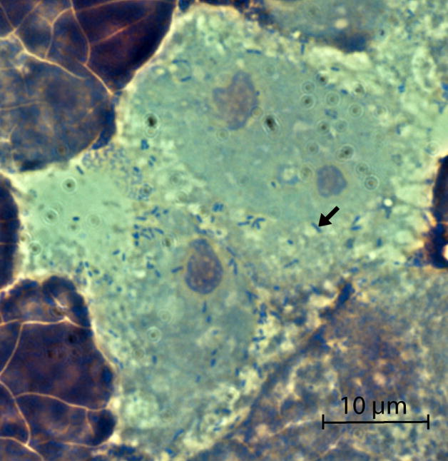
Observe the samples under oil immersion. The rickettsiae (approximately 0.2–0.5 μm by 0.3–2.0 μm in size) will appear bright red/purple against a light blue/green background (Figure 1). These preparations can allow for an estimation of the percentage of cells infected and the rickettsial burden per infected cell; however, if the cells are heavily infected, many cells may lyse during the scraping and centrifugation processes, and free rickettsiae will be visible on the cytospin slides.
Figure 1.
Giménez staining of rickettsiae-infected Vero 76 cells. The black arrow points to an individual bacterium.
ALTERNATE PROTOCOL 2
MONITORING RICKETTSIA RICKETTSII INFECTION IN VERO CELLS USING THE GIEMSA STAIN
Although the Giménez stain (BASIC PROTOCOL 3) is considered the best rickettsial stain for light microscopy work, it is time-consuming to prepare fresh stain mixture every time the status of rickettsiae-infected cells needs to be checked. An acceptable alternative to the Giménez stain is the Giemsa stain, which was first described for the staining of blood parasites in 1904 (Giemsa 1904). This stain is a mixture of methylene blue and eosin and binds to phosphate groups of DNA where there are high levels of adenine-thymine (A-T) bonding. Because rickettsial DNA is very A-T rich (67.53% A-T for the R. rickettsii strain Sheila Smith genome (Ellison et al. 2008)), the Giemsa stain can be used to distinguish rickettsiae from host cell material. The rickettsiae will stain dark blue in contrast to the light pink staining of surrounding host cell material. Stock solutions for the Giemsa stain are widely commercially available, making this stain a quick and easy alternative to the Giménez stain. Modifications of the classic Giemsa stain have also been used for the staining of rickettsiae-infected cells. Specifically, the Diff-Quik Stain Set manufactured by Dade Behring is often used in rickettsiology laboratories. Rickettsiae-infected Vero 76 cells stained with the Diff-Quik stain are shown in Figure 2.
Figure 2.
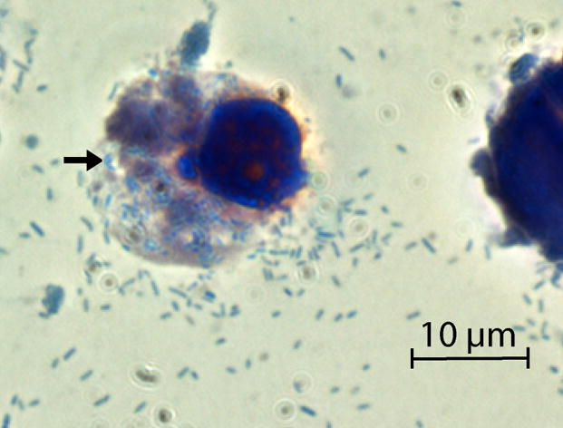
Diff-Quik staining of rickettsiae-infected Vero 76 cells. The black arrow points to an individual bacterium.
BASIC PROTOCOL 4
PARTIAL PURIFICATION OF RICKETTSIA RICKETTSII FROM VERO CELLS BY SONICATION
Several applications that require cell-free rickettsiae, such as transferring in vitro infections between cell types, mammalian and arthropod infections, and rickettsial nucleic acid purification, are not adversely affected by the presence of limited host cell contamination. When highly purified rickettsiae are not required, a partial purification may be a good option. Partial purification of rickettsiae is the first step for further purification by density gradient centrifugation (See BASIC PROTOCOL 5). In addition, the partial purification procedure is ideal for applications requiring freshly isolated rickettsiae, with as little time from cell culture to experiment as possible. This protocol describes the partial purification for R. rickettsii from Vero cells, but can be applied to any rickettsial species grown in any cell line.
Materials
R. rickettsii-infected Vero cells in 150cm2 flask, maintained at 34°C with 5% CO2
DMEM supplemented with 5% heat-inactivated FBS, filter-sterilized (see recipe)
Sucrose-phosphate-glutamate (SPG; see recipe)
Sonicator with hand-held probe, such as the Fisher Scientific Sonic Dismembrator Model 100 (must fit inside biosafety cabinet)
Cell scraper, sterile
15mL and 50mL high-speed conical tubes, sterile
Serological pipets, sterile
1.5mL microcentrifuge tubes, sterile
0.22μm syringe-driven membrane filter and 10mL syringe, sterile (optional)
CAUTION: The sonication process creates aerosols, so all sonication must be done inside a class II biosafety cabinet within a BSL-3 facility.
NOTE: All equipment and solutions coming into contact with cells must be sterile prior to use, and proper sterile technique should be used. All work should be done in a class II biosafety cabinet within a BSL-3 laboratory.
Lyse host cells
1. In biosafety cabinet, remove media from the infected Vero cells.
2. Gently scrape down cells using a sterile cell scraper.
-
3. Wash down cells and resuspend in 10mL fresh DMEM with 5% FBS.
If most of the Vero cells are detaching from the flask (indicating an overgrown rickettsial infection), wash down cells in existing media in the flask. Divide the cell/media suspension into 50mL high-speed conical tubes and centrifuge at 17,000 × g for 10 minutes at 4°C. Discard the supernatant and resuspend pellets in a total volume of 10–20mL DMEM supplemented with 10% FBS, 2mM L-glutamine and 2mM sucrose.
4. Transfer the cell suspension into two 50 mL conical tubes, with 5mL of cell suspension into each tube. Keep tubes on ice.
-
5. Sonicate until host cell membranes have been lysed, while rickettsiae remain intact.
The sonication conditions will depend on the type of sonicator used. For the Fisher Scientific Sonic Dismembrator Model 100, two 20-second pulses at setting 6.5 is sufficient to lyse the Vero cells. It may be necessary to try different pulse times and strengths to optimize host cell disruption while the rickettsial cells remain intact. The cells can be monitored by fixing a drop of the sonicated cell suspension to a glass slide, staining (see BASIC PROTOCOL 3), and visualizing under a light microscope.
The sonication process will generate heat, so it is very important to do all sonication with the conical tubes embedded in ice.
Separate rickettsiae from host cell debris
-
6. Spin the sonicated cell suspension at 1,000 × g for 5 minutes at 4°C.
This low-speed spin will pellet any remaining host cells and heavy cellular debris, leaving the small rickettsiae (as well as smaller cellular fragments) suspended in the supernatant.
-
7. Remove supernatant (containing rickettsiae) and transfer to sterile 15mL high-speed conical tubes. Be careful not to disturb the pellet of host-cell debris.
If using a non-swing out bucket centrifuge, the cellular debris will be deposited in a line up and down the side of the tube rather than forming a single pellet.
-
8. If using the purified rickettsiae to start an infection in a different cell line (other than Vero cells) or for density gradient centrifugation (to obtain highly purified rickettsiae), pass the supernatant through a 0.22μm syringe-driven membrane filter, attached to a 10mL syringe.
The membrane filter will remove any remaining host cells and larger host cell debris; the rickettsiae will pass through the filter.
Pellet rickettsiae
-
9. Spin the supernatant at 17,000 × g for 10 minutes at 4°C.
This high-speed spin will pellet the rickettsiae (and any small cellular debris).
-
10. Carefully remove and discard the supernatant; do not dislodge the rickettsial pellet.
Depending on the application (e.g. transferring the R. rickettsii infection from Vero cells into a new cell type), the rickettsiae may be used at this point.
11. Resuspend/wash pellet in 1mL SPG; transfer suspension to 1.5mL tube.
12. Spin the suspension at 17,000 × g for 10 minutes at 4°C.
-
13. Carefully remove and discard the supernatant; keep pellets on ice.
The partially purified rickettsiae can be visualized by fixing a few drops of the bacterial suspension on a glass slide and staining as described in BASIC PROTOCOL 3. After this partial purification process, there will be a lot of visible cell debris, but none (or very few) intact host cells, and a lot of free rickettsiae (see Figure 3).
The next steps and the resuspension solution will depend on the application. For example, if using the rickettsiae for nucleic acid or protein isolation, resuspend the pellet in the appropriate lysis solution (such as Trizol for RNA extraction). If highly purified rickettsiae are required, proceed with density gradient centrifugation (BASIC PROTOCOL 5). The rickettsiae can be maintained on ice in SPG for several hours if necessary. For freezing, resuspend pellets in SPG (see BASIC PROTOCOL 8). The rickettsiae can also be heat-killed (10–20 minutes at 90°C), at which point they can safely be removed from the BSL-3 laboratory.
Figure 3.
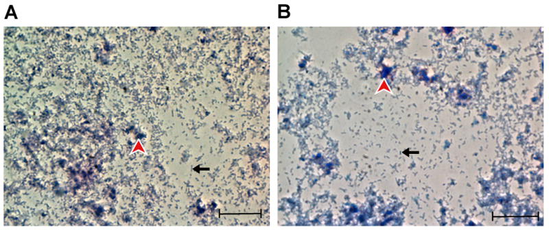
Rickettsiae, partially purified from Vero 76 cells. The black arrow points to an individual bacterium, and the red arrowhead points to cellular debris. (A) Giménez staining. (B) Diff-Quik staining. Scale bar = 10 μm
ALTERNATE PROTOCOL 3
PARTIAL PURIFICATION OF RICKETTSIAE FROM VERO CELLS USING NEEDLE AND SYRINGE
If a sonicator is not available for the disruption of host cells, the rickettsiae can be released using a small-bore (27–30 gauge) needle. This method is not recommended for R. rickettsii or other rickettsiae that are handled in BSL-3 facilities, where the use of needles should be avoided when possible. However, this method is commonly used to free non-pathogenic rickettsiae from host cells in BSL-2 laboratories.
Materials
R. rickettsii-infected Vero cells in 150cm2 flask, maintained at 34°C with 5% CO2
DMEM supplemented with 5% heat-inactivated FBS, filter-sterilized (see recipe)
SPG (see recipe)
27 gauge needles, sterile
5mL or 10mL syringes, sterile
Cell scraper, sterile
15mL and 50mL high-speed conical tubes, sterile
Serological pipets, sterile
1.5mL microcentrifuge tubes, sterile
NOTE: All equipment and solutions coming into contact with cells must be sterile, and proper sterile technique should be used. All work should be done in a class II biological safety (biosafety) cabinet.
In biosafety cabinet, remove spent media from the infected Vero cells.
Gently scrape down cells using a sterile cell scraper.
-
Wash down cells and resuspend in 10mL fresh DMEM with 5% FBS.
If most of the Vero cells are sloughing off of the flask (indicating an overgrown rickettsial infection), wash down cells in existing media in the flask. Divide the cell/media suspension into 50mL high-speed conical tubes and centrifuge at 17,000 × g for 10 minutes at 4°C. Discard the supernatant and resuspend pellets in a total volume of 10–20mL DMEM supplemented with 10% FBS, 2mM L-glutamine and 2mM sucrose.
Transfer the cell suspension into 50mL conical tubes, with 5–10mL of cell suspension into each tube. Keep tubes on ice.
Carefully attach the 27 gauge needle to the syringe and pass the cell suspension through 5 times.
Centrifuge the disrupted cell lysates at 1,000 × g for 5 minutes at 4°C.
Continue from this point as described in BASIC PROTOCOL 4.
BASIC PROTOCOL 5
PURIFICATION OF RICKETTSIA RICKETTSII BY ISOPYCNIC DENSITY GRADIENT CENTRIFUGATION
For those applications where highly purified rickettsiae are required, the bacteria can be separated from host cell materials by isopycnic density gradient centrifugation. Renografin (sodium and meglumine diatrizoate, Bracco Diagnostics) has traditionally been used as the gradient medium to isolate viable, purified rickettsiae. Renografin (also called Urografin) is a non-toxic supporting medium with a high density yet low viscosity, which makes it ideal for the purification of rickettsiae from host cell debris. This protocol describes a discontinuous gradient made with three different Renografin concentrations, used for the purification of viable R. rickettsii from host (Vero) cell material; however, the protocol can be applied to any rickettsial species partially purified from any host cells.
Materials
Partially purified R. rickettsii in SPG, on ice (see BASIC PROTOCOL 4)
32%, 36% and 42% Renografin-60 solutions (see recipe)
SPG, ice-cold (see recipe)
Pasteur pipets, sterile
Microcentrifuge (1.5mL) tubes, sterile
Ultracentrifuge tubes (with at least 11mL capacity), sterile
Swinging-bucket ultracentrifuge rotor (such as Beckman Coulter SW 41 Ti or equivalent)
Ultracentrifuge, inner chamber at 4°C
NOTE: Renografin-60 (Bracco Diagnostics) is a controlled substance. To use Renografin-60 for research purposes, permission must be obtained from the United Stated Department of Justice Drug Enforcement Administration (DEA).
NOTE: All equipment and solutions coming into contact with cells must be sterile prior to use, and proper sterile technique should be used. All work should be done in a class II biosafety cabinet within a BSL-3 laboratory.
Prepare discontinuous Renografin gradient
-
1. In sterile ultracentrifuge tube, carefully add 3mL 42% Renografin to bottom of tube.
If using the Beckman Coulter SW 41 Ti swinging-bucket rotor, the Ultra-Clear tubes with 13.2mL capacity (from Beckman-Coulter) are both suitable for the rotor and are clear enough to easily view the rickettsial bands(post-centrifugation) in the gradient. These tubes cannot be autoclaved and therefore must be cold-sterilized prior to use.
2. Gently add 4mL 36% Renografin on top of the 42% Renografin layer.
3. Gently add 3mL 32% Renografin on top of the 36% layer.
Apply rickettsiae to gradient
-
4. Bring volume of partially purified R. rickettsii up to 1mL in SPG and gently add to the top of the Renografin layers. Add additional SPG to fill the ultracentrifuge tube to within 2–3mm from the top of the tube.
It is very important to fill the ultracentrifuge tubes to within 2–3mm from the top of the tube. If the tubes are not full, they can bend and deform during centrifugation, which can lead to sample leakage. It is also critical that samples are balanced in the ultracentrifuge. For the swinging bucket rotors, the samples must be balanced to within 0.2g.
-
5. Very carefully, place ultracentrifuge tube containing gradient and rickettsiae into an ultracentrifuge swinging bucket rotor (such as the Beckman Coulter SW 41 Ti).
If using the Beckman Coulter SW 41 Ti rotor, the buckets can be removed from the rotor. The buckets should be loaded and covered in the biosafety cabinet.
6. Centrifuge at 90,000 × g at 4°C for 90 minutes.
7. When centrifugation is complete, carefully remove the ultracentrifuge tube and transport it back into the biosafety cabinet.
Collect purified rickettsiae
-
8. Use a sterile Pasteur pipet to collect the band of viable rickettsiae, which will sediment at the 36%–32% Renografin interface, and transfer into a sterile microcentrifuge tube.
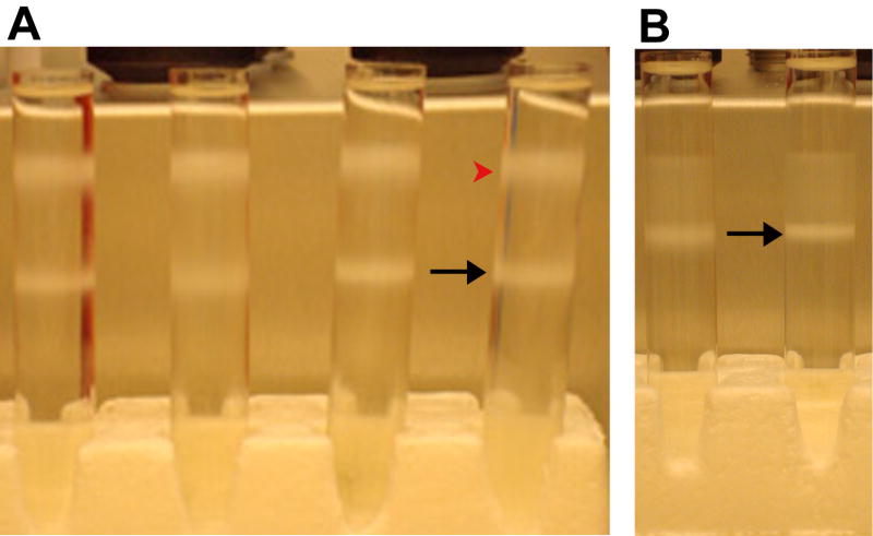
After ultracentrifugation, there will be a band of low density debris that sediments at the interface between the SPG at the top of the gradient and the 32% Renografin layer. The viable, infective rickettsiae, will sediment in a band at an approximate density of 1.20 g/mL, at the interface between the 32% and 36% Renografin layers. There may also be a “heavy” rickettsial band in the 42% Renografin layer (at an approximate density of 1.23 g/mL), which will contain mostly damaged bacteria (see Hanson et al. 1981 and Figure 4A).
-
9. Add ice-cold SPG to the rickettsiae to a final volume of 1mL.
If desired, the purity of the rickettsiae can be assessed by fixing a few drops of the suspension on a glass slide and staining as described in BASIC PROTOCOL 3. Depending on the application, it may be possible to use the rickettsiae at this point.
-
10. Subject the rickettsiae to a second round of Renografin density gradient centrifugation (repeat steps 1–8).
After the second round of centrifugation, the rickettsial band at the 32%–36% Renografin interface should be greatly enriched (see Figure 4B).
-
11. Wash the rickettsiae 1–3 times in ice-cold SPG.
To pellet the rickettsiae after washes, centrifuge at 17,000 × g for 10 minutes. It may be necessary to dilute the Renografin with up to 10mL of SPG in order to generate a pellet during centrifugation.
-
12. Keep the purified rickettsial pellet on ice.
The next steps and the resuspension solution will depend on the application. For example, if using the rickettsiae for nucleic acid or protein isolation, resuspend the pellet in the appropriate lysis solution. The rickettsiae can be maintained on ice in SPG for several hours if necessary. If desired, the purity of the rickettsiae can be assessed by fixing a few drops of the suspension on a glass slide and staining as described in BASIC PROTOCOL 3 (Figure 5). For freezing, resuspend pellets in SPG (see BASIC PROTOCOL 8). The rickettsiae can also be heat-killed (10–20 minutes at 90°C), at which point they can safely be removed from the BSL-3 laboratory.
Figure 4.
Ultracentrifuge tubes with rickettsiae purified from Vero 76 cells by Renografin density gradient ultracentrifugation. The rickettsiae were purified from six 150cm2 flasks of heavily infected cells. (A) Tubes after the first run through the Renografin gradient. The black arrow points to the band of viable rickettsiae at the 32%–36% Renografin interface. The red arrowhead points to the debris at the SPG-32% Renografin interface. (b) Tubes after the second run through the Renografin gradient. The black arrow points to the band of viable rickettsiae at the 32%–36% Renografin interface.
Figure 5.
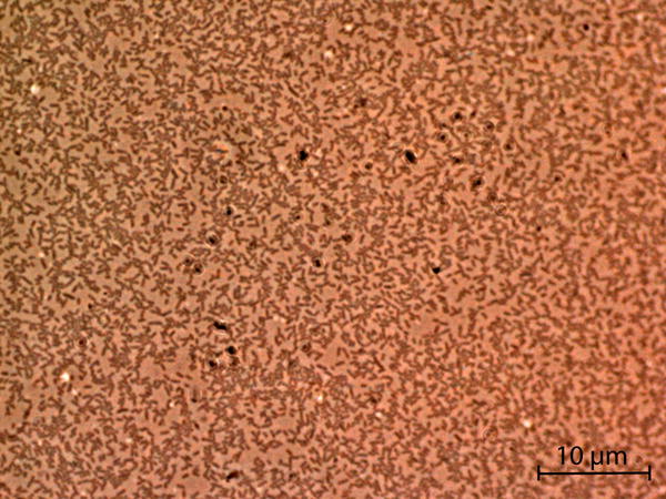
Giménez staining of rickettsiae purified from Vero 76 cell by two rounds of Renografin density gradient ultracentrifugation.
ALTERNATE PROTOCOL 4
PURIFICATION OF RICKETTSIA RICKETTSII USING A RENOGRAFIN “CUSHION”
The Renografin density gradient centrifugation process described in BASIC PROTOCOL 5 takes several hours to complete and requires an ultracentrifuge. If purified rickettsiae are needed more quickly (for applications that require as little processing time as possible, such as RNA extraction), running the partially purified rickettsiae through a Renografin “cushion” provides an alternative that will remove a significant amount of host cell debris with a much shorter processing time (less than one hour). This protocol can also be used if an ultracentrifuge is not available in the BSL-3 facility.
Materials
Partially purified R. rickettsii in SPG, on ice (see BASIC PROTOCOL 4)
25% Renografin-60 solution (see recipe)
SPG, ice-cold (see recipe)
Microcentrifuge (1.5mL) tubes, sterile
NOTE: Renografin-60 (Bracco Diagnostics) is a controlled substance. To use Renografin-60 for research purposes, permission must be obtained from the United Stated Department of Justice Drug Enforcement Administration (DEA).
NOTE: All equipment and solutions coming into contact with cells must be sterile prior to use, and proper sterile technique should be used. All work should be done in a class II biosafety cabinet within a BSL-3 laboratory.
Resuspend partially purified R. rickettsii pellet in 200μL SPG and keep suspension on ice.
-
Place 500μL of the 25% Renografin-60 solution into the bottom of a microcentrifuge tube.
The 25% Renografin solution comprises the “cushion.”
Gently pipet the 200μL of rickettsial suspension on top of the Renografin cushion.
-
Centrifuge at 17,000 × g at 4°C for 10 minutes.
The rickettsiae and limited host cell debris will pellet at the bottom of the Renografin cushion, while most host cell debris will gather at the SPG/Renografin interface.
Carefully aspirate the host cell debris and Renografin, being careful not to disturb the rickettsiae pellet.
-
Wash the pellet 3 times in 1mL ice-cold SPG.
To pellet the rickettsiae after washes, centrifuge at 17,000 × g for 10 minutes.
-
Keep the purified rickettsial pellet on ice.
The next steps and the resuspension solution will depend on the application. If highly purified rickettsiae are necessary, and an ultracentrifuge is not available, this procedure can be repeated 2–3 times to eliminate most host cell material. The rickettsiae can be maintained on ice in SPG for several hours if necessary. If desired, the purity of the rickettsiae can be assessed by fixing a few drops of the suspension on a glass slide and staining as described in BASIC PROTOCOL 3. For freezing, resuspend pellet in SPG (see BASIC PROTOCOL 8). The rickettsiae can also be heat-killed (10–20 minutes at 90°C), at which point they can safely be removed from the BSL-3 laboratory.
BASIC PROTOCOL 6
QUANTIFICATION OF RICKETTSIA RICKETTSII BY PLAQUE ASSAY
The plaque assay is a method to measure the amount of infectious rickettsiae in a sample by determining the number of plaque forming units (PFU) formed on a monolayer of Vero cells. At a high dilution of rickettsial stock, each plaque represents a zone of cells whose infection was initiated with a single bacterium. Therefore, the titer of a rickettsial stock can be calculated in PFU per milliliter. If performing a plaque assay to determine the PFU titer of a stock, the titration should be performed in duplicate. Plaque assays can also be used to obtain single (clonal) rickettsial isolates. This protocol is written for R. rickettsii stocks grown in Vero cells but can be applied to other spotted fever group rickettsiae.
Materials
Purified (cell-free) R. rickettsii stock
DMEM, supplemented with 10% FBS, filter-sterilized, kept at 37°C (see recipe)
1% agarose in water, autoclaved and maintained at 50°C
6-well cell culture plates, with Vero cells, about 90% confluent (see APPENDIX 4E)
Neutral red solution, filter-sterilized (see recipe)
Inverted microscope
NOTE: All equipment and solutions coming into contact with cells and agarose overlay must be sterile, and proper sterile technique should be used. All work should be done in a class II biological safety (biosafety) cabinet in a BSL-3 laboratory.
Warm media and agarose solutions
-
1. Before beginning the plaque assay, warm supplemented DMEM to 37°C and agarose solution to 50°C.
The DMEM should be warm enough so as not to solidify the agarose solutions when mixed together, and it should not be too hot, which will kill the Vero cells. The agarose solution should be kept at 50°C to prevent the solution from solidifying before use. When both solutions are mixed together, the temperature of the resulting solution will be appropriate for immediate addition to the Vero cell monolayer, maintaining cell viability.
Prepare rickettsial dilutions
-
2. Thaw vial of purified R. rickettsii on ice, or quickly thaw in a 32°C water bath.
It is necessary to use cell-free rickettsiae to ensure that each plaque represents a single bacterium rather than a single infected cell, which could contain multiple rickettsiae. BASIC PROTOCOL 4 describes a technique to separate rickettsiae from their host cells.
-
3. Perform a ten-fold dilution series (in supplemented DMEM) starting at 10−1 and diluting rickettsiae down to 10−10.
Change disposable pipet tips between tubes to decrease the chance of sample carryover.
Infect cells
-
4. Bring the 6-well plates with Vero cell monolayers into the biosafety cabinet.
Use Vero cells when monolayer is approximately 90% confluent. Seeding 5 × 10⁵ to 7 × 10⁵ cells per well 24 hours prior to starting the plaque assay should results in an appropriate Vero cell monolayer.
5. Remove growth medium from Vero cells.
6. Inoculate 6-well tissue culture wells with 100μL diluted rickettsial sample and rotate tissue culture plate so that dilution sample covers the monolayer.
7. Incubate inoculated plates for one hour at 37°C.
8. Wash wells twice with warm supplemented DMEM.
Overlay cells with agarose
-
9. For each 6-well plate, combine 7mL warm DMEM with 7mL 1% agarose, mix thoroughly, and immediately add 2mL of mixture over the cells in each well.
Carefully add the agarose/media mixture down the sides of the wells, being careful not to disturb the Vero cell monolayer.
10. Let agarose solidify, then add 1mL supplemented DMEM over the top.
Monitor cells
11. Incubate plaque assay samples at 34°C with 5% CO2.
-
12. Check cells daily and replenish media overlay as necessary. Use an inverted microscope to observe the Vero cell monolayer for plaque formation.
Plaques will appear as small (~1mm) clear areas in the monolayer and will begin to form around the fifth day of incubation.
-
13. When plaques are detected, replace media overlay in wells with 2mL of the neutral red solution and let incubate overnight at 34°C.
The addition of neutral red will aid in the visualization of the plaques. As only live cells can take up neutral red stain, the plaques ill appear clear against a red background.
Count plaques
14. Observe plaques using inverted microscope; select one set of duplicate wells with between 10 and 30 plaques and count them.
-
15. Calculate the average number of plaques for that dilution and calculate the rickettsial stock titer.
Rickettsial titer (PFU/mL) = average plaque count × 10mL−1 (because 0.1mL of each dilution was added to the well)× dilution factor (the inverse of the dilution used on the plate used to count the plaques).
BASIC PROTOCOL 7
PREPARATION OF FROZEN STOCKS OF RICKETTSIA RICKETTSII-INFECTED VERO CELLS
When actively growing rickettsial cultures are not needed, freezing aliquots of rickettsiae-infected cells is a convenient way to maintain stocks. These frozen samples can easily be used to infect cell cultures when needed (See BASIC PROTOCOL 1). This protocol is written for R. rickettsii-infected Vero cells (see BASIC PROTOCOL 2) but can be applied to rickettsiae grown in any type of cell line, with media requirements determined by the specific cell lines used.
Materials
R. rickettsii-infected Vero cells in 150cm2 flask, maintained at 34°C with 5% CO2
DMEM supplemented with 10% heat-inactivated FBS, 2mM L-glutamine and 2mM sucrose, filter-sterilized (see recipe)
Dimethyl sulfoxide (DMSO)
Cryovials suitable for freezing at −80°C or in liquid nitrogen
Serological pipets, sterile
Cell scraper, sterile
CAUTION: DMSO is hazardous; see UNIT 1A.3 for guidelines on handling, storage, and disposal.
NOTE: All equipment and solutions coming into contact with cells must be sterile, and proper sterile technique should be used. All work should be done in a class II biosafety cabinet in a BSL-3 facility
-
In biosafety cabinet, add DMSO to the supplemented DMEM for a final concentration of 10% DMSO.
DMSO will dissolve cellulose acetate membranes commonly used for filter-sterilization, so it should be added after the DMEM has been supplemented with FBS, L-glutamine, and sucrose. The DMSO can be filtered using nylon membrane filters; alternatively, only open the DMSO bottle under sterile conditions in the biosafety cabinet.
Remove media from flask of infected Vero cells.
Gently scrape down cells with sterile cell scraper.
-
Wash down cells with 10–20mL DMEM supplemented with 10% FBS, 2mM L-glutamine and 2mM sucrose.
If most of the Vero cells are detaching from the flask (indicating an overgrown rickettsial infection), wash down cells in existing media in the flask. Divide the cell/media suspension into 50mL conical tubes and centrifuge at 17,000 × g for 10 minutes at 4°C. Discard the supernatant and resuspend pellets in a total volume of 10–20mL DMEM supplemented with 10% FBS, 2mM L-glutamine and 2mM sucrose.
-
Aliquot 500μL to 1mL of resuspended cells into each cryovial.
The DMSO and FBS help preserve the host cells during the freezing and thawing processes, respectively.
-
Freeze cells slowly to −80°C, then continue to store vials at −80°C or in liquid nitrogen.
It is ideal to freeze the cells with the temperature decreasing at a rate of −1°C per minute. This can be achieved using freezing containers such as the Nalgene Cryo 1°C Freezing Container. Alternatively, the cells can be put at 4°C for several hours, then at −20°C overnight, then at −80°C overnight, and then either kept at −80°C or transferred into liquid nitrogen storage. The samples can also be quickly frozen using an alcohol/dry ice bath. If this method is used, the host cells will be destroyed, but the rickettsiae will remain viable.
BASIC PROTOCOL 8
PREPARATION OF FROZEN STOCKS OF PURIFIED (CELL-FREE) RICKETTSIA RICKETTSII
Purified (e.g. cell-free) rickettsiae are necessary for a variety of applications, including transferring in vitro infections between cells types, mammalian and arthropod in vivo infections, and a variety of biochemical and molecular biology applications. Because purification can be labor-intensive, it is often useful to perform rickettsial purifications in large batches, and then store the cell-free rickettsiae for future applications. This protocol describes the process for preparing frozen stock of partially- or Renografin-purified R. rickettsii grown in Vero cells (see BASIC PROTOCOL 4 or BASIC PROTOCOL 5) but can be applied to any species of rickettsiae purified from any cell line.
Materials
Cell-free R. rickettsii, purified as described in BASIC PROTOCOL 4 or BASIC PROTOCOL 5, resuspended in SPG, kept on ice
SPG, filter-sterilized and ice-cold (see recipe)
Cryovials suitable for freezing at −80°C or in liquid nitrogen, chilled on ice
NOTE: All equipment and solutions coming into contact with cells must be sterile, and proper sterile technique should be used. All work should be done in a class II biosafety cabinet in a BSL-3 laboratory.
-
Prepare dilutions of purified R. rickettsii as desired using SPG.
The rickettsiae will be frozen in 500μL aliquots, so prepare dilutions accordingly.
Sucrose has been shown to be effective for the preservation of rickettsial viability during freezing and thawing (Bovarnick et al. 1950).
Transfer rickettsiae suspension into cryotubes in 500μL aliquots.
-
Freeze rickettsiae to −80°C, then store at −80°C or transfer to liquid nitrogen storage.
For cell-free rickettsiae, freeze the bacteria quickly using an alcohol/dry ice bath or place the aliquotted dilutions directly into the −80°C freezer.
RECIPES AND SOLUTIONS
Carbol fuchsin stock
5% aqueous phenol (v/v)
10% ethyl alcohol, absolute (v/v)
1% basic fuchsin (w/v)
Bring to desired volume with distilled water
Store at 4°C
To make 50mL carbol fuchsin stock, add 2.5mL phenol, 5mL alcohol and 0.5g basic fuchsin to 42.5mL distilled water.
Dulbecco’s modification of Eagle Medium (DMEM) supplemented with heat-inactivated fetal bovine serum (FBS)
5% or 10% heat-inactivated FBS (see APPENDIX 2A)
Bring to desired volume in DMEM (available from ATCC or other suppliers of cell culture materials)
Filter-sterilize
Store at 4°C
To make 500mL DMEM with 5% FBS, add 25mL heat-inactivated FBS to 475mL DMEM.
To make 500mL DMEM with 10% FBS, add 50mL heat-inactivated FBS to 450mL DMEM.
DMEM supplemented with heat-inactivated FBS and 2mM L-glutamine and/or 2mM sucrose
2mM L-glutamine
2mM sucrose
Bring to desired volume in DMEM with 5% or 10% heat-inactivated FBS (see above)
Filter-sterilize
Store at 4°C
To make 500mL media, add 0.146g L-glutamate and 0.34g sucrose to 500mL DMEM with desired percentage of heat-inactivated FBS.
0.8% malachite green oxalate
0.8% malachite green oxalate (w/v)
Bring to desired volume in distilled water
Store at room temperature
To make 50mL dye, add 0.4g malachite green oxalate to 50mL distilled water.
Neutral Red solution
1% neutral red (w/v)
1% DMEM (v/v)
Bring to desired volume in distilled water.
Filter-sterilize
Make fresh for each application
To make 50mL solution, add 0.5g neutral red and 1mL DMEM to 49mL distilled water.
Renografin-60 solutions
NOTE: Renografin-60 (Bracco Diagnostics) is a controlled substance. To use Renografin-60 for research purposes, permission must be obtained from the United Stated Department of Justice Drug Enforcement Administration (DEA).
60% Renografin solution (Renografin-60, Bracco Diagnostics)
SPG (see recipe below) Make all solutions fresh for each application
To make 5mL of the various Renografin-60 solution listed below, combine the following:
25% solution: 2.1mL Renografin-60 and 2.9mL SPG
32% solution: 2.7mL Renografin-60 and 2.3mL SPG
36% solution: 3mL Renografin-60 and 2mL SPG
42% solution: 3.5mL Renografin-60 and 1.5mL SPG
Sucrose-phosphate-glutamate (SPG)
218mM sucrose
3.76mM potassium phosphate monobasic
7.1 mM potassium phosphate dibasic
4.9 mM potassium glutamate
Bring to desired volume in distilled water
Filter-sterilize
Store at 4°C
To make 50ml SPG, add 3.73g sucrose, 0.03g KH2PO4, 0.06g K2HPO4, 0.05g potassium glutamate to 50mL distilled water.
COMMENTARY
Background Information
Historically, and in the present, the importance of rickettsial diseases, in terms of morbidity and mortality, has been underestimated worldwide due to misdiagnosis. The advent of molecular diagnostics and the importance of selected rickettsial pathogens as biothreat agents have kindled new interest in rickettsial diseases. Recent serosurveys have demonstrated a high prevalence of rickettsial infections worldwide, particularly in warm and humid climates. Additionally, new molecular diagnostic technology has revealed very narrow gaps in worldwide rickettsial distributions. For example, several rickettsial species including R. rickettsii that were known only in the United States are now reported in Central and South America. Indeed, in the U.S. the rickettsial diseases are on the rise as evident by recent 2004 CDC data that reported over 1,800 cases of Rocky Mountain spotted fever (RMSF) and 1,070 combined cases of human granulocytic and monocytic ehrlichioses (see Section 3A). In many cases, nonspecific early symptoms and the lack of a precise diagnosis of rickettsial disease result in misdiagnosis with potentially dire outcomes. Because of the diversity of rickettsial pathogens throughout the world and the variable clinical manifestations attributed to these pathogens, we limit our coverage to tick-born Rickettsia rickettsii, the causative agent of Rocky Mountain spotted fever. RMSF, the most severe tick-borne rickettsiosis, like all other rickettsial infections, is classified as a zoonosis, which requires a biological vector such as tick in order to be transmitted between animal hosts and accidentally to human. Human infection occurs via the bite of infected ixodid ticks (U.S.A.: Dermacentor variabilis and D. andersoni; Central and South America: Amblyomma cajennense; Mexico: Rhipicephalus sanguineus).
Rickettsiae are obligate intracellular gram-negative bacteria which enter their eukaryotic host cells through clathrin-coated pits by way of induced phagocytosis and evade destruction by exiting the phagosome before phagolysosomal fusion occurs. They are able to grow only within the cytoplasm and occasionally the nucleus (as has been noted for R. rickettsii and some spotted fever group rickettsiae) of a variety of eukaryotic host cells. Living freely in the cytoplasm of the host cell allows rickettsiae access to host nutrients as well as protection from the host’s humoral immune response. R. rickettsii move between neighboring cells by inducing host actin polymerization. R. rickettsii infects a wide range of eukaryotic host cells such as vertebrate fibroblast, endothelial and epithelial cells, as well as invertebrate epithelial cells. Although this nonspecific rickettsial ability to infect diverse cell lines from reptilian, avian and mammalian hosts allows rickettsial propagations under different physiological conditions to simulate intact host infection, we have only detailed in vitro culture using the Vero (Green Monkey fibroblast) cell line. In this unit, we have limited the protocols to basic techniques most appropriate for growing R. rickettsii in a BSL-3 laboratory. Other cell culture methodologies traditionally used for rickettsiae, such as shell vial culture, embryonated chicken eggs, isolation of rickettsiae from infected ticks, animal and human blood and tissues, are not described in this unit due to the added complexity of using these techniques with a Select Agent in a BSL-3 facility.
The biology of R. rickettsii is inextricably linked to its complex vertebrate host/tick system. R. rickettsii have several unique rickettsial biological characteristics, such as environmental stability, small size, aerosol transmission, low infectious dose, and high morbidity and mortality. These attributes have been the contributory factors behind laboratory-acquired infections. Thus, the real concern of working with R. rickettsii in the laboratory, and therefore consideration should be given, particularly when high yielding propagation is required, is that there are limited effective antibiotic choices and no licensed rickettsial vaccine available. As a result, throughout this unit, we have reemphasized that all work with live R. rickettsia should be performed in class II biological safety cabinets within a BSL-3 laboratory, and safety procedures for the propagation of this pathogen should be strictly followed. Of particular importance is to follow the approved standard operating procedure (SOP) step-by-step to avoid laboratory-acquired infections. We also recommend decontamination of work areas before and after the routine tissue culture and propagation/purification procedures, following your approved decontamination SOP.
Critical Parameters and Troubleshooting
Despite their reduced genome size (≤1.5 Mbp), rickettsiae are highly evolved and exquisitely adapted to the intracytoplasmic environment of their eukaryotic host cells. However, working with rickettsiae requires patience because of difficulties associated with growing these organisms due to their slow growth (doubling time of 9–12 hours) and obligate intracellular nature. The major issues to consider while working with rickettsiae are as follows:
Personal safety: Rickettsiae have caused many laboratory-acquired infections both via parenteral and aerosol routes. Thus, working with rickettsiae not only requires high-level containment but also careful attention to hazard prevention and decontamination techniques during sample handling, pipeting, and media transfer. All waste, liquid or solid, must be disposed of properly as required in your BSL-3 laboratory SOP.
Rickettsial viability: Rickettsiae are quite labile, and therefore the specimens should be properly frozen and stored. Vials containing rickettsiae, must be kept on ice at all times to prevent loss of viability.
Contamination: As noted in many of the protocols in this unit, the use of antibiotics in cell culture media is not recommended when the cells will be used for propagating rickettsiae. However, this puts the cultures at extra risk for contamination with adventitious agents. Contamination can be a serious problem, both for current rickettsial growth and experiments, and also if the propagated rickettsial samples are frozen and added to the laboratory inventory. To remedy the contamination problems, rickettsial samples should be monitored via direct microscopy of Gram stained smears of cells, and/or via molecular approaches. Antibiotics (100 mg streptomycin sulfate/50 mg potassium penicillin G/5 mg neomycin sulfate) per ml of the culture medium could be used to control bacterial contamination in tissue culture. Vero cells then can be maintained with reduced or without antibiotics thereafter if the contamination is resolved. If premature cytopathic effects continue to persist rickettsiae should be harvested with centrifugation and re-plated in Vero cells. The quality of the harvested rickettsiae could be evaluated using the shell vial culture for fast results at 24 and or 48 hours (See Raoult et al. 1991).
The integrity of the rickettsial samples should be continuously monitored using molecular methods (PCR/sequencing) and/or immunofluoresent antibody assays. To prevent cross-contamination or different rickettsial species or strains, regular verification is important, especially if multiple species or strains of rickettsiae are being propagated in the same BSL-3 facility.
The host cell line used should be regularly monitored for Mycoplasma infections and treated as recommended by cell line supplier if contamination is detected. Several Mycoplasma detection and treatment kits are commercially available.
Anticipated Results
The doubling time for R. rickettsii is between 9–12 hours; therefore, depending on the multiplicity of infection (MOI), infections can take several days to over a week to thoroughly infect the host cell monolayer. R. rickettsii infections in Vero cells need to be closely monitored, as the infection can become overwhelming for the host cells. As the rickettsial begin to outgrow the available host cells in the flask, the tissue culture medium (DMEM) will decrease in pH and turn color from red to orange to yellow. When this occurs, the rickettsiae must be passaged, purified, or frozen immediately.
Time Considerations
Laboratory maintenance of rickettsiae is very time-consuming. Experiments requiring rickettsiae need to be planned carefully in advance to coordinate preparation of uninfected host cells and rickettsiae-infected cells. As mentioned above, rickettsiae have a doubling time of many hours; therefore, obtaining a large sample of rickettsiae for laboratory applications could take several days to weeks. Further, depending on the application, the purification process can take several hours as well. Because all work with live R. rickettsii must be done in a BSL-3 facility, the time commitment for working in such a laboratory (including extensive decontamination procedures) must be considered.
Internet Resources
http://www.cdc.gov/ncidod/dvrd/rmsf/index.htm
The CDC Rocky Mountain spotted fever website. This website serves as a good general reference about R. rickettsii natural history, epidemiology, laboratory detection, treatment, prevention and control.
The CDC Select Agent Program website. This website lists all select agents and provides information on public laws and regulations regarding research with select agents.
http://www.selectagents.gov/securitydoc.html
The National Select Agent Registry (NSAR) website. This website has posted guidelines for compliance with the security requirements of the select agent regulations.
The Belgian Biosafety Server website. This website has posted guidelines for biosafety in Belgium and the European Union, as well as links to other international biosafety guidelines.
http://internationalbiosafety.org
The International Biosafety Working Group website. This website provides an international forum for the discussion of biosafety issues.
http://www.who.int/csr/resources/publications/biosafety/WHO_CDS_CSR_LYO_2004_11/en/
The World Health Organizations Epidemic and Pandemic Alert Response Laboratory Biosafety Manual (third edition) website. This website provides links for the WHO Laboratory Biosafety manual in English, French, Spanish, Portuguese, Chinese, Russian, Italian, Japanese, Serbian in Vietnamese.
Contributor Information
Nicole C. Ammerman, Email: namme001@umaryland.edu.
Magda Beier-Sexton, Email: mbeie001@umaryland.edu.
Abdu F. Azad, Email: aazad@umaryland.edu.
Literature Cited
- Bovarnick MR, Miller JC, Snyder JC. The influence of certain salts, amino acids, sugars, and proteins on the stability of rickettsiae. J Bacteriol. 1950;59:509–522. doi: 10.1128/jb.59.4.509-522.1950. [DOI] [PMC free article] [PubMed] [Google Scholar]
- Dumler JS, Walker DH. Rocky Mountain spotted fever changing ecology and persisting virulence. New Engl J Med. 2005;353:551–3. doi: 10.1056/NEJMp058138. [DOI] [PubMed] [Google Scholar]
- Ellison DW, Clark TR, Sturdevant DE, Virtaneva K, Porcella SF, Hackstadt T. Genomic comparison of virulent Rickettsia rickettsii Sheila Smith and avirulent Rickettsia rickettsii Iowa. Infect Immun. 2008;76:542–550. doi: 10.1128/IAI.00952-07. [DOI] [PMC free article] [PubMed] [Google Scholar]
- Giemsa G. Eine vereinfachung und vervollkommnung meiner methylenazur-methylenblau-eosin-farbemethode zur erzielung der Romanowsky-Nocht’schen chromatinfarbung. Zentabl Bakteriol Parasitenkd Infectkrankh. 1904;37:308. [Google Scholar]
- Giménez DF. Staining rickettsiae in yolk-sac cultures. Stain Technol. 1964;39:135–140. doi: 10.3109/10520296409061219. [DOI] [PubMed] [Google Scholar]
- Hanson BA, Wisseman CL, Jr, Waddell A, Silverman DJ. Some characteristics of heavy and light bands of Rickettsia prowazekii on Renografin gradients. Infect Immun. 1981;34:596–604. doi: 10.1128/iai.34.2.596-604.1981. [DOI] [PMC free article] [PubMed] [Google Scholar]
- Raoult D, Torres H, Drancourt M. Shell-vial assay: evaluation of a new technique for determining antibiotic susceptibility, tested in 13 isolates of Coxiella burnettii. Antimicrob Agents Chemother. 1991;1991:2070–2077. doi: 10.1128/aac.35.10.2070. [DOI] [PMC free article] [PubMed] [Google Scholar]
Key References
- Weiss E, Coolbaugh JC, Williams JC. Separation of viable Rickettsia typhi from yolk sac and L cell host components by Renografin density gradient centrifugation. Appl Microbiol. 1975;30:456–463. doi: 10.1128/am.30.3.456-463.1975. This is the seminal article describing the use of a Renografin density gradient for the isolation of pure, viable rickettsiae. [DOI] [PMC free article] [PubMed] [Google Scholar]
- Kimman TG, Smit E, Klein MR. Evidence-based biosafety: a review of the principles and effectiveness of micrbiological containment measures. Clin Microbiol Rev. 2008;21:403–425. doi: 10.1128/CMR.00014-08. The review article discusses the principles and methods of biosafety regulations as determined by the NIH, the CDC, the WHO and the European Union. [DOI] [PMC free article] [PubMed] [Google Scholar]


