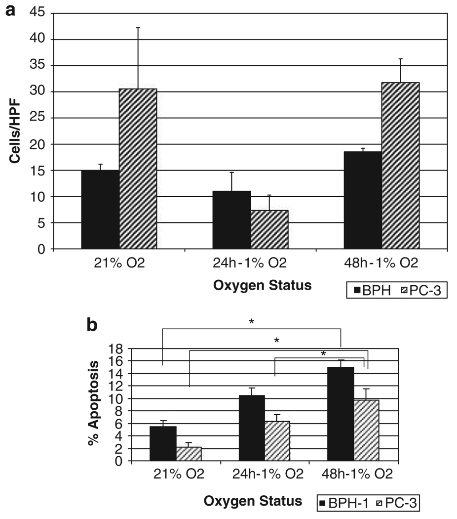
Figure 1.
Response of benign prostate epithelial cells benign prostate epithelial (BPH)-1 and malignant prostate cells PC-3 to hypoxia. (a) The effect of hypoxia on prostate cell migration. Wounding assays were performed on benign and malignant prostate epithelial cells and the number of migratory cells was quantified. Three random fields (× 400) were counted under light microscopy and the average values per field were determined. Under normoxia, PC-3 cells show enhanced migration potential compared to BPH-1 cells and following hypoxia exposure (24 h), both cell lines exhibited a diminished ability to migrate in a transient manner. (b) The apoptotic response of the two cell lines to hypoxia; exposure of PC-3 and BPH-1 cells to hypoxia leads to enhanced apoptosis. *P < 0.001.

