Abstract
Study Design
Hyperreflexia occurs after spinal cord injury and can be assessed by measuring low frequency-dependent depression of the H-reflex in the anesthetized animal.
Objective
To determine the effects of Modafinil (MOD), given orally, following a complete SCI compared with animals receiving MBET and transected untreated animals and examine if changes exist in Connexin 36 (Cx-36) protein levels in the lumbar enlargement of animals for the groups described.
Setting
CTN, Little Rock, AR, USA.
Methods
Adult female rats underwent complete transection (Tx) at T10 level. H-reflex testing was performed 30 days following Tx in one group, and after initiation of treatment with MOD in another group, and after MBET training in the third group. The Lumbar enlargement tissue was harvested and western blots were performed after immunoprecipitation techniques to compare Cx-36 protein levels.
Results
Statistically significant decreases in low frequency-dependent depression of the H-reflex were observed in animals that received MOD and those that were treated with MBET compared with the Tx, untreated group. Statistically significant changes in Cx-36 protein levels were not observed in animals treated with MOD compared with Tx, untreated animals.
Conclusion
Normalization of the loss of low frequency -dependent depression of the H-reflex was demonstrated in the group receiving MOD and the group receiving MBET compared with the Tx, untreated group. Further work is needed to examine if Cx-36 protein changes occur in specific subregions of the spinal cord.
Keywords: spinal cord transection, electrical coupling, gap junctions, modafinil, Cx-36, H-reflex
Introduction
The deficits resulting from spinal cord injury (SCI) include hyperreflexia and spasticity below the level of the lesion. The mechanisms of hyperreflexia and spasticity are unknown, although numerous theories have been postulated.1 One of the mechanisms that has been proposed is that spinal cord injury leads to the loss of descending pathways that provide presynaptic inhibition to the motor system. However, Hasegawa and Ono2 suggest that noradrengergic descending pathway tonically suppresses spinal presynaptic inhibition and may not contribute to hyperreflexia. The H-reflex or Hoffman reflex has been used to quantify hyperreflexia3, and several investigators have utilized frequency-dependent depression of the H-reflex to examine changes in spinal cord circuitry after SCI.4–6 We previously examined the use of motorized exercise bicycle training (MBET) in normalizing the loss of frequency-dependent depression of the H-reflex that occurs following complete spinal cord transection (Tx) in the rat5,7 as well as the effects of passive exercise in the acute as well as the chronic phase of injury.8
Recently, we found that Tx transiently decreased levels of the neuronal gap junction protein Connexin 36 (Cx-36).9 Cx-36 levels decreased 30% 7 days after injury, and returned to control levels over the next 2–4 weeks. The onset of hyperreflexia was co-incident with recovery of Cx-36 to control levels. We hypothesized that a change in electrical coupling occurs after Tx that contributes to the hyperreflexive state, although the nature of this change is unknown.
The stimulant modafinil is approved for the treatment of excessive sleepiness in narcolepsy, obstructive sleep apnea and shift work disorder. A recent landmark study found that the mechanism of action of modafinil is to increase electrical coupling between cortical interneurons, thalamic reticular neurons, and inferior olivary neurons.10 The literature revealed limited studies of modafinil as a treatment of hyperreflexia or spasticity induced from SCI. Mukai and Costa11 reported positive effects from modafinil on self esteem in 2 patients with SCI. Hurst et al.12 described a retrospective study of the use of modafinil in a population of children diagnosed with cerebral palsy. These authors reported 76% of the patients studied (n=30) reported decreased spasticity after treatment with modafinil, and showed decreased tone after physical examination. Hurst and Lajara-Nanson13 conducted a pilot study to examine the benefit of modafinil on spasticity and went on to hypothesize that modafinil reduces spasticity of central origin. An additional study in 2006 by Hurst et al.14 reported that 29/59 pediatric patients with spastic cerebral palsy that were treated by modafinil demonstrated improvements in gait during the treatment.
The present study was undertaken to determine if modafinil, administered orally, would normalize the loss of frequency-dependent depression of the H-reflex that is observed in spinally transected rats, and how this treatment compares to passive exercise of the hindlimbs that is initiated in the acute phase of exercise. We also wanted to examine the changes in Cx-36 protein in the lumbar tissue following Tx and after treatment. Preliminary results were presented in abstract form.15
Methods
The methods employed have been previously published.5,7,8–9 All animal procedures were approved by the Institutional Animal Care and Use Committee at UAMS.
Surgery
Adult female Sprague-Dawley rats (n=48, 200 to 300 g, Harlan) underwent a lower thoracic laminectomy under ketamine (60 mg/kg, i.m.) and xylazine (10 mg/kg, i.m.) anesthesia. A complete transection (Tx) of the spinal cord was made by aspiration and the transected ends of the cord retracted, producing a 2–3 mm cavity. Surgery and postsurgical care was performed as previously described.5 We certify that all applicable institutional and governmental regulations concerning the ethical use of animals were followed during the course of this research.
One group of intact rats served as non-transected controls (n=10) and another group of Tx animals (Tx 30D, n=16) underwent no further treatment until H-reflex testing was carried out 30 days after complete Tx. The remaining rats were divided into 2 groups. One group (n= 16) was treated with modafinil beginning 7days after Tx. The modafinil was given orally at a dose of 4 mg/Kg. The second group (n=16) began passive exercise training 7 days after Tx and exercised 5 days per week for 30 days. This group exercised for two thirty minute exercise sessions per day with a ten minute rest period in between the two sessions. Specific methods for MBET have been previously described.5,7
Reflex Testing
H-reflex testing was measured as previously described.7,9 Recordings were made using amplifier (Grass P511) filter settings of 3 Hz to 3 KHz with the 60 Hz notch filter in use. Responses to the stimulus were digitized and averaged using a GW Instruments (Somerville, MA) digitizer module and SuperScopeR software.
The reflex first was tested at 0.2 Hz to determine threshold and maximal response levels. After discarding the first 5 responses at each frequency in order to obtain an average of the stabilized reflex, averages of 10 responses were obtained. Averages were compiled following stimulation at 0.2, 1, 5, and 10 Hz. The change in the response at various frequencies was calculated as the percent of the response at 0.2 Hz in order to determine depression of the H-reflex as a function of stimulation frequency. Following the frequency series testing, the H-reflex amplitude was confirmed at 0.2 Hz for consistency. If the amplitude at recheck was less than 90% of the initial amplitude, the data was discarded.
At the end of the experiment, animals were euthanized with an overdose of barbiturate (Nembutal) and the Tx confirmed either visually or histologically following transcardial perfusion with paraformaldehyde (4%) and sucrose (20%).
Measurement and statistics
The amplitude of the H-wave was measured from peak to peak of the two components. For comparison of data between the different groups in each experiment, measures were tested using one factor, two factor or multifactor analysis of variance (ANOVA) to conclude whether any of the factors had a significant effect on the magnitude of the variable and also whether the interaction of the factors significantly affected the variable. Differences were considered significant at values of p<0.05. If statistical significance was present, posthoc tests were used to compare between groups.
Cx-36 protein analysis
The methods employed for Cx-36 protein analysis have been previously published.9,16 At the end of recording, cores (3–4 cu mm, 300–600 mg) from the lumbar enlargement were removed using a 3–4 mm dermal biopsy punch after performing laminectomies in anesthetized rats. Tissue was homogenized in 600 μl ice cold RIPA buffer with HALT protease inhibitors (Pierce) and centrifuged to remove debris. Tissue and lysate was kept on ice at all times. 5 μg of anti-Cx-36 antibody (37–4600, Zymed, Invitrogen) per sample was covalently coupled to a gel support (Seize Primary, Pierce). 800 μg of protein from spinal cord lysates was mixed with antibody-coupled gel in a total of 1 ml RIPA (with protease inhibitors) and incubated 4°C, overnight, with gentle end-over-end mixing. Immunoprecipitates were washed twice with RIPA buffer and resolved by SDS-PAGE.9 The amount of Cx-36 immunoprecipitated was determined by western blot as previously described.9
Results
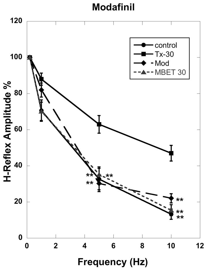
The habituation of the H-reflex was examined following stimulation at 0.2, 1, 5 and 10 Hz in the following groups of animals: Tx and untreated for 30 days (Tx only 30D), Tx and exercised for 30 days (Tx+Ex 30 D), Tx and treated with modafinil (MOD), and intact Control. ANOVA of these groups showed statistically significant differences across experimental groups for stimulation at 5 Hz (df=,3 F= 7.662, p<0.0002), and 10 Hz (df= 3, F= 18.647, p< 0.0001). Post hoc comparisons between all groups were undertaken using the Newman-Keuls test and those against the Tx only 30 D (transected, untreated) group were considered the most relevant at 5 Hz and 10 Hz.
Comparison against the Tx only 30 D group revealed significant differences to the intact control group at 5 Hz (p<0.01) and 10 Hz (p< 0.01), the passive exercise (MBET) group (Tx + Ex 30D) at 5 Hz (p<0.01) and 10 Hz (p< 0.01), and the MOD group at 5 Hz (p<0.01) and 10 Hz (p<0.01). The Control group revealed significant differences compared to the unexercised, untreated group (Tx only 30 D) at 5 Hz (p< 0.01) and 10 Hz (p< 0.01). Figure 1 is a graph of the habituation of the H-reflex following stimulation at 0.2,1,5 and 10 Hz for all groups described.
Figure 1.

H-reflex amplitude at 0.2, 1, 5 and 10 Hz for intact animals (Control, black circles), Tx only 30 days (Tx-30, square), MBET 30 days (Tx+ Ex 30D, triangle), and Modafinil 30 days (Mod, diamond). Frequency-dependent depression of the H-reflex at 0.2 Hz was designated 100%, and statistical comparisons made against the Tx only 30 day group. At 5 Hz and 10 Hz, the Tx Only 30 D group(Tx-30) differed from the MBET group ( triangle) (p<0.01), the Mod group (diamond) (p< 0.01), and the control group (black circle) (p<0.01).
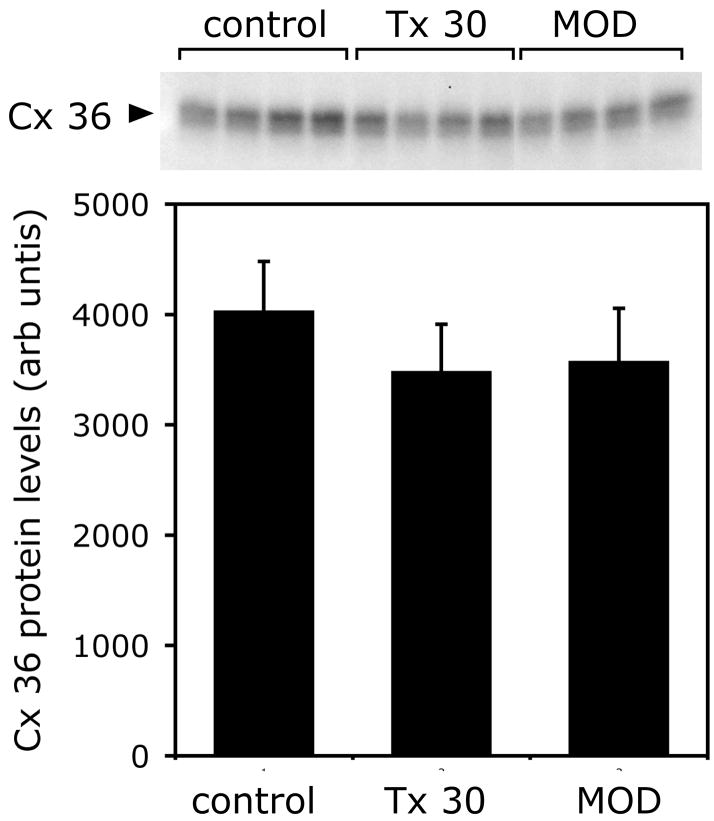
We hypothesized that the improvements in the H-reflex after MOD treatment were due to increased electrical coupling. To investigate this we determined the amount of Cx-36 protein in the lumbar enlargement of the spinal cord. We measured total Cx-36 in control animals that had not undergone transection or exercise, animals that had undergone transection 30 days previously but remained untreated (Tx only 30) and animals that had undergone Tx and were then treated with MOD, starting 7 days after surgery. Previous studies have shown that total Cx-36 protein transiently decreased 7 days after Tx, and then returned to control levels over the next few weeks.9 The results of this study (Figure 2) showed that in animals that received MOD, total Cx-36 protein levels are not different to animals that did not receive MOD.
Figure 2.
MOD does not change total Cx 36 levels. Upper panel, western blot of immunoprecipitated Cx 36 from spinal cord. Lower panel, quantification of Cx 36 from western blot. Bars show mean + SD. Tissue was taken from control animals, animals 30 days after transection (Tx 30), and transected animals after 30 days of modafinil treatment (MOD) that was initiated 10 days after injury.
Discussion
The results of this study indicate that both passive exercise (MBET), if initiated in the acute phase of injury for thirty days, and treatment with MOD (without exercise), normalized the loss of frequency-dependent depression of the H-reflex that occurs after SCI.
Some methodological considerations and limitations of this study should be considered. It has been shown that MOD increases electrical coupling between GABA neurons.10 Therefore, improvements in H-reflex attenuation at high frequencies in animals that had undergone MOD treatment could be explained by increased coupling of spinal neurons. We found that there was no change in total Cx-36 protein in animals that had MOD treatment compared to animals that had not, indicating that MOD did not change Cx-36 gene expression in whole spinal cord tissue. It is not known how MOD increases electrical coupling but it has been suggested that MOD might induce translocation of Cx-36 from intracellular stores to the plasma membrane.10 In this study, we used western blots to measure total tissue protein. As this technique is unable to detect translocation events, we cannot support or refute a translocation hypothesis.
In a previous study, we showed that after Tx Cx-36 protein transiently decreased by 7 days post-Tx, but returned to control levels within 30 days. The return of Cx-36 control levels paralleled the onset of hyperreflexia.9 In another study, we showed that when Tx rats are passively exercised, hyperreflexia was normalized and Cx-36 levels were slightly higher than in unexercised rats, suggesting that exercise may change Cx-36 protein levels.8 Hyperreflexia can also be normalized with MOD, as demonstrated in this study, although there was no evidence for changes in protein levels. Therefore, while passive exercise and MOD could both target electrical coupling, they may do so in different ways.
The focus of this study was on measuring how the treatments tested influenced hyperreflexia as manifested in frequency-dependent depression of the H-reflex. This study did not address the symptom of spasticity that is observed in the rat following spinal cord transection. Spasticity is classically referred to as resistance to passive limb movement in proportion to the velocity of movement.17 This velocity-dependent resistance is thought to be due to increased stretch reflex (SR) responses in the lengthened muscle.18 Additional studies are ongoing that include SR measures that would allow insights into the effects of passive exercise or MOD on spasticity compared with hyperreflexia in the transected rat.
An additional consideration is that this study utilized MOD treatment to rats beginning 7 days following Tx (in the acute phase of injury). The effects of MOD on normalization of the H-reflex in the chronic phase of injury (after hyperreflexia has been established) is unknown. Further studies are needed to address the effects of MOD in the chronic phase of SCI and the long term effects of normalization of the H-reflex in the acute model.
A final issue is the potential site of action of MOD at the level of the spinal cord. It is known that motoneurons are extensively coupled during development, but coupling decreases by 14 days postnatally in the rat.19 There are a number of interneurons that were found to be electrically coupled in the ventromedial region of the spinal cord and appear related to locomotor control.20 It is not known if MOD directly affects electrical coupling in the adult Tx rat, but if it does, it may do so by influencing motoneurons, locomotion-related interneurons or perhaps even GABAergic spinal interneurons, as it does in the brain.10 While a number of new questions are raised by this study, our results do strongly suggest that MOD may represent a valuable therapeutic adjunct to the treatment of SCI, and supports previous results in cerebral palsy, but suggest that some of the gains made may have been due to direct effects on spinal circuitry rather than cerebral in origin.
Another potential mechanism is the possibility that MOD influences monoamine regulation of persistent inward currents (PICs) in motoneurons. Recent results suggest that motoneurons in the adult sacral cord of the rat reacquire the ability to generate PICs and plateau potentials within 1–2 months after spinal transection.21 MOD appears to modulate monoamine transporters and receptors. 22–24 Therefore, additional studies are needed to verify how exactly might MOD have a salutary effect on hyperreflexia, but this does not preclude clinical testing.
Supplementary Material
Acknowledgments
NIH-NCRR Grant P20 RR020146 to the Center for Translational Neuroscience Additional support for this study was provided by NINDS P30 NS047546
Sponsors: NIH-NCRR Grant P20 RR020146 to the CTN and NINDS P30 NS047546
References
- 1.Hultborn H. Spinal reflexes, mechanisms and concepts: From Eccles to Lundberg and beyond. Progress in Neurobiology. 2006;78:215–232. doi: 10.1016/j.pneurobio.2006.04.001. [DOI] [PubMed] [Google Scholar]
- 2.Hasegawa Y, Ono H. Descending noradrenergic neurons tonically suppress spinal presynaptic inhibition in rats. Neuro report. 1995;7 (29):262–266. [PubMed] [Google Scholar]
- 3.Angel RW, Hofmann WW. The H-reflex in normal, spastic, and rigid subjects. Arch Neurol. 1963;8:591–596. doi: 10.1001/archneur.1963.00460060021002. [DOI] [PubMed] [Google Scholar]
- 4.Thompson FJ, Reier PJ, Lucas CC, Parmer R. Altered Patterns of Reflex Excitability Subsequent to Contusion Injury of the Rat Spinal Cord. J of Neurophysiol. 1992;68:1473–86. doi: 10.1152/jn.1992.68.5.1473. [DOI] [PubMed] [Google Scholar]
- 5.Skinner RD, Houle JD, Reese NB, Berry CL, Garcia-Rill E. Effects of exercise and fetal spinal cord implants on the H-Reflex in chronically spinalized adult rats. Brain Res. 1996;729:127–31. [PubMed] [Google Scholar]
- 6.Chen XY, Feng-Chen KC, et al. Short-Term and medium-term effects of spinal cord tract transections on soleus H-reflex in freely moving rats. J Neurotrauma. 2001;18:313–27. doi: 10.1089/08977150151070973. [DOI] [PubMed] [Google Scholar]
- 7.Reese NB, Skinner RD, Mitchell D, Yates C, Barnes CN, Kiser TS, Garcia-Rill E. Restoration of frequency-dependent depression of the H-reflex by passive exercise in spinal rats. Spinal Cord. 2006;44:28–34. doi: 10.1038/sj.sc.3101810. [DOI] [PubMed] [Google Scholar]
- 8.Yates C, Charlesworth A, Reese NB, Skinner RD, Garcia-Rill E. The effects of passive exercise therapy initiated prior to or after the development of hyperreflexia following spinal transection. Exp Neurology. 2008 Oct;213(2):405–9. doi: 10.1016/j.expneurol.2008.07.002. [DOI] [PMC free article] [PubMed] [Google Scholar]
- 9.Yates C, Charlesworth A, Allen SR, Reese NB, Skinner RD, Garcia-Rill E. The Onset of Hyperreflexia in the Rat Following Complete Spinal Cord Transection. Spinal Cord. 2008;46:798–803. doi: 10.1038/sc.2008.49. [DOI] [PMC free article] [PubMed] [Google Scholar]
- 10.Urbano FJ, Leznik E, Llinas R. Modafinil enhances thalamocortical activity by increasing neuronal electrotonic coupling. Proc Natl Acad Sci. 2007;104:12554–12559. doi: 10.1073/pnas.0705087104. [DOI] [PMC free article] [PubMed] [Google Scholar]
- 11.Mukai A, Costa Jonathan L. The effect of Modafinil on Self-Esteem in Spinal Cord Injury Patients: A Report of 2 Cases and Review of the Literature. Arch Phys Med Rehab l. 2005;86:1887–1889. doi: 10.1016/j.apmr.2005.01.009. [DOI] [PubMed] [Google Scholar]
- 12.Hurst DL, Lajara-Nanson WA, Dinakar P, Schiffer RB. Retrospective review of modafinil use for cerebral palsy. J Child Neurology. 2004 Dec;19(12):948–51. doi: 10.1177/08830738040190120701. [DOI] [PubMed] [Google Scholar]
- 13.Hurst DL, Lajara-Nanson W. Use of Modafinil in spastic cerebral palsy. J Child Neurology. 2002 Mar;17(3):169–72. doi: 10.1177/088307380201700303. [DOI] [PubMed] [Google Scholar]
- 14.Hurst DL, Lajara-Nanson WA, Lance-Fish ME. Walking with Modafinil and its use in diplegic cerebral palsy: retrospective review. J Child Neurology. 2006 Apr;21(4):294–7. doi: 10.1177/08830738060210042001. [DOI] [PubMed] [Google Scholar]
- 15.Yates C, Reese NB, Kiser T, Skinner RD, Garcia-Rill E. Modafinil (MOD) normalizes hyperreflexia induced by spinal cord transection in the rat. Neurosci Abst . 2007;33:405.21. [Google Scholar]
- 16.Heister D, Hayar A, Charlesworth A, Yates C, Zhou YH, Garcia-Rill E. Evidence for electrical coupling in the SubCoeruleus(SubC) nucleus. J of Neurophysiol. 2007 Apr;97(4):3142–7. doi: 10.1152/jn.01316.2006. [DOI] [PMC free article] [PubMed] [Google Scholar]
- 17.Lance JW. In: Symposium synopsis in Spasticity: Disordered Motor Control. Feldman RG, Young RR, Koella WP, editors. Chicago Press; Year Book: 1980. pp. 485–494. [Google Scholar]
- 18.Powers RK, Rymer WZ. Effects of acute dorsal spinal hemisection on motoneuron discharge in the medial gastrocnemius of the decerebrate cat. J Neurophysiol. 1988;59:1540–1556. doi: 10.1152/jn.1988.59.5.1540. [DOI] [PubMed] [Google Scholar]
- 19.Walton KD, Navarrete R. Postnatal Changes in Motoneurone Electronic Coupling Studied in the in Vitro Rat Lumbar Spinal Cord. J Physiol. 1991;433:283–305. doi: 10.1113/jphysiol.1991.sp018426. [DOI] [PMC free article] [PubMed] [Google Scholar]
- 20.Hinckley CA, Ziskind-Conhaim L. Electrical coupling between locomotor-related excitatory interneurons in the mammalian spinal cord. J of Neurosci. 2006;26:8477–8483. doi: 10.1523/JNEUROSCI.0395-06.2006. [DOI] [PMC free article] [PubMed] [Google Scholar]
- 21.Bennett DJ, Li Y, Siu M. Plateau potentials in sacrocaudal motomeurons of chronic spinal rats, recorded in vitro. J Neurophysiol. 2001;86:1955–1971. doi: 10.1152/jn.2001.86.4.1955. [DOI] [PubMed] [Google Scholar]
- 22.Madras BK, Xie Z, Lin Z, Jassen A, Panas H, Lynch L, Johnson R, Livni E, Spencer TJ, Bonab AA, Miller GM, Fischman AJ. Modafinil occupies dopamine and norepinephrine transporters in vivo and modulates the transporters and trace amine activity in vitro. J Pharmacol Exp Ther. 2006;319:561–569. doi: 10.1124/jpet.106.106583. [DOI] [PubMed] [Google Scholar]
- 23.Mitchell HA, Bogenpohl JW, Liles LC, Epstein MP, Bozyczko-Coyne D, Williams M, Weinshenker D. Behavioral responses of dopamine beta-hydroxylase knockout mice to modafinil suggest a dual noradrenergic-dopaminergic mechanism of action. Pharmacol Biochem Behav. 2008 doi: 10.1016/j.pbb.2008.07.014. epub ahead of print. [DOI] [PMC free article] [PubMed] [Google Scholar]
- 24.Qu WM, Huang ZL, Xu XH, Matsumoto N, Urade Y. Dopaminergic D1 and D2 receptors are essential for the arousal effect of modafinil. J Neurosci. 2008;28:8462–8469. doi: 10.1523/JNEUROSCI.1819-08.2008. [DOI] [PMC free article] [PubMed] [Google Scholar]
Associated Data
This section collects any data citations, data availability statements, or supplementary materials included in this article.


