Abstract
Expression of the chemokine receptor CXCR4, a G protein-coupled receptor, and HER2, a receptor tyrosine kinase, strongly correlates with the aggressive and metastatic potential of breast cancer cells. We studied estrogen regulation of CXCR4 in estrogen receptor (ER)-positive MCF-7 breast cancer cells overexpressing HER2 (MCF7-HER2). Although estrogen evoked no change in CXCR4 mRNA levels, CXCR4 protein was significantly up-regulated after estrogen treatment of these cells, whereas estrogen had no effect on CXCR4 protein level in parental MCF7 cells that are low in HER2. Use of the CXCR4 specific inhibitor, AMD 3100, indicated that this increase in CXCR4 protein was partially responsible for the increase in estrogen-induced migration of these cells. The estrogen-induced increase in CXCR4 protein in MCF-7-HER2 cells was abrogated by the antiestrogen ICI 182780 and by gefitinib (Iressa; a phosphotyrosine kinase inhibitor), indicating an ER-mediated effect and confirming involvement of receptor tyrosine kinases, respectively. Using specific pathway inhibitors, we show that the estrogen-induced increase in CXCR4 involves PI3K/AKT, MAPK and mTOR pathways. PI3K/AKT and MAPK pathways are known to result in the phosphorylation and functional inactivation of tuberin (TSC2) of tuberous sclerosis complex thereby negating its inhibitory effects on mTOR, which in turn stimulates the translational machinery. Small interfering RNA (siRNA) mediated knockdown of tuberin elevated the level of CXCR4 protein in MCF7-HER2 cells and also nullified further estrogen up-regulation of CXCR4. This study suggests a pivotal role of PI3K, MAPK and mTOR pathways, via tuberin, in post-transcriptional control of CXCR4, initiated through estrogen-stimulated crosstalk between ER and HER2. Thus, post-transcriptional regulation of CXCR4 by estrogens acting through ER via kinase pathways may play a critical role in determining the metastatic potential of breast cancer cells.
Keywords: Estrogen, Estrogen Receptor, HER2, CXCR4, Breast Cancer
INTRODUCTION
Metastasis is the major cause of mortality and morbidity in breast cancers. Expression of several genes has been shown to impart metastatic potential and enhanced growth characteristics to the breast cancer. The chemokine receptor CXCR4, a G protein-coupled receptor (GPCR), is one of the proteins that has been shown to play key roles in tissue-specific metastasis of the cancer cells [1]. Overexpression of erbB2 (HER2), a receptor tyrosine kinase of the epidermal growth factor receptor family, has also been associated with enhanced growth and the metastatic phenotype of breast cancers. Amplification or overexpression of HER2 is found in 20–25% of breast cancers and is frequently associated with metastasis and poor prognosis [2–4].
Using siRNA or selective synthetic peptides against CXCR4, several recent studies have shown that CXCR4 is responsible not only for metastasis but also for primary growth of the breast cancer [5–9]. Interestingly, studies aimed at investigating expression of specific genes in breast cancer responsible for metastasis to bone and lung found CXCR4 to be significantly up-regulated [10, 11]. Furthermore, a large tissue microarray study from pre-invasive and invasive breast carcinomas indicated that higher expression of CXCR4 protein is associated with tumor progression [12].
Inhibition of the HER2 oncogene using various blocking strategies has been implemented to restrict the aberrant signaling cascades initiated by HER2. Herceptin, the recombinant humanized monoclonal antibody against the extracellular domain of HER2, has been used therapeutically to treat HER2 overexpressing breast cancer with beneficial clinical outcome [13]. Overexpression of HER2 in breast cancers is often negatively correlated with estrogen receptor (ER) expression, and estrogen has also been shown to down-regulate HER2 expression in ER-positive breast cancer cells [14].
Despite this overall inverse association between the expression of these proteins, a subset of patients overexpressing HER2 are also ER positive [2]. A recent study revealed that 49% of HER2 positive breast cancers also express ER [15]. Studies also indicate that cross talk between HER2 and ER in breast cancer cells can lead to resistance to endocrine therapies like tamoxifen [16, 17]. Although various signaling pathways are known to function aberrantly in ER positive, HER2 overexpressing cells, the underlying molecular mechanisms responsible for tumor progression and metastasis are far from completely understood.
Studies have conclusively stressed the importance of CXCR4 in tumor growth and metastasis, but our knowledge of how CXCR4 itself is regulated in these cells is still inadequate. Although a recent study established that HER2-mediated up-regulation of CXCR4 protein is responsible for the invasiveness of HER2-overexpressing cells [18], there is no information available regarding the regulation of CXCR4 in ER positive, HER2 overexpressing breast cancer cells. Therefore, the aim of these studies was to examine regulation of CXCR4 in breast cancer cells expressing both ER and HER2 and to better understand the downstream processes following cross talk of ER and HER2 and the impact of estrogen-ER signaling pathways on CXCR4 and cell motility activities. We find that, in the presence of HER2 over-expression, the estrogen-occupied ER exerts regulation of CXCR4 at a post-transcriptional level, with critical involvement of PI3 kinase/AKT, MAPK pathways and their subsequent effect on the mTOR pathway, via tuberin, in the post-transcriptional regulation of this chemokine receptor.
MATERIALS AND METHODS
Cell Culture and Reagents
Cell culture media were purchased from Life Technologies, Inc. (Grand Island, NY). Calf serum (CS) was obtained from HyClone Laboratories (Logan, UT), and fetal calf serum (FCS) was from Atlanta Biologicals (Atlanta, GA). The parental MCF7 cell line which contains high levels of ER and low HER2, and MCF7-HER2 cells which have high levels of ER and high levels of HER2/neu expression, were maintained in culture exactly as described previously [19, 20]. At 6 days prior to use in experiments, cells were plated in 10 cm plates and switched to phenol red-free media containing 5% charcoal dextran treated calf serum (CD-CS). Media were changed on day 2 and day 4 of culture. Cells were treated with ligand as described in figure legends and all treatment groups were harvested at the same time.
Total RNA Isolation and Real Time PCR
Total RNA was isolated using TRIzol reagent (Invitrogen, Carlsbad, CA) according to the manufacturer’s instructions. Real-time PCR was performed as described [19]. Briefly, one microgram of total RNA was reverse transcribed in a total volume of 20 µl using 200 U reverse transcriptase, 50 pmol random hexamer, and 1mM deoxy-NTP (New England Biolabs, Beverly, MA). The cDNA was then diluted to 100 µl with sterile water. The real-time PCR was performed in 20 µl reaction which included 1X SYBR green PCR master mix (Applied Biosystems, Foster City, CA), 62.5 nM each of forward and reverse primers and 5 µl of diluted cDNA. Reactions were carried out in an ABI Prism 7900 HT Sequence Detection System (Applied Biosystems, Foster City, CA) for 40 cycles (95 °C for 15 seconds, 60 °C for 1 min) following an initial 10 min incubation at 95 °C. The change in expression of transcripts was determined as described previously and used the ribosomal protein 36B4 mRNA as the internal control [21].
Cell Migration Assay
Cells were grown in 75cm2 flasks in charcoal-stripped media for 4 days before treating them with vehicle or 10nM estradiol for 72 h. Before plating for the migration assay, the cells were grown in serum-free medium overnight. The transwell chambers (Corning Inc., Corning, NY) with 8 micron pore size membrane were equilibrated overnight with media according to the manufacturer’s recommendation. Cells were harvested and 300,000–500,000 cells/ml were added to the upper chamber. The lower chamber had 10% fetal bovine serum in 1x MEM media as chemoattractant. Cells were allowed to migrate for 24 h and, thereafter, non-migrated cells on the upper surface of the membrane were cleaned with a cotton swab. The migrated cells on the lower surface of the membrane were fixed in methanol and stained with Diff Quik solutions I and II (Baxter Scientific Corp., Miami, FL). The cells were then counted in at least four microscopic fields at 100X magnification, and experiments were conducted three times.
Fluorescence Microscopy
Cells were grown in 75cm2 flasks in phenol red-free media containing charcoal dextran-treated serum and media were changed on days 2 and 4 of culture. On day 4, cells were detached and 12,000 cells were plated per well of a 24-well plate, with each well containing a round cover slip. Cells were allowed to adhere to the cover slip for 16 h, and treatments with vehicle or estradiol (10 nM) were then begun. At the conclusion of treatment, cells were washed with phosphate buffered saline (PBS) and subsequently fixed with 4% paraformaldehyde in PBS for 20 min at room temperature. Cells were next permeabilized with 0.2% Triton X-100 in PBS for 20 min at room temperature. Cells were then washed with PBS and blocked with 3% BSA in PBS for 2 h at room temperature, followed by overnight incubation with anti-CXCR4 primary antibody (Santa Cruz Biotechnology, Santa Cruz CA) at 1:500 dilution, in a humidified chamber at 4°C. The cells on the cover slips were washed with PBS containing 0.05% Tween-20 and incubated with an FITC-labeled goat anti-rabbit IgG secondary antibody (Santa Cruz Biotechnology, Santa Cruz CA), at a 1:400 dilution, for 1 h at room temperature. After extensive washing, the cover slips were mounted on the slides with Vectashield Hard Set Mounting Medium with 4,6-diamidino-2-phenylindole (Vector Laboratories, Burlingame, CA) to identify the nuclei. Samples were observed through a Nikon TE2000-5 inverted microscope. Fluorescence images were collected using a CoolSnap HQ camera (Photometrics, Tuscon, AZ) and Metamorph software v6.1.
Western Immunoblotting
MCF7-HER2 and MCF7 parental cells were plated in 10 cm-plates (3×10⁵ cells per plate). Following plating, cells were incubated in phenol red-free media containing charcoal stripped serum, and were grown in serum-free medium for 16–24 h prior to treatment. After treatment with the indicated compounds for various times, cells were rinsed with cold phosphate buffered saline and then lysed by incubation with 400 µl per plate of cell lysis buffer (Cell Signaling Technology, Beverly, MA) supplemented with PMSF (10 µM). Cell lysates were collected by scraping, and were then sonicated (3× for 10 seconds on ice) and centrifuged at 14,000 rpm for 20 min at 4°C. Cell supernatants were aliquoted and stored at −80°C. Protein concentration was determined using BCA Protein Assay Kit (Pierce, Rockford, IL). Proteins (20–40 µg) were separated by electrophoresis using 10% polyacrylamide gels containing sodium dodecyl sulfate (SDS-PAGE) and transferred onto nitrocellulose membranes (Pall, Pensacola, FL). Primary antibodies used for Western blotting were raised against phospho-p44/42 MAP Kinase (Th202/Tyr204), p44/p42 MAP Kinase, phospho-AKT (Cell Signaling Technology, Beverly, MA), CXCR4 and tuberin (Santa Cruz Biotechnology, Santa Cruz, CA) and β-actin (Sigma-Aldrich Corp., St. Louis, MO). Western blotting was performed according to the manufacturer’s instructions.
Small interfering RNA (siRNA) Experiments
For siRNA experiments, MCF7-HER2 cells were grown in 6 well plates, in antibiotic-free media. Cells were transfected with 50 nM TSC2 siRNA (smart pool, cat# M-003029; Dharmacon, Inc.) using 5µl of Dharmafect transfection reagent as per manufacturer’s instructions, for 48 h. Cells were then allowed to recover in complete medium for 24 h, followed by 16 h of serum-free media incubation, and then 24 h of vehicle or estradiol treatment. Protein was extracted using RIPA buffer in the presence of protease inhibitors and western blotting was performed.
RESULTS
Estrogen induces up-regulation of CXCR4 protein but does not change the CXCR4 mRNA level in MCF7-HER2 cells
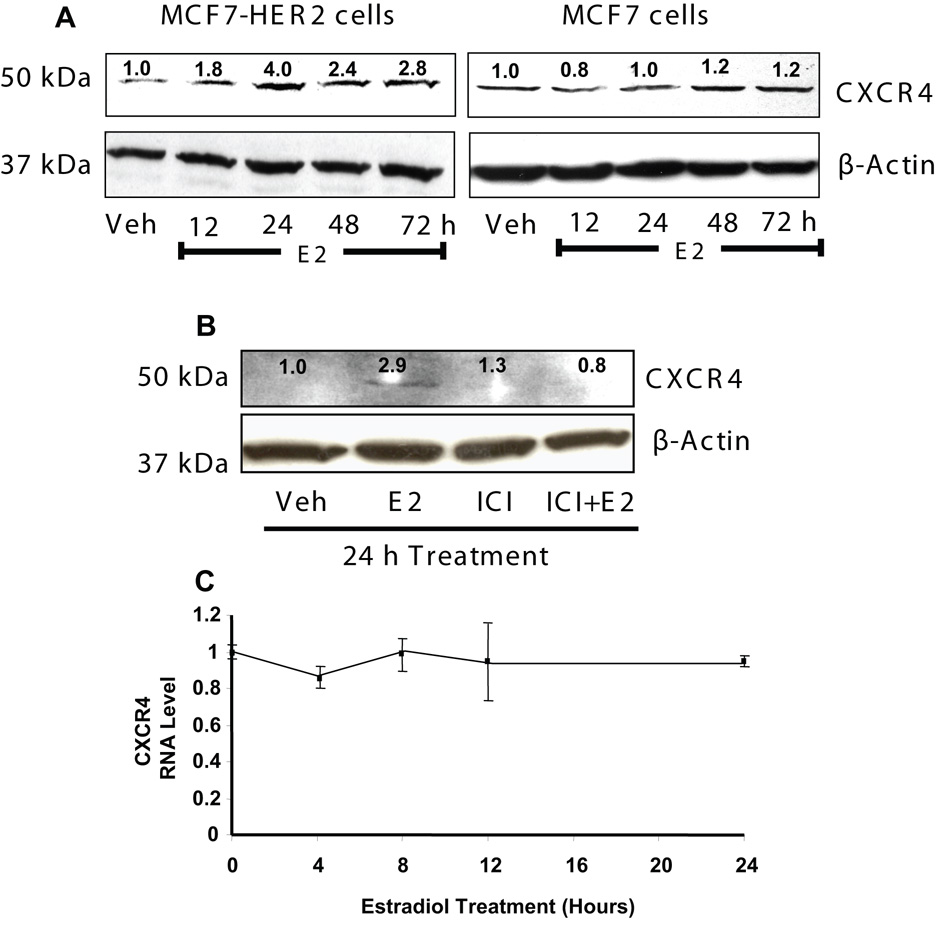
We first examined the effect of estrogen on the levels of CXCR4 protein in MCF7-HER2 cells and in parental MCF7 cells after different times of hormone treatment. In MCF7-HER2 cells, but not MCF7 parental cells, estradiol (E2) elicited a marked increase in CXCR4 protein by 12–24 h that remained elevated over the 72 h of estrogen treatment, as seen by western blot analyses (Fig 1A). This increase in CXCR4 protein in MCF7-HER2 cells was mediated by the estrogen receptor as ICI182,780 (ICI), a pure estrogen antagonist, was able to block the estrogen-induced CXCR4 protein up-regulation (Fig 1B). We also monitored the mRNA level of CXCR4 in these cells by real-time PCR, after different times of estrogen treatment and found that E2 treatment did not change the CXCR4 mRNA level in these cells (Fig 1C). The up-regulation of CXCR4 protein in MCF7-HER2 cells was further confirmed by immunofluorescence (Fig 2), which showed a marked increase in CXCR4 immunofluorescence in these cells after estrogen treatment.
Figure 1.
Estrogen mediated up-regulation of CXCR4 protein in MCF7-HER2 cells, but not in MCF7 parental breast cancer cells. (A) Time course of E2 treatment of MCF7-HER2 cells or MCF7 cells and its effect on CXCR4 protein levels. Cells were treated with vehicle (0.1% ethanol) or E2 (10nM) for times up to 72 h. (B) Effect of the antiestrogen ICI182,780 (ICI) on E2 induced up-regulation of CXCR4 protein. MCF7-HER2 cells were treated with control (0.1% ethanol) vehicle, E2 (10nM), ICI (10−6M) or E2+ICI for 24 h. (C) CXCR4 mRNA levels in MCF7-HER2 cells after treatment with control vehicle or E2 (10nM) for times up to 24 h. mRNA was monitored using quantitative real time PCR. Values are mean ± SD from 3 experiments. In panels A and B, the western immunoblots were scanned and quantified. Levels of CXCR4 normalized for β-actin, relative to vehicle treatments, are indicated above the individual bands of the western blots. The experiments were repeated three times with similar findings.
Figure 2.
Immunofluorescent cytochemistry showing the increase in CXCR4 protein in MCF7-HER2 cells after E2 treatment. Cells were treated with either vehicle or E2 (10nM) for 72 h, fixed and processed for immunofluorescence as described in Materials and Methods. Cells were also stained with DAPI to visualize the nucleus of the cells.
Estrogen increases migration of MCF7-HER2 cells in part through CXCR4 up-regulation
The chemokine receptor CXCR4 is well known to impart migratory potential to cancer cells. To investigate whether the migration of the cells changes after estrogen treatment, we pretreated the MCF7 parental and MCF7-HER2 cells with estrogen or vehicle for 72 h and then studied the migration of the cells, using transwell chambers, towards the attractant, 10% FBS in 1X minimal essential media without phenol red. A large increase (~ 6-fold) in migration of MCF7-HER2 cells was observed after E2 treatment (Fig 3A), whereas no increase in migration was observed in parental MCF7 cells (not shown). We further investigated whether this estrogen-induced increase in migration of MCF7-HER2 cells was due to the increase in CXCR4, by using a specific inhibitor of CXCR4, a small bicyclam molecule AMD3100[22]. In the presence of this inhibitor, estrogen treated MCF7-HER2 cells exhibited significantly less migration than observed in the absence of AMD3100 (Fig 3C and D), implying that the increased migration stimulated by estrogen was partly attributable to the enhanced CXCR4 in the cells.
Figure 3.
Up-regulation of CXCR4 by estrogen is partly responsible for enhanced migration of MCF7-HER2 cells. (A) Cells were treated with either vehicle or E2 (10nM) for 72 h and were grown in serum free medium for 16 h before plating them on the upper chambers of transwell chambers (Corning Inc.). Cells plated on the upper chambers were allowed to migrate towards 10% FBS in the lower chamber for 24 h. (B) Western blot showing the level of CXCR4 protein in the cells under identical treatment conditions. (C) Cells were treated as mentioned in (A) and were allowed to migrate for 24h towards 10% FBS in the lower chamber in the absence or presence of AMD 3100 (1.25µM), a CXCR4 specific inhibitor. (D) Representative photographs of the lower surface of membranes showing the migrated E2 treated cells in absence or presence of AMD 3100. Cells were counted in four microscopic fields per membrane, in duplicate for each condition and repeated three times, with similar results. Values in panels A and C are mean ± SD.
Effect of inhibitors of receptor tyrosine kinase, PI3 kinase, Phospho MAP kinase and mTOR on estrogen-mediated up-regulation of CXCR4 protein in MCF7-HER2 cells
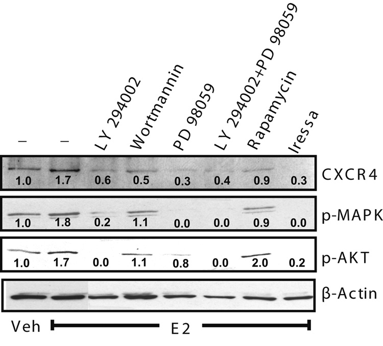
In order to further dissect the involvement of downstream processes, we treated the MCF7-HER2 cells with estrogen in the presence of the EGFR tyrosine kinase inhibitor Iressa (ZD1839), the phosphatidylinositol 3-kinase inhibitors, LY294002 and wortmannin, the MEK1/2 inhibitor PD 98059 or the mTOR inhibitor rapamycin. Cells were also co-treated with both LY294002 and PD 98059. Assessment of CXCR4 protein in these samples showed involvement of PI3K as well as MAPK in the estrogen-mediated up-regulation of CXCR4 in MCF7-HER2 cells. As evident from Figure 4, treatment with the EGFR tyrosine kinase inhibitor, Iressa, completely abrogated the estrogen-induced CXCR4 up-regulation, amd inhibitors of PI3 kinase and MAPK individually or together drastically inhibited the increase in CXCR4 and also reduced the basal level of CXCR4 below that of the vehicle treated cells. Noticeably, treatment with Iressa also considerably inhibited the phosphorylation of MAP kinase and AKT (PKB). This observation agrees with an earlier report where Iressa blocked estrogen-mediated phosphorylation of AKT and MAPK in MCF7-HER2 cells [20].
Figure 4.
Effect of HER2/MAPK/AKT/mTOR pathway inhibitors on estrogen mediated up-regulation of CXCR4 protein levels in MCF7-HER2 cells. Cells were treated with E2 (10nM) for 24 h in the absence or presence of HER2 inhibitor (Iressa, 5µM), PI-3 kinase inhibitors (LY294002, 50µM; wortmannin, 500nM), MAPK inhibitor (PD 98052, 50µM) or mTOR inhibitor (rapamycin, 100nM). Cells were pretreated with inhibitors for 1h before vehicle or E2 was added. The blots were scanned and quantified. Beta-actin normalized values, relative to vehicle treatment, are indicated above the individual bands of the western blots. The experiment was repeated three times with similar findings.
Silencing of tuberin (TSC2) increases CXCR4 in MCF7-HER2 cells
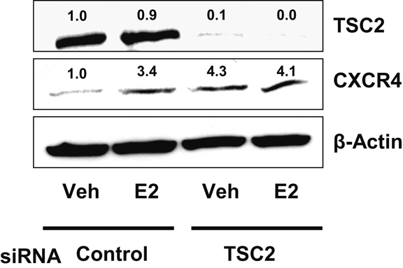
Recent studies have noted that tuberin is just upstream of mTOR and mediates the effects of PI-3 kinase and MAP kinase pathways on mTOR [23]. To examine whether the effect of estrogen on CXCR4 also involves tuberin (TSC2), we used siRNA against tuberin (Smart pool from Dharmacon Inc.) to deplete it from cells and evaluated the CXCR4 expression levels in these MCF7-HER2 cells. As seen in Fig. 5, knockdown of tuberin was very effective (ca. 95% knockdown). Of note, we found that tuberin silencing was enough to up-regulate CXCR4 protein levels in control MCF7-HER2 cells (Fig 5) and estrogen treatment of cells depleted of tuberin did not further increase this 4-fold elevated level of CXCR4, suggesting that the usual estrogen-induced increase in CXCR4 in MCF7-HER2 cells may involve tuberin. These findings imply that TSC2 is normally suppressing CXCR4 and that the rise in CXCR4 after estrogen exposure derives from abrogation of the CXCR4 suppression by TSC2. Because E2 does not affect the level of TSC2 protein (Fig. 5, control siRNA, Veh versus E2 lanes), the observations suggest that E2 treatment may instead change TSC2 activity.
Figure 5.
Effect of siRNA mediated silencing of tuberin (TSC2) on expression of CXCR4 protein in MCF7-HER2 cells. Cells were treated with siRNA against TSC2 or control siRNA (RISC-free, from Dharmacon Inc) before treating with vehicle or E2 (10nM) for 24h. Western blotting was performed for TSC2 and CXCR4. Levels of β-actin were used to assess equal loading. β-actin normalized quantified values, relative to vehicle treatment, are indicated above the individual bands of the western blots. The experiment was repeated three times with similar findings.
DISCUSSION
Amplification or over-expression of HER2 in breast cancers represents a clinically unfavorable condition and is associated with increased proliferation, invasiveness and poor prognosis [24]. Although expression of estrogen receptor is inversely associated with HER2 expression in breast cancers, a subset of HER2 over-expressing breast cancers also express estrogen receptor. One of the largest recent studies has reported that 49% of HER2 positive breast cancers also express estrogen receptor [15]. Poor responsiveness to tamoxifen therapy is also associated with increased HER2 expression in ER-positive breast cancers [25], and this is presumably due to increased cross talk between ER and HER2 [20]. However, the molecular mechanisms and the effector molecules underlying the interactions of these signaling pathways are incompletely understood. The results of our study help in further understanding the estrogen mediated crosstalk between ER and HER2. Our observations show that this crosstalk increases the level of the chemokine receptor CXCR4 protein, by a post-transcriptional mechanism, and that this increase was inhibited by the EGFR tyrosine kinase inhibitor Iressa (gefitinib, ZD 1839) which can block signaling from EGFR and HER2 [20] and by pharmacological inhibitors of PI3-kinase, phospho-MAPK and mTOR.
The ER is expressed in ca. 70% of breast cancers and it is thought to influence the phenotypic properties of these ER-positive tumors, in large part, by broadly impacting the transcription of genes in many functional categories [19, 26–31]. Our findings, reported here, show that, in the presence of HER2, crosstalk between these two receptor pathways allows estrogen to regulate cell behavior by post-transcriptional mechanisms that involve several protein kinase pathways in the up-regulation of the chemokine receptor CXCR4. The involvement of these protein kinase pathways in CXCR4 regulation is of particular interest because of the extranuclear, as well as nuclear, localization and actions of estrogen receptors in breast cancer, and the increasing evidence for involvement of these pathways in contributing to the endocrine therapy refractoriness of breast cancers expressing ER in the context of HER2 over-expression [17, 18, 24, 32–36].
Recent studies have shown that PI3K/AKT and MAPK pathways can coordinately phosphorylate tuberin (TSC2) of tuberous sclerosis complex, and functionally inactivate it by not allowing formation of a complex with hamartin (TSC1). This dissociation of TSC2 leads to the activation of mTOR, which further stimulates the mRNA translational apparatus by phosphorylating its two major substrates, ribosomal p70S6 kinase (S6K) and eukaryotic initiation factor 4E binding protein-1 (4E-BP1) [23]. Our finding that knockdown of TSC2 elevated the level of CXCR4 protein in MCF7-HER2 cells and also nullified further estrogen stimulation of CXCR4, implies involvement of the TSC complex.
Clinically, CXCR4 expression strongly correlates with the degree of lymph node metastasis in breast cancers [37]. In a large breast cancer tissue microarray study, positive staining of CXCR4 in the cytoplasm was also associated with invasiveness as well as HER2 expression [12]. Another study likewise found significant positive correlation between HER2 and CXCR4 expression in primary breast tumor tissues, which was also associated with poor patient survival [18]. These findings strongly imply a role for HER2 over-expression in CXCR4 regulation. However, the exact mechanism of CXCR4 regulation by HER2 is not known. One of the studies showed that PI3K/AKT/mTOR pathway is involved in HER2 mediated translational regulation of CXCR4, and also in inhibiting ligand-induced CXCR4 degradation [18]. The results of our present study support these findings as we show that HER2 is required in estrogen up-regulation of CXCR4 protein levels, without a concurrent increase in mRNA levels and also that this involves the PI3K/AKT/mTOR pathway. In addition, our results indicate that the MAPK pathway is also involved, and we furthermore show that in the case of HER2 over-expression in ER-positive breast cancer cells, CXCR4 protein levels are under additional control of estrogen receptor activation.
Interestingly, some recent studies have also implicated the MAPK pathway, in addition to the PI3K/AKT pathway, in the regulation of translational machinery [23, 38]. Our findings also strongly support this notion because we found that blocking either MAPK or AKT pathways individually or simultaneously resulted in a very marked inhibition of the estrogen-mediated increase in CXCR4 protein.
It is known from previous studies by us and others that SDF1, the ligand for the CXCR4 receptor, is upregulated by estrogen in parental MCF7 breast cancer cells [19, 27, 39]. We find that estrogen also upregulates expression of this ligand in MCF7-HER2 cells. The fact that estrogen upregulates the ligand in both low and high HER2 expressing MCF7 breast cancer cells while the increase in cell migration, indicative of a more aggressive and invasive phenotype is observed only in the MCF7-HER2 cells, implies that up-regulation of the ligand alone is not sufficient for the invasive phenotype. Therefore, we explored the unique estrogen-dependent up-regulation of the CXCR4 receptor that we observed only in the HER2 overexpressing cells and was found to be associated with a post-transcriptional increase in CXCR4 protein. The effect of estrogen in high HER2-expressing, ER-positive cells suggests that crosstalk between ER and HER2 impacts CXCR4 levels, up-regulating them, which is critical in activating the pathways associated with cell migration and invasiveness. Hence, our data reveals a second level of regulation by estrogen beyond the CXCR4 ligand SDF1 that involves crosstalk of HER2 with ER and results in up-regulation of the CXCR4 receptor itself.
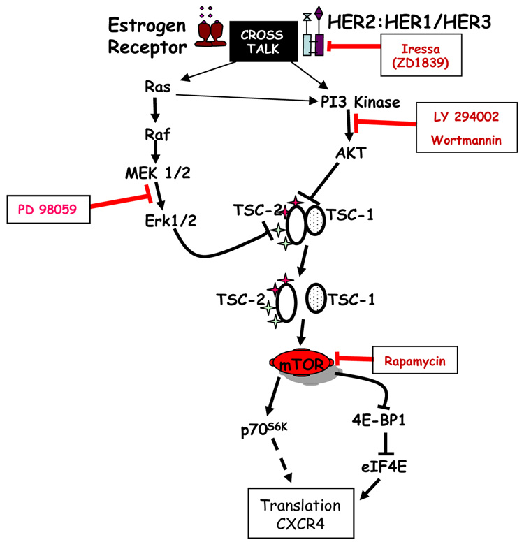
Our present model suggests that PI3K/AKT and MAPK pathways can regulate the translational machinery by converging at tuberous sclerosis complex, which is upstream of mTOR (Fig 6). Activated PI3K/AKT and MAPK pathways phosphorylate TSC2 (tuberin), a component of tuberous sclerosis complex, which leads to its dissociation from TSC1 (hamartin). This dissociation is known to impair the ability of TSC2 to inhibit mTOR activity [23]. Activated mTOR can phosphorylate its two major substrates, ribosomal protein p70S6 kinase (S6K) and eukaryotic initiation factor 4E binding protein-1 (4E-BP1), which are responsible for 5’ TOP mRNAs and 5’ cap dependent translation, respectively [40]. We found the estrogen-mediated, translational up-regulation of CXCR4 in this study to be inhibited by rapamycin, suggesting involvement of mTOR.
Figure 6.
Schematic diagram depicting our findings on the post-transcriptional regulation of CXCR4 in MCF7-HER2 cells as a result of estrogen and HER2 cross-talk and the involvement of activated PI3K and MAPK pathways and TSC2 (tuberin). Disruption of TSC1 and TSC2 interaction is known to activate the mTOR pathway which phosphorylates two key downstream proteins, 4E-BP1 and p70 S6 kinase, which are responsible for increased translation and protein synthesis. Inhibitors used to assess involvement of different components of these pathways are indicated. See Discussion for further details.
Since the PI3K/AKT and MAPK signaling pathways converge at TSC2, and its dissociation from TSC1 is a key event in the activation of mTOR, we further confirmed the involvement of TSC2 in estrogen-mediated up-regulation of CXCR4 protein, by silencing TSC2 with siRNA. Indeed, TSC2 knockdown increased the CXCR4 protein level in these MCF7-HER2 cells, and estrogen was not able to further increase this elevated level of CXCR4, suggesting that estrogen-driven CXCR4 protein up-regulation likely involves this translational pathway.
Importantly, a recent xenograft study using the same MCF7-HER2 cells has revealed that these cells are initially responsive to estrogen deprivation or fulvestrant treatment, but eventually develop resistance to both. Treating the animals with Iressa (gefitinib) along with estrogen deprivation or fulvestrant delayed the occurrence of resistance but was not able to prevent it completely [41]. Interestingly, these resistant cells also have elevated levels of p-MAPK and p-AKT, implying that these pathways may contribute in the acquisition of resistance.
In summary, this study reports estrogen-driven post-transcriptional up-regulation of CXCR4 in HER2-overexpressing, ER-positive breast cancer cells. This process critically involves mTOR signaling, activated by PI3K/AKT and MAPK pathways via functional inactivation of TSC2 (tuberin). Thus, estrogen-mediated elevation of CXCR4 protein may play a key role in determining the metastatic potential of ER-positive HER2-overexpressing breast cancer cells. The findings suggest that mTOR and CXCR4 blocking strategies may have a promising therapeutic potential in HER2-overexpressing, ER-positive breast cancers showing de novo or acquired resistance to endocrine therapies.
ACKNOWLEDGEMENTS
This work was supported by NIH grant CA 18119 and a grant from The Breast Cancer Research Foundation (to B.S.K.), and by NIH Breast Cancer SPORE grant P50 CA8183 (to R.S.).
Abbreviations
- E2
17β-estradiol
- ER
estrogen receptor
- ICI
ICI 182,780
- CXCR4
chemokine receptor (CXC) 4
- PI3K
phosphatidylinositol-3-kinase
- MAPK
mitogen-activated protein kinase
- mTOR
mammalian target of rapamycin
REFERENCES
- 1.Muller A, Homey B, Soto H, Ge N, Catron D, Buchanan ME, McClanahan T, Murphy E, Yuan W, Wagner SN, Barrera JL, Mohar A, Verastegui E, Zlotnik A. Involvement of chemokine receptors in breast cancer metastasis. Nature. 2001;410:50–56. doi: 10.1038/35065016. [DOI] [PubMed] [Google Scholar]
- 2.Konecny G, Pauletti G, Pegram M, Untch M, Dandekar S, Aguilar Z, Wilson C, Rong HM, Bauerfeind I, Felber M, Wang HJ, Beryt M, Seshadri R, Hepp H, Slamon DJ. Quantitative association between HER-2/neu and steroid hormone receptors in hormone receptor-positive primary breast cancer. J Natl Cancer Inst. 2003;95:142–153. doi: 10.1093/jnci/95.2.142. [DOI] [PubMed] [Google Scholar]
- 3.Slamon DJ, Clark GM, Wong SG, Levin WJ, Ullrich A, McGuire WL. Human breast cancer: correlation of relapse and survival with amplification of the HER-2/neu oncogene. Science. 1987;235:177–182. doi: 10.1126/science.3798106. [DOI] [PubMed] [Google Scholar]
- 4.Yu D, Hung MC. Overexpression of ErbB2 in cancer and ErbB2-targeting strategies. Oncogene. 2000;19:6115–6121. doi: 10.1038/sj.onc.1203972. [DOI] [PubMed] [Google Scholar]
- 5.Chen Y, Stamatoyannopoulos G, Song CZ. Down-regulation of CXCR4 by inducible small interfering RNA inhibits breast cancer cell invasion in vitro. Cancer Res. 2003;63:4801–4804. [PubMed] [Google Scholar]
- 6.Lapteva N, Yang AG, Sanders DE, Strube RW, Chen SY. CXCR4 knockdown by small interfering RNA abrogates breast tumor growth in vivo. Cancer Gene Ther. 2005;12:84–89. doi: 10.1038/sj.cgt.7700770. [DOI] [PubMed] [Google Scholar]
- 7.Smith MC, Luker KE, Garbow JR, Prior JL, Jackson E, Piwnica-Worms D, Luker GD. CXCR4 regulates growth of both primary and metastatic breast cancer. Cancer Res. 2004;64:8604–8612. doi: 10.1158/0008-5472.CAN-04-1844. [DOI] [PubMed] [Google Scholar]
- 8.Liang Z, Wu T, Lou H, Yu X, Taichman RS, Lau SK, Nie S, Umbreit J, Shim H. Inhibition of breast cancer metastasis by selective synthetic polypeptide against CXCR4. Cancer Res. 2004;64:4302–4308. doi: 10.1158/0008-5472.CAN-03-3958. [DOI] [PubMed] [Google Scholar]
- 9.Liang Z, Yoon Y, Votaw J, Goodman MM, Williams L, Shim H. Silencing of CXCR4 blocks breast cancer metastasis. Cancer Res. 2005;65:967–971. [PMC free article] [PubMed] [Google Scholar]
- 10.Kang Y, Siegel PM, Shu W, Drobnja kM, Kakonen SM, Cordon-Cardo C, Guise TA, Massague J. A multigenic program mediating breast cancer metastasis to bone. Cancer Cell. 2003;3:537–549. doi: 10.1016/s1535-6108(03)00132-6. [DOI] [PubMed] [Google Scholar]
- 11.Minn AJ, Gupta GP, Siegel PM, Bos PD, Shu W, Giri DD, Viale A, Olshen AB, Gerald WL, Massague J. Genes that mediate breast cancer metastasis to lung. Nature. 2005;436:518–524. doi: 10.1038/nature03799. [DOI] [PMC free article] [PubMed] [Google Scholar]
- 12.Salvucci O, Bouchard A, Baccarelli A, Deschenes J, Sauter G, Simon R, Bianchi R, Basik M. The role of CXCR4 receptor expression in breast cancer: a large tissue microarray study. Breast Cancer Res Treat. 2006;97:275–283. doi: 10.1007/s10549-005-9121-8. [DOI] [PubMed] [Google Scholar]
- 13.Slamon DJ, Leyland-Jones B, Shak S, Fuchs H, Paton V, Bajamonde A, Fleming T, Eiermann W, Wolter J, Pegram M, Baselga J, Norton L. Use of chemotherapy plus a monoclonal antibody against HER2 for metastatic breast cancer that overexpresses HER2. N Engl J Med. 2001;344:783–792. doi: 10.1056/NEJM200103153441101. [DOI] [PubMed] [Google Scholar]
- 14.Read LD, Keith D, Jr, Slamon DJ, Katzenellenbogen BS. Hormonal modulation of HER-2/neu protooncogene messenger ribonucleic acid and p185 protein expression in human breast cancer cell lines. Cancer Res. 1990;50:3947–3951. [PubMed] [Google Scholar]
- 15.Lal P, Tan LK, Chen B. Correlation of HER-2 status with estrogen and progesterone receptors and histologic features in 3,655 invasive breast carcinomas. Am J Clin Pathol. 2005;123:541–546. doi: 10.1309/YMJ3-A83T-B39M-RUT9. [DOI] [PubMed] [Google Scholar]
- 16.Osborne CK, Shou J, Massarweh S, Schiff R. Crosstalk between estrogen receptor and growth factor receptor pathways as a cause for endocrine therapy resistance in breast cancer. Clin Cancer Res. 2005;11:865s–870s. [PubMed] [Google Scholar]
- 17.Benz CC, Scott GK, Sarup JC, Johnson RM, Tripathy D, Coronado E, Shepard HM, Osborne CK. Estrogen-dependent, tamoxifen-resistant tumorigenic growth of MCF-7 cells transfected with HER2/neu. Breast Cancer Res Treat. 1992;24:85–95. doi: 10.1007/BF01961241. [DOI] [PubMed] [Google Scholar]
- 18.Li YM, Pan Y, Wei Y, Cheng X, Zhou BP, Tan M, Zhou X, Xia W, Hortobagyi GN, Yu D, Hung MC. Upregulation of CXCR4 is essential for HER2-mediated tumor metastasis. Cancer Cell. 2004;6:459–469. doi: 10.1016/j.ccr.2004.09.027. [DOI] [PubMed] [Google Scholar]
- 19.Frasor J, Danes JM, Komm B, Chang KC, Lyttle CR, Katzenellenbogen BS. Profiling of estrogen up- and down-regulated gene expression in human breast cancer cells: insights into gene networks and pathways underlying estrogenic control of proliferation and cell phenotype. Endocrinology. 2003;144:4562–4574. doi: 10.1210/en.2003-0567. [DOI] [PubMed] [Google Scholar]
- 20.Shou J, Massarweh S, Osborne CK, Wakeling AE, Ali S, Weiss H, Schiff R. Mechanisms of tamoxifen resistance: increased estrogen receptor-HER2/neu cross-talk in ER/HER2-positive breast cancer. J Natl Cancer Inst. 2004;96:926–935. doi: 10.1093/jnci/djh166. [DOI] [PubMed] [Google Scholar]
- 21.Livak KJ, Schmittgen TD. Analysis of relative gene expression data using real-time quantitative PCR and the 2(-Delta Delta C(T)) Method. Methods. 2001;25:402–408. doi: 10.1006/meth.2001.1262. [DOI] [PubMed] [Google Scholar]
- 22.De Clercq E. The bicyclam AMD3100 story. Nat Rev Drug Discov. 2003;2:581–587. doi: 10.1038/nrd1134. [DOI] [PubMed] [Google Scholar]
- 23.Ma L, Chen Z, Erdjument-Bromage H, Tempst P, Pandolfi PP. Phosphorylation and functional inactivation of TSC2 by Erk: implications for tuberous sclerosis and cancer pathogenesis. Cell. 2005;121:179–193. doi: 10.1016/j.cell.2005.02.031. [DOI] [PubMed] [Google Scholar]
- 24.Berger MS, Locher GW, Saurer S, Gullick WJ, Waterfield MD, Groner B, Hynes NE. Correlation of c-erbB-2 gene amplification and protein expression in human breast carcinoma with nodal status and nuclear grading. Cancer Res. 1988;48:1238–1243. [PubMed] [Google Scholar]
- 25.Gago FE, Fanelli MA, Ciocca DR. Co-expression of steroid hormone receptors (estrogen receptor alpha and/or progesterone receptors) and Her2/neu (c-erbB-2) in breast cancer: clinical outcome following tamoxifen-based adjuvant therapy. J Steroid Biochem Mol Biol. 2006;98:36–40. doi: 10.1016/j.jsbmb.2005.07.002. [DOI] [PubMed] [Google Scholar]
- 26.Carroll JS, Meyer CA, Song J, Li W, Geistlinger TR, Eeckhoute J, Brodsky AS, Keeton EK, Fertuck KC, Hall GF, Wang Q, Bekiranov S, Sementchenko V, Fox EA, Silver PA, Gingeras TR, Liu XS, Brown M. Genome-wide analysis of estrogen receptor binding sites. Nat Genet. 2006;38:1289–1297. doi: 10.1038/ng1901. [DOI] [PubMed] [Google Scholar]
- 27.Frasor J, Stossi F, Danes JM, Komm B, Lyttle CR, Katzenellenbogen BS. Selective estrogen receptor modulators: Discrimination of agonistic versus antagonistic activities by gene expression profiling in breast cancer cells. Cancer Res. 2004;64:1522–1533. doi: 10.1158/0008-5472.can-03-3326. [DOI] [PubMed] [Google Scholar]
- 28.Green KA, Carroll JS. Oestrogen-receptor-mediated transcription and the influence of co-factors and chromatin state. Nat Rev Cancer. 2007;7:713–722. doi: 10.1038/nrc2211. [DOI] [PubMed] [Google Scholar]
- 29.Lin CY, Vega VB, Thomsen JS, Zhang T, Kong SL, Xie M, Chiu KP, Lipovich L, Barnett DH, Stossi F, Yeo A, George J, Kuznetsov VA, Lee YK, Charn TH, Palanisamy N, Miller LD, Cheung E, Katzenellenbogen BS, Ruan Y, Bourque G, Wei CL, Liu ET. Whole-genome cartography of estrogen receptor alpha binding sites. PLoS Genet. 2007;3:e87. doi: 10.1371/journal.pgen.0030087. [DOI] [PMC free article] [PubMed] [Google Scholar]
- 30.Barnett DH, Sheng S, Howe Charn T, Waheed A, Sly WS, Lin C-Y, Liu ET, Katzenellenbogen BS. Estrogen receptor regulation of carbonic anhydrase XII through a distal enhancer in breast cancer. Cancer Res. 2008;68:3505–3515. doi: 10.1158/0008-5472.CAN-07-6151. [DOI] [PubMed] [Google Scholar]
- 31.Katzenellenbogen BS, Frasor J. Therapeutic targeting in the estrogen receptor hormonal pathway. Semin Oncol. 2004;31:28–38. doi: 10.1053/j.seminoncol.2004.01.004. [DOI] [PubMed] [Google Scholar]
- 32.Harrington WR, Kim SH, Funk CC, Madak-Erdogan Z, Schiff R, Katzenellenbogen JA, Katzenellenbogen BS. Estrogen dendrimer conjugates that preferentially activate extranuclear, nongenomic versus genomic pathways of estrogen action. Mol Endocrinol. 2006;20:491–502. doi: 10.1210/me.2005-0186. [DOI] [PubMed] [Google Scholar]
- 33.Madak-Erdogan Z, Kieser KJ, Kim SH, Komm B, Katzenellenbogen JA, Katzenellenbogen BS. Nuclear and Extranuclear Pathway Inputs in the Regulation of Global Gene Expression by Estrogen Receptors. Mol Endocrinol. 2008;22:2116–2127. doi: 10.1210/me.2008-0059. [DOI] [PMC free article] [PubMed] [Google Scholar]
- 34.Massarweh S, Schiff R. Unraveling the mechanisms of endocrine resistance in breast cancer: new therapeutic opportunities. Clin Cancer Res. 2007;13:1950–1954. doi: 10.1158/1078-0432.CCR-06-2540. [DOI] [PubMed] [Google Scholar]
- 35.Osborne CK, Schiff R. Estrogen-receptor biology: continuing progress and therapeutic implications. J Clin Oncol. 2005;23:1616–1622. doi: 10.1200/JCO.2005.10.036. [DOI] [PubMed] [Google Scholar]
- 36.Pietras RJ, Marquez-Garban DC. Membrane-associated estrogen receptor signaling pathways in human cancers. Clin Cancer Res. 2007;13:4672–4676. doi: 10.1158/1078-0432.CCR-07-1373. [DOI] [PubMed] [Google Scholar]
- 37.Kato M, Kitayama J, Kazama S, Nagawa H. Expression pattern of CXC chemokine receptor-4 is correlated with lymph node metastasis in human invasive ductal carcinoma. Breast Cancer Res. 2003;5:R144–R150. doi: 10.1186/bcr627. [DOI] [PMC free article] [PubMed] [Google Scholar]
- 38.Kelleher RJ, 3rd, Govindarajan A, Jung HY, Kang H, Tonegawa S. Translational control by MAPK signaling in long-term synaptic plasticity and memory. Cell. 2004;116:467–479. doi: 10.1016/s0092-8674(04)00115-1. [DOI] [PubMed] [Google Scholar]
- 39.Rae JM, Johnson MD, Scheys JO, Cordero KE, Larios JM, Lippman ME. GREB1 is a critical regulator of hormone dependent breast cancer growth. Breast Cancer Res Treat. 2005;92:141–149. doi: 10.1007/s10549-005-1483-4. [DOI] [PubMed] [Google Scholar]
- 40.Astrinidis A, Henske EP. Tuberous sclerosis complex: linking growth and energy signaling pathways with human disease. Oncogene. 2005;24:7475–7481. doi: 10.1038/sj.onc.1209090. [DOI] [PubMed] [Google Scholar]
- 41.Massarweh S, Osborne CK, Jiang S, Wakeling AE, Rimawi M, Mohsin SK, Hilsenbeck S, Schiff R. Mechanisms of tumor regression and resistance to estrogen deprivation and fulvestrant in a model of estrogen receptor-positive, HER-2/neu-positive breast cancer. Cancer Res. 2006;66:8266–8273. doi: 10.1158/0008-5472.CAN-05-4045. [DOI] [PubMed] [Google Scholar]


