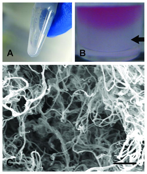
Figure 3.
Myocardial matrix gelation and characterization. (A) At room temperature the solubilized matrix was a liquid. (B) At 37 °C, the myocardial matrix self-assembled into a hydrogel, as indicated by the arrow. Pink media is shown on top as a contrast to the solidified gel. (C) Scanning electron micrograph of a cross-section of the myocardial matrix gel with nanofibers approximately 40-100 nm. Scale bar is 1 μm.

