Abstract
Purpose
The ovarian carcinoma subline A2780/C10B (C10B) is an oxaliplatin resistant clone derived from the human ovarian carcinoma cell line A2780. The C10B cells are characterized by mesenchymal phenotype, decreased platinum uptake and increased glutathione levels (Hector et al. in Cancer Lett 245:195–204, 2007; Varma et al. in Oncol Rep 14:925–932, 2005). Na,K-ATPase-β subunit (Na,K-β1) functions as a cell–cell adhesion molecule in epithelial cells and is reduced in a variety of carcinoma cells that show mesenchymal phenotype. The purpose of this study is to evaluate the relationship between Na,K-β expression and sensitivity to oxaliplatin.
Methods
Cell lines used include A2780, C10B, C10B transfected with Na,K-β1 (C10B-Na,K-β) and a canine kidney carcinoma cell line MSV-MDCK also transfected with Na,K-β1 (MSV-MDCK-β subunit). Cytotoxicity studies were performed by sulforhodamine-blue assay. The Na,K-α1 and Na,K-β1 subunit localization and expression were by immunofluorescence microscopy and Western blot analysis. Platinum accumulation measurements were by atomic absorption spectrophotometry.
Results
C10B cells express highly reduced levels of Na,K-β1 subunit. Exogenous expression of Na,K-β1 increased platinum accumulation and sensitized C10B cells to oxaliplatin. The pharmacological inhibitor of Na,K-ATPase ouabain did not alter the oxaliplatin accumulation indicating that Na,K-β1 sensitizes cells in a Na,K-ATPase enzyme activity independent manner. These findings were also confirmed in MSV-MDCK-β subunit cells.
Conclusions
This study for the first time reveals that reduced expression of the Na,K-β1 protein is associated with oxaliplatin resistance in cancer cells and demonstrates a novel role for this protein in sensitizing the cells to oxaliplatin. This study suggests a potentially important role for Na,K-β1 in both prognosis and therapy of oxaliplatin resistant malignancies.
Introduction
Platinum drugs are central to many chemotherapy regimens in cancer treatment. Oxaliplatin is active in many cisplatin responsive cancers including ovarian [24], and approved for colon cancer, a cisplatin non-responsive cancer, because of its unique activity when administered in combination with 5-fluorouracil [35]. Like other forms of chemotherapy while responsive initially, platinum drug treatment can eventually result in tumor resistance. Resistance to platinum drugs, has been extensively studied especially for cisplatin and is multifactorial in nature [2, 12]. Mechanisms of platinum drug resistance include drug accumulation deficiencies, intracellular detoxification, increased repair of DNA-Platinum adducts, increased tolerance to the drug and alterations in downstream signaling affecting cell death pathways [2, 12, 25]. A variety of drug resistant carcinoma cell models including of human ovarian, colon, bladder and lung have been used in these studies. Studies from our laboratory using A2780 human ovarian carcinoma cells indicated that the mechanisms of oxaliplatin resistance are similar to those for cisplatin, and primarily include decreased drug accumulation and increased detoxification by conjugation with glutathione [15, 16]. These studies suggested that reduced DNA-Platinum adduct formation was a consequence of the reduced drug accumulation [15, 16]. Additionally, recent studies from our laboratory and those of others using A2780 ovarian carcinoma cells suggest that the extracellular matrix (ECM) may play a role in the resistance to oxaliplatin and cisplatin [16, 30, 34].
While mechanisms of drug resistance are extensively studied, factors that sensitize cancer cells to cytotoxic drugs are less developed and poorly understood. Epithelial to mesenchymal transition (EMT) is one of the mechanisms involved in carcinoma cell progression to metastatic and drug resistant cancer [32]. During this transition, epithelial cells lose morphology and expression of epithelial markers such as cytokeratins, and show fibroblastic phenotype characterized by increased expression of mesenchymal markers such as vimentin and fibronectin [13]. In colorectal carcinoma cell lines KM12L4 and HT-29, induction of EMT was reported to be associated with oxaliplatin resistance [36]. The A2780/C10B (C10B) is a clonal subline, selected for resistance to oxaliplatin from the parental A2780 human ovarian carcinoma cells [16]. As reported previously [16], the C10B clone was isolated using limit-dilution procedure from A2780/C10, an oxaliplatin resistant cell line derived from A2780 [9] by plating of single cell suspension and isolating a colony of >50 cells that was expanded. The C10B cells are spindle shaped and fibroblast-like in morphology as described in EMT with a lack of epithelial membrane antigen (EMA), whereas the parental cells exhibit epithelial phenotype with the presence of EMA [16]. Consistent with the phenotype, C10B cells have elevated levels of vimentin, and reduced levels of cytokeratins [16]. Affymetrix gene expression analysis demonstrated that the C10B cells have high expression of collagen 6, matrix metalloproteinases and TGF-β2 [34]. While it is reported that chemoresistance to paclitaxel induces EMT in ovarian carcinoma cells [21], this phenomenon is not well documented in this tumor type for oxaliplatin resistance.
The Na,K-ATPase, also known as the sodium pump is a plasma membrane bound, oligomeric enzyme localized to the basolateral plasma membrane in most epithelial cells. It is composed of two essential, non-covalently bound subunits, α1 (Na,K-α1) and β1 (Na,K-β1). Na,K-α1 is the catalytic subunit, whereas Na,K-β1 is involved in the translation, stability and transport of Na,K-α1 on the plasma membrane [27]. Na,K-ATPase transports two K+ in and three Na+ out at the expense of one molecule of ATP. It not only maintains intracellular ion homeostasis but is also critical for the maintenance of polarized phenotype of epithelial cells [26]. Ouabain is a well studied pharmacological inhibitor of Na,K-ATPase which specifically binds to Na,K-α with an IC50 of 10 nM in most human cell types [19]. Well differentiated epithelial cells express high levels of Na,K-β1, whereas poorly differentiated carcinoma cells with mesenchymal phenotype express reduced levels of this protein [11, 28]. Na,K-β1 levels were drastically reduced in highly motile and invasive carcinoma cell lines [11] and correlate with their invasiveness and metastatic potential [18, 28]. Restoration of Na,K-β1 in Moloney Sarcoma virus transformed Madin–Darby canine kidney cells (MSV-MDCK) suppressed motility, invasion [6, 28] and tumorigenicity [18]. These studies strongly indicated a functional link between reduced Na,K-β1 expression and cancer progression.
In this study, we tested the hypothesis that reduced expression of Na,K-β1 in C10B is associated with oxaliplatin resistance by evaluating the relationship between oxaliplatin sensitivity, Na,K-β1 expression, and platinum accumulation in C10B cells in comparison with the parental A2780 and C10B cells transfected with Na,K-β1 subunit (C10B-Na,K-β). Results presented here reveal that the resistant C10B cells have reduced expression of Na,K-β1. Exogenous expression of Na,K-β1 results in increased oxaliplatin uptake and cellular sensitivity to the drug, independent of Na,K-ATPase enzyme activity.
Materials and methods
Drugs
Oxaliplatin was a gift from Dr. Paul Juniewicz of Sanofi-Synthelabo (Malvern, PA).
Cell lines
The ovarian carcinoma cell line A2780 was a gift from Dr. Ozols (Fox Chase Cancer Center, Philadelphia, PA). The oxaliplatin resistant C10B subline was derived as a single cell clone from a previously described A2780/C10 cell line [9]. A pooled clone of C10B cells stably expressing Na,K-β1 (C10B-Na,K-β) was generated by transfecting the C10B cells with human Na,K-β1 cDNA in a mammalian expression vector with neomycin resistance (pcDNA3.1 from Invitrogen, Carlsbad, CA). C10B cells transfected with the empty vector (C10B-Vector) were used as a control. The ovarian carcinoma cell lines and the C10B-Na,K-β cells were maintained in RPMI 1640 supplemented with 10% heat inactivated fetal bovine serum (FBS) and 1% l-glutamine (Invitrogen, Grand Island, NY). Other cell lines used in the study included MSV-MDCK cells stably transfected with pcDNA3.1 vector (MSV-MDCK-vector) or Na,K-β1 (MSV-MDCK-β subunit). Generation of these cell lines has been described previously [28]. These cells were grown in DMEM with 10% FBS, 1% penicillin and streptomycin and 200 μg/ml neomycin.
Cytotoxicity assays
Cytotoxicity analysis was carried out using sulforhodamine-blue (SRB) assay as described previously [16]. Cells were continuously exposed to oxaliplatin for 72 h, fixed and subjected to the SRB assay. The optical density (OD) was read at 570 nm (EL309 Automated Microplate Reader, Bio-Tek Instruments, Winooski, VT) and percent survival was determined as [OD570 (treated cells)/OD570 (untreated cells)] × 100.
Platinum accumulation
Cells were exposed to oxaliplatin (100 μM) for 2 h, washed thoroughly with PBS and total intracellular platinum was measured as previously described [15]. For this drug exposed cells were trypsinized and centrifuged into a pellet and lysed in 0.5 ml of 0.1% Triton X-100 and 0.2% nitric acid overnight and sonicated for 1 min. Platinum in the cell lysate was measured by graphite furnace atomic absorption spectrophotometry with Zeeman background correction (PE 4100ZL, Norwalk, CT). Protein was measured using the Bradford assay. In experiments aimed at Na,K-ATPase inhibition, cells were pretreated with ouabain (50 μM) for 15 min prior to oxaliplatin exposure with a continued incubation with ouabain (50 μM) also during the exposure to oxaliplatin.
Immunofluorescence
Cells grown on cover slips were fixed with chilled methanol, incubated with antibodies against Na,K-β1 or Na,K-α1 and stained with Alexa Fluor 488 conjugated goat anti-mouse antibody (Invitrogen). Samples were excited with an argon laser, and the light emitted around 519 nm was recorded. Images were obtained using a confocal microscope Leica TCS SP5 (Leica Microsystems Inc., Bannockburn, IL).
Reverse transcription and quantitative real-time PCR
Total RNA was isolated from A2780, C10B and C10B-Na,K-β cells using the RNA Aqueous kit (Ambion, Austin, TX). RNA concentration was measured using the Nanodrop Spectrophotometer (Thermo Fisher Scientific, Waltham, MA). First strand cDNA was generated from total RNA using the High Capacity cDNA kit (Applied Biosystems, Foster City, CA). One microgram of RNA was reverse transcribed to cDNA according to the manufacturer’s protocol. Quantitative RT-PCR analysis of ATP1B1 was performed using the ATP1B1 gene specific TaqMan MGB assay and TaqMan universal master mix on an ABI 7900HT System from Applied Biosystems. The ‘comparative threshold’ method was used to calculate relative gene expression. Values were normalized against 18S rRNA.
Western blot analysis
Immunoblot analysis was performed as described previously [28]. Cells were lysed in a buffer containing 20 mM Tris–HCl, pH 7.4, 100 mM NaCl, 1% Triton X-100, 1 mM EDTA, 1 mM EGTA, 1 mM sodium glycerolphosphate, 1 mM sodium orthovanadate, 1 mM PMSF, and 5 μg/ml each of antipain, leupeptin and pepstatin. The lysates were sonicated, and clarified by centrifugation at 13,000 rpm for 10 min at 4°C. Total protein was estimated from the supernatants using the Bio-Rad DC reagent (Bio-Rad, Hercules, CA) as per manufacturer’s instructions. Total protein (50 μg) was resolved on SDS-PAGE and transferred to nitrocellulose membrane (Schleicher & Schuell, Keene, NH). Immunoblotting was performed using a primary antibody, and HRP-conjugated secondary antibody diluted in tris-buffered saline containing 5% non-fat dried milk and 0.1% Tween20. The proteins were detected by using the enhanced chemiluminescence lighting system according to the manufacturer’s recommendations (PerkinElmer Life and Analytical Sciences, Boston, MA). The bands from two independent experiments were quantified using GeneTools software from PerkinElmer, Waltham, MA. Na,K-β1 and Na,K-α1 antibodies were a gift from Dr. William James Ball Jr., University of Cincinnati, Cincinnati. Anti-fibronectin and E-cadherin antibodies were from BD Biosciences, San Jose, CA. β-Actin (Sigma, St. Louis, MO) was used as a loading control.
Statistical analysis
Significance of differences in cytotoxicity and platinum accumulation was evaluated using Student’s t test (Systat Software Inc., Richmond, CA).
Results
Expression of Na,K-β1 in A2780, C10B and C10B-Na,K-β cells
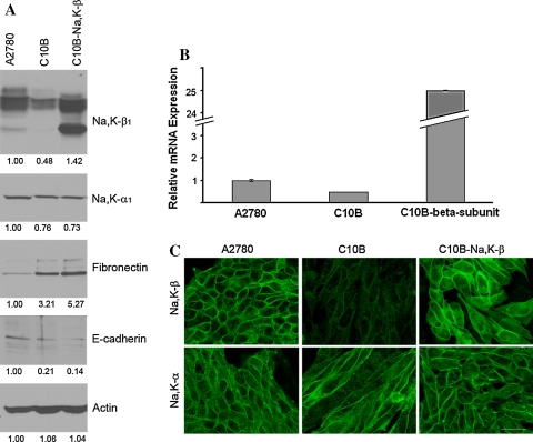
A2780 cells express high levels of Na,K-β1 compared to C10B (Fig. 1a). Exogenous expression of Na,K-β1 in C10B cells increased Na,K-β1 levels comparable to its expression in A2780 (Fig. 1a). There was a 50% reduction in Na,K-β1 transcript levels in C10B cells compared to A2780 cells (Fig. 1b) consistent with the reduction at the protein level. There was no significant change in the Na,K-α1 levels in all these cell lines (Fig. 1a). However, they were similar between C10B and C10B-Na,K-β cells (Fig. 1a). As expected both Na,K-α1 and Na,K-β1 were localized to the plasma membrane in A2780 and C10B-NaK-β1 cells (Fig. 1c). Consistent with the immunoblot analysis, C10B cells revealed highly reduced levels of Na,K-β1 (Fig. 1c). Morphologically, C10B cells showed a fibroblastic phenotype, whereas, the A2780 were adherent to each other and showed epithelial like morphology (Fig. 1c). Interestingly, following Na,K-β1 expression C10B cells grew adherent to each other and appeared morphologically similar to A2780 cells (Fig. 1c). Expression of mesenchymal marker fibronectin is one of the hallmarks of EMT [17]. Consistent with the fibroblastic morphology, the C10B cells expressed high levels of fibronectin which was present at a lower level in parental A2780 cells (Fig. 1a). On the other hand, the expression of the epithelial marker, E-cadherin was greatly reduced in C10B cells (Fig. 1a). Although Na,K-β1 expression induced morphological changes there was no change in the levels of fibronectin and E-cadherin compared to C10B cells (Fig. 1b). This result indicates that exogenous expression of Na,K-β1 in these cells did not reverse the EMT.
Fig. 1.
a Immunoblot analysis of Na,K-ATPase subunits, fibronectin, E-cadherin and actin in parental A2780, C10B and C10B-Na,K-β cells. Densitometric quantitations are included under each panel. b Relative mRNA expression using quantitative real-time PCR. c Morphological characterization and localization of Na,K-ATPase subunits by phase contrast and epifluorescence microscopy. Bar 20 μm
Increased sensitivity to oxaliplatin in Na,K-β1 transfected C10B cells
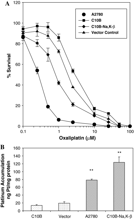
The sensitivity profiles of the parental A2780 cells to oxaliplatin along with the resistant C10B, the C10B-Na,K-β and the vector control are shown in Fig. 2a. The C10B-Na,K-β cells (IC50 = 1 μM) were significantly sensitized to oxaliplatin as compared to the resistant C10B cells (IC50 = 3 μM) or the vector control (IC50 = 2 μM) although their sensitivity was not as high as the parental A2780 cells (IC50 = 0.2 μM). The enhanced sensitivity of C10B-Na,K-β cells to oxaliplatin was statistically significant compared to C10B cells or the vector transfected cells (P < 0.01).
Fig. 2.
a Growth inhibition dose response curves for A2780, C10B, C10B-Na,K-β and vector control following a 72 h exposure to oxaliplatin. Data represents percent growth as determined by (OD 570 of treated cells/OD570 of untreated cells) × 100. Data represent mean ± SD, n = 3. IC50’s A2780: 0.2 μM; C10B: 3 μM; C10B-Na,K-β: 1 μM and Vector: 2 μM. b Accumulation of Pt in C10B, Vector control, A2780 and C10B-Na,K-β following oxaliplatin treatment. Platinum accumulation following exposure to 100 μM oxaliplatin for 2 h. Data represent mean ± SD, n = 3. The ** represent statistically highly significant difference compared to C10B or vector (P < 0.001). The difference between A2780 and C10B-Na,K-β is also statistically significant (P = 0.005)
Increased drug accumulation in Na,K-β1 transfected C10B cells
We previously reported that C10B cells are deficient in drug accumulation compared to the parental A2780 cells [16]. Since the C10B-Na,K-β cells exhibit increased sensitivity to oxaliplatin compared to the resistant, C10B cells, we investigated if there was an altered drug accumulation in these cells. C10B-Na,K-β cells showed about sixfold higher platinum accumulation as compared to the C10B or the vector cells (Fig. 2b). This increase was highly statistically significant (P < 0.001). The amount of platinum accumulated by C10B-Na,K-β cells was even higher than the parental cells under identical conditions of treatment (P = 0.005). These results suggest that there is a positive correlation between Na,K-β1 expression and increased platinum accumulation.
Confirmation of the sensitization and drug accumulation phenomenon in MSV-MDCK-β subunit cells
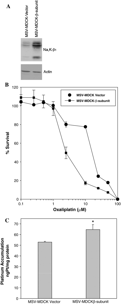
Sensitization to oxaliplatin by exogenous expression of Na,K-β1 was investigated in another cell line to test whether this phenomenon is specific to ovarian cells or occurs in other cell type as well. MSV-MDCK cells expresses low levels of Na,K-β1 and are highly invasive and metastatic (Fig. 3a). Exogenous expression of Na,K-β1 in MSV-MDCK cells (MSV-MDCK-β subunit) reduced their motility, invasiveness and tumorigenic potential [28]. Cytotoxicity and platinum accumulation studies indicated that MSV-MDCK-β subunit cells were more sensitive to oxaliplatin as compared to the parental cells carrying the vector and do not express this protein (Fig. 3b). Further studies confirmed a statistically significant (P < 0.05) increase in the accumulation of platinum in MSV-MDCKβ cells (Fig. 3c) indicating that Na,K-β1 expression facilitates oxaliplatin uptake and increases sensitivity to this drug.
Fig. 3.
a Immunoblot showing levels of Na,K-β1 in MSV-MDCK cells. b Growth inhibition dose response curves for MSV-MDCK vector and MSV-MDCK-β-subunit following 72 h exposure to oxaliplatin. Data represents percent growth as determined by (OD 570 of treated cells/OD570 of untreated cells) × 100. Data expressed as mean ± SE where n = 3. IC50’s Vector: 18 μM, MSV-MDCK-β: 2.5 μM. c Platinum accumulation following exposure to 100 μM oxaliplatin for 2 h in MSV-MDCK vector and MSV-MDCK-β cells. Data represent mean ± SD, n = 3. The * represent statistically significant difference (P = 0.016)
Oxaliplatin accumulation and resulting sensitization to oxaliplatin is independent of the pump function of Na,K-ATPase
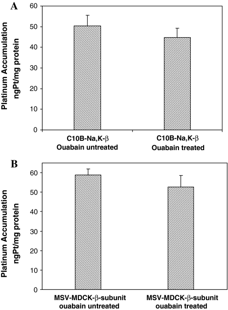
In order to determine whether the Na,K-ATPase enzyme activity determines the changes in altered drug accumulation, both C10B-Na,K-β cells and the MSV-MDCK-β subunit cells were treated with ouabain, a pharmacological inhibitor of Na,K-ATPase. Ouabain at 50 μM concentration that we used in our experiments is known to completely inhibit Na,K-ATPase activity in human and canine cells [19]. The results show that there is no significant difference in platinum accumulation in ouabain treated versus untreated cells (Fig. 4a, b), suggesting that the pump function of Na,K-ATPase might not be involved in the observed increases in drug uptake and sensitization to oxaliplatin in these cells.
Fig. 4.
a Accumulation of platinum in C10B-Na,K-β cells exposed to oxaliplatin (100 μM for 2 h) with and without ouabain (50 μM) treatment. Data represent mean ± SD, n = 3. b Accumulation of platinum in MSV-MDCK-β cells exposed to oxaliplatin (100 μM for 2 h) with and without ouabain (50 μM) treatment. Data represent mean ± SD, n = 3
Discussion
The exogenous expression of Na,K-β1 in the oxaliplatin resistant C10B cells increased the uptake and cell sensitivity to the drug. Increased uptake and sensitization of cells to the drug were independently confirmed in kidney carcinoma cells expressing Na,K-β1. Our data also suggests that Na,K-β1 expression increases sensitivity to oxaliplatin independent of Na,K-ATPase pump function. Thus, these results for the first time reveal that reduced expression of Na,K-β1 protein is associated with oxaliplatin resistance and that increasing its levels enhances oxaliplatin sensitivity of carcinoma cells.
Reduced drug accumulation is one of the commonly described mechanisms for platinum drug resistance in many studies with a strong correlation between platinum accumulation and relative resistance as demonstrated in human hepatoma BEL7404 cells with increasing acquired resistance to cisplatin [20], and primary bladder cancer cell lines with intrinsic resistance to cisplatin [23]. While there is a consensus that drug accumulation is a major determinant of platinum drug sensitivity, the mechanisms involved in uptake are still not well understood. In addition, much of the work to date is on the prototype drug cisplatin and relatively fewer reports exist on the uptake of other platinum analogs including oxaliplatin. In a recent review, Hall et al. [14] discuss the current evidence for how cisplatin enters the cells, its potential extrusion out of cells and cellular changes in resistance including information that is available for the platinum analogs. Most of the evidence suggests that cisplatin uptake and possibly of other analogs involves multiple mechanisms including diffusion, CTR1 copper influx transporter mediated uptake (of cisplatin but not oxaliplatin) [29], and organic cation transporters (SLC family members OCT1 and 2) which may be involved in oxaliplatin uptake [37]. It was suggested that the potential speciation in various media of platinum analogs that imparts the charged versus neutral characteristics to these drugs could in addition dictate which mechanism of transport may play a major role [14]. In the absence of evidence for changes in physicochemical properties of membranes accounting for altered diffusion in resistant cells, the available evidence only points to alterations in transport proteins [14]. A potential role for copper efflux transporters ATP7A/7B and glutathione conjugate pump (GS-X family) in the efflux of cisplatin and resistance to the drug has been described [8, 22].
Na,K-ATPase enzyme function mediated transport has also been suggested as a potential mechanism in cisplatin uptake [4, 14]. While the experiments with ouabain inhibition indicated a potential role for Na,K-ATPase in cisplatin uptake into 2008 human ovarian carcinoma cells and their cisplatin resistant variant A2780/DDP [4], no differences in Na,K-ATPase activity between cisplatin sensitive and resistant cells have been observed [3]. Their studies suggested for a Na+-dependent transporter for cisplatin. In separate experiments primarily aimed at disrupting the sodium gradient they concluded that it is the sodium gradient dissipation, which is also resulting from the inhibition of Na,K-ATPase activity by ouabain that is affecting the cisplatin accumulation [3]. In a panel of human small cell lung cancer (SCLC) and human non-small cell lung cancer (NSCLC) cell lines all expressing Na,K-ATPase activity, ouabain inhibition lowered cisplatin accumulation in NSCLC cell lines, but not in SCLC, suggesting that other factors distinct from the pump activity may have an impact on cisplatin uptake into cells [5]. A more recent report connects Na,K-ATPase activity and not ATP-7A and ATP-7B in cisplatin sensitivity in oral squamous cell carcinoma cells [1]. At the moment it is unclear what the exact role of Na,K-ATPase function is in cisplatin uptake, or a role if there is any for oxaliplatin uptake. In the two cell models transfected with Na,K-β1, we did not observe any change in platinum uptake following ouabain treatment. In our experiments, by increased expression of Na,K-β1 in ovarian carcinoma cells, restoration to sensitivity was achieved partially, but not completely indicating the multi-factorial nature of resistance such as high levels of glutathione in C10B cells that we described earlier [16].
The parental A2780 cells are adherent to each other and show epithelial phenotype. The C10B cells on the other hand were of mesenchymal phenotype indicating that during the selection of oxaliplatin resistance the A2780 cells have undergone EMT. This is further confirmed by the increased levels of fibronectin in C10B cells (present study), positivity for vimentin [16] and up-regulated expression of genes of extracellular matrix, such as matrix metalloproteinases, integrin alpha and TGF-β2 [34], genes described to be either part of EMT and/or part of the extracellular matrix mediated drug resistance [10]. Interestingly, although expression of Na,K-β1 induced an adherent phenotype as revealed by the change in the morphology of C10B-Na,K-β1 cells, there was no change in the levels of fibronectin compared to C10B cells. This result suggests that the reversal of EMT, i.e., mesenchymal to epithelial transition (MET) has not taken place in these cells at least at the molecular level. This is also the case with MSV-MDCK cells expressing Na,K-β1. These cells have not reverted their mesenchymal phenotype but are more adherent to each other, show reduced motility [6, 28] and tumorigenicity [18]. We and others recently showed that Na,K-β1 is a cell–cell adhesion molecule and increases cell–cell contact when expressed in carcinoma cells [7, 31, 33]. Although the mechanism/s by which Na,K-β1 increases sensitivity to oxaliplatin is not known, it is tempting to speculate that the cell–cell adhesion function of Na,K-β1 might be involved in this process. It is possible that cells that are contact naive are less sensitive to drugs compared to cells adherent to each other. Future experiments are necessary to unravel mechanisms by which Na,K-β1 is increasing the drug uptake and sensitizing cells for oxaliplatin. However, the current findings have immense therapeutic potential. First, Na,K-β1 levels could be used as a biomarker for screening patients that would respond better to platinum based chemotherapy. Secondly, it may be possible to target the cancer cells which have become drug-insensitive by increasing the levels of Na,K-β1 in these cells using targeted gene therapy approaches. We suggest that ability to increase the expression of Na,K-β1 should have significant therapeutic value for treating drug-insensitive malignancies.
Acknowledgments
This work was supported in part by NCICA109619 (LP) and NIHDK56216 (AR). We acknowledge the support of Comprehensive Cancer Center grant NCICA10656 for the Pharmacokinetics/Pharmacodynamics Core Facility usage.
Open Access This article is distributed under the terms of the Creative Commons Attribution Noncommercial License which permits any noncommercial use, distribution, and reproduction in any medium, provided the original author(s) and source are credited.
References
- 1.Ahmed Z, Deyama Y, Yoshimura Y, Suzuki K (2008) Cisplatin sensitivity of oral squamous carcinoma cells is regulated by Na(+), K(+)-ATPase activity rather than copper-transporting P-type ATPases, ATP7A and ATP7B. Cancer Chemother Pharmacol 63(4):643–650 [DOI] [PubMed]
- 2.Andrews PA (1994) Mechanisms of acquired resistance to cisplatin. Cancer Treat Res 73:217–248 [DOI] [PubMed]
- 3.Andrews PA, Mann SC, Huynh HH, Albright KD (1991) Role of the Na+,K(+)-adenosine triphosphatase in the accumulation of cis-diamminedichloroplatinum(II) in human ovarian carcinoma cells. Cancer Res 51:3677–3681 [PubMed]
- 4.Andrews PA, Velury S, Mann SC, Howell SB (1988) cis-Diamminedichloroplatinum(II) accumulation in sensitive and resistant human ovarian carcinoma cells. Cancer Res 48:68–73 [PubMed]
- 5.Bando T, Fujimura M, Kasahara K, Matsuda T (1998) Significance of Na+,K(+)-ATPase on intracellular accumulation of cis-diamminedichloroplatinum(II) in human non-small-cell but not in small-cell lung cancer cell lines. Anticancer Res 18:1085–1089 [PubMed]
- 6.Barwe SP, Anilkumar G, Moon SY, Zheng Y, Whitelegge JP, Rajasekaran SA, Rajasekaran AK (2005) Novel role for Na,K-ATPase in phosphatidylinositol 3-kinase signaling and suppression of cell motility. Mol Biol Cell 16:1082–1094 [DOI] [PMC free article] [PubMed]
- 7.Barwe SP, Kim S, Rajasekaran SA, Bowie JU, Rajasekaran AK (2007) Janus model of the Na,K-ATPase beta-subunit transmembrane domain: distinct faces mediate alpha/beta assembly and beta-beta homo-oligomerization. J Mol Biol 365:706–714 [DOI] [PMC free article] [PubMed]
- 8.Borst P, Kool M, Evers R (1997) Do cMOAT (MRP2), other MRP homologues and LRP play a role in MDR? Semin Cancer Biol 8:205–213 [DOI] [PubMed]
- 9.El-akawi Z, Abu-hadid M, Perez R, Glavy J, Zdanowicz J, Creaven PJ, Pendyala L (1996) Altered glutathione metabolism in oxaliplatin resistant A2780 ovarian carcinoma cells. Cancer Let 105:5–14 [DOI] [PubMed]
- 10.Elliott T, Sethi T (2002) Integrins and extracellular matrix. Expert Rev Anticancer Ther 2:449–459 [DOI] [PubMed]
- 11.Espineda CE, Chang JH, Twiss J, Rajasekaran SA, Rajasekaran AK (2004) Repression of Na,K-ATPase beta1-subunit by the transcription factor snail in carcinoma. Mol Biol Cell 15:1364–1373 [DOI] [PMC free article] [PubMed]
- 12.Gosland M, Lum B, Schimmelpfennig J, Baker J, Doukas M (1996) Insights into mechanisms of cisplatin resistance and potential for its clinical reversal. Pharmacotherapy 16:16–39 [PubMed]
- 13.Guarino M, Rubino B, Ballabio G (2007) The role of epithelial-mesenchymal transition in cancer pathology. Pathology (Phila) 39:305–318 [DOI] [PubMed]
- 14.Hall MD, Okabe M, Shen DW, Liang XJ, Gottesman MM (2008) The role of cellular accumulation in determining sensitivity to platinum-based chemotherapy. Annu Rev Pharmacol Toxicol 48:495–535 [DOI] [PubMed]
- 15.Hector S, Bolanowska-Higdon W, Zdanowicz J, Hitt S, Pendyala L (2001) In vitro studies on the mechanisms of oxaliplatin resistance. Cancer Chemother Pharmacol 48:398–406 [DOI] [PubMed]
- 16.Hector S, Nava ME, Clark K, Murpy M, Pendyala L (2007) Characterization of a clonal isolate of an oxaliplatin resistant ovarian carcinoma cell line A2780/C10. Cancer Lett 245:195–204 [DOI] [PubMed]
- 17.Huber MA, Kraut N, Beug H (2005) Molecular requirements for epithelial-mesenchymal transition during tumor progression. Curr Opin Cell Biol 17:548–558 [DOI] [PubMed]
- 18.Inge LJ, Rajasekaran SA, Yoshimoto K, Mischel PS, McBride W, Landaw E, Rajasekaran AK (2008) Evidence for a potential tumor suppressor role for the Na,K-ATPase beta1-subunit. Histol Histopathol 23:459–467 [DOI] [PMC free article] [PubMed]
- 19.Jewell EA, Lingrel JB (1991) Comparison of the substrate dependence properties of the rat Na,K-ATPase alpha 1, alpha 2, and alpha 3 isoforms expressed in HeLa cells. J Biol Chem 266:16925–16930 [PubMed]
- 20.Johnson SW, Shen D, Pastan I, Gottesman MM, Hamilton TC (1996) Cross-resistance, cisplatin accumulation, and platinum-DNA adduct formation and removal in cisplatin-sensitive and -resistant human hepatoma cell lines. Exp Cell Res 226:133–139 [DOI] [PubMed]
- 21.Kajiyama H, Shibata K, Terauchi M, Yamashita M, Ino K, Nawa A, Kikkawa F (2007) Chemoresistance to paclitaxel induces epithelial-mesenchymal transition and enhances metastatic potential for epithelial ovarian carcinoma cells. Int J Oncol 31:277–283 [PubMed]
- 22.Katano K, Kondo A, Safaei R, Holzer A, Samimi G, Mishima M, Kuo YM, Rochdi M, Howell SB (2002) Acquisition of resistance to cisplatin is accompanied by changes in the cellular pharmacology of copper. Cancer Res 62:6559–6565 [PubMed]
- 23.Koga H, Kotoh S, Nakashima M, Yokomizo A, Tanaka M, Naito S (2000) Accumulation of intracellular platinum is correlated with intrinsic cisplatin resistance in human bladder cancer cell lines. Int J Oncol 16:1003–1007 [DOI] [PubMed]
- 24.Mathe G, Kidani Y, Segiguchi M, Eriguchi M, Fredj G, Peytavin G, Misset JL, Brienza S, de Vassals F, Chenu E, Bourut C (1991) Oxalato-platinum or l-OHP, a third generation platinum complex: an experimental and clinical appraisal and preliminary comparison with cis-platinum and carboplatinum. Biomed Pharmacother 43:237–250 [DOI] [PubMed]
- 25.Rabik CA, Dolan ME (2007) Molecular mechanisms of resistance and toxicity associated with platinating agents. Cancer Treat Rev 33:9–23 [DOI] [PMC free article] [PubMed]
- 26.Rajasekaran SA, Beyenbach KW, Rajasekaran AK (2008) Interactions of tight junctions with membrane channels and transporters. Biochim Biophys Acta 1778:757–769 [DOI] [PubMed]
- 27.Rajasekaran SA, Gopal J, Willis D, Espineda C, Twiss JL, Rajasekaran AK (2004) Na,K-ATPase beta1-subunit increases the translation efficiency of the alpha1-subunit in MSV-MDCK cells. Mol Biol Cell 15:3224–3232 [DOI] [PMC free article] [PubMed]
- 28.Rajasekaran SA, Palmer LG, Quan K, Harper JF, Ball WJ Jr, Bander NH, Peralta SA, Rajasekaran AK (2001) Na,K-ATPase beta-subunit is required for epithelial polarization, suppression of invasion, and cell motility. Mol Biol Cell 12:279–295 [DOI] [PMC free article] [PubMed]
- 29.Safaei R (2006) Role of copper transporters in the uptake and efflux of platinum containing drugs. Cancer Lett 234:34–39 [DOI] [PubMed]
- 30.Sherman-Baust CA, Weeraratna AT, Rangel LB, Pizer ES, Cho KR, Schwartz DR, Shock T, Morin PJ (2003) Remodeling of the extracellular matrix through overexpression of collagen VI contributes to cisplatin resistance in ovarian cancer cells. Cancer Cell 3:377–386 [DOI] [PubMed]
- 31.Shoshani L, Contreras RG, Roldan ML, Moreno J, Lazaro A, Balda MS, Matter K, Cereijido M (2005) The polarized expression of Na+,K+-ATPase in epithelia depends on the association between beta-subunits located in neighboring cells. Mol Biol Cell 16:1071–1081 [DOI] [PMC free article] [PubMed]
- 32.Tse J, Kalluri R (2007) Mechanisms of metastasis: epithelial to mesenchymal transition and contribution of tumor microenvironment. J Cell Biochem 101:816–829 [DOI] [PubMed]
- 33.Vagin O, Tokhtaeva E, Sachs G (2006) The role of the beta1 subunit of the Na,K-ATPase and its glycosylation in cell-cell adhesion. J Biol Chem 281:39573–39587 [DOI] [PubMed]
- 34.Varma RR, Hector S, Clark K, Greco WR, Hawthorn L, Pendyala L (2005) Gene expression profiling of a clonal isolate of oxaliplatin resistant ovarian carcinoma cell line A2780/C10. Oncol Rep 14:925–932 [PubMed]
- 35.Wiseman LR, Adkins JC, Plosker GL, Goa K (1999) Oxaliplatin. A review of its use in the management of metastatic colorectal cancer. Drugs Aging 14:459–475 [DOI] [PubMed]
- 36.Yang AD, Fan F, Camp ER, Van BG, Liu W, Somcio R, Gray MJ, Cheng H, Hoff PM, Ellis LM (2006) Chronic oxaliplatin resistance induces epithelial-to-mesenchymal transition in colorectal cancer cell lines. Clin Cancer Res 12:4147–4153 [DOI] [PubMed]
- 37.Zhang S, Lovejoy KS, Shima JE, Lagpacan LL, Shu Y, Lapuk A, Chen Y, Komori T, Gray JW, Chen X, Lippard SJ, Giacomini KM (2006) Organic cation transporters are determinants of oxaliplatin cytotoxicity. Cancer Res 66:8847–8857 [DOI] [PMC free article] [PubMed]


