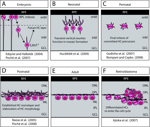
Fig. 2.
Unusual features of HCs. (A) During embryonic development, retinal progenitor cells (RPCs) undergo mitosis near the outer limiting membrane and subsequently migrate to their appropriate retinal layer. HC precursors do not migrate directly to the prospective HC layer, but rather bypass this layer and migrate basally to the ganglion cell layer (GCL) before changing direction to migrate apically towards the HC layer. This second phase of HC migration has been shown to depend on Lim1. (B) Upon the completion of HC migration, the HCs are organized in a non-random spatial arrangement (mosaic) within the outer retina. HC spacing has been shown to be regulated by homotypic repulsive interactions mediated by transient, apically directed neurites (arrowheads). (C) Zebrafish and chick retinae contain committed progenitor cells that divide to produce only HCs, and in the zebrafish do so within the HC stratum. (D,E) Subsequent to cell cycle exit, homotypic interactions restrain dendritic overlap, as these processes stratify and form synaptic contacts with their afferents in the outer plexiform layer (OPL). (F) In a mouse model of retinoblastoma, fully differentiated HCs re-enter the cell cycle and give rise to aggressively metastatic tumors. ONL, outer nuclear layer; INL, inner nuclear layer; IPL, inner plexiform layer; onbl, outer neuroblastic layer; inbl, inner neuroblastic layer; RPE, retinal pigment epithelium.

