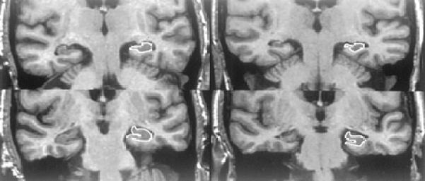
Figure 1. Neuroanatomic Boundaries.

The column of images on the left are cropped oblique coronal MR images through the temporal lobes of a 75-year-old woman. The upper image is through the body of the hippocampus and lower image is through the head of the hippocampus. This MCI patient remained stable over 49 months of clinical followup. At baseline her hippocampal W score was 0.21. On the right are matched imaging sections of a 70-year-old woman, who was initially categorized as MCI, but become demented after 43.5 months of followup. Her hippocampal W score was −2.48 at entry into the study. The hippocampi of the patient who became demented (right) are visibly atrophic relative to the stable patient (left) despite the fact that the crossover patient was 5 years younger. The anatomic outlines of the left hippocampus are indicated.
