
Fig. 7.
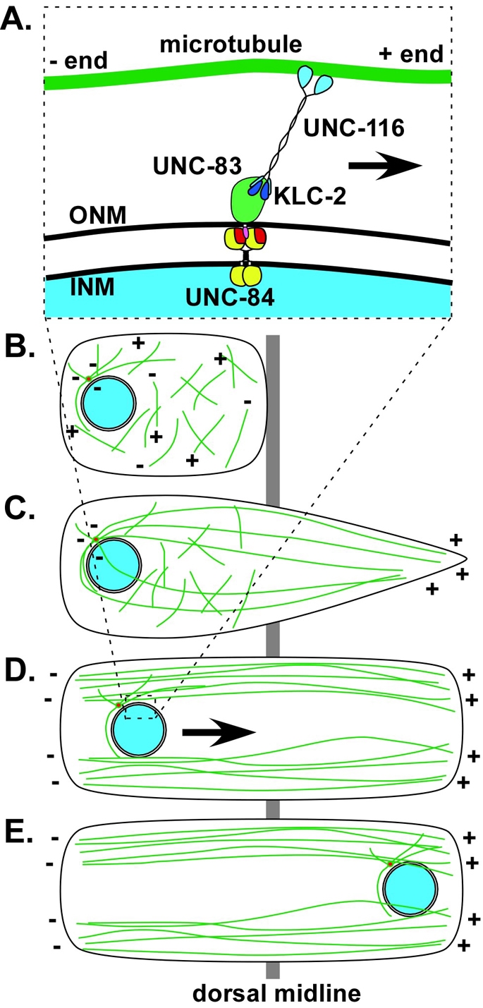
A model of nuclear migration in C. elegans hyp7 cells. (A) The nuclear envelope during nuclear migration. UNC-84 (red and yellow) localizes to the inner nuclear membrane (INM) and UNC-83 (green and pink) localizes to the outer nuclear membrane (ONM). The UNC-84 SUN domain (red) interacts with the UNC-83 KASH domain (pink) in the perinuclear space. The cytoplasmic domain of UNC-83 (green) interacts with KLC-2 (dark blue). The motor activity of UNC-116 (light blue) moves hyp7 nuclei toward the plus ends of polarized microtubules (green). (B) Microtubules (green) resemble a meshwork at the beginning of cell intercalation (plus and minus ends of microtubules marked). (C) As intercalation proceeds across the dorsal midline (gray), microtubules nucleated from the centrosome (red) at the stationary end of the cell (left) extend into the growing tip (right) of the cell. (D) At the completion of intercalation, most microtubules are arranged in polarized, centrosome-independent bundles parallel to the cell boundaries. (E) Nuclei (blue) move along these polarized microtubule bundles (in the direction of the arrow in D).